. 2014 Sep 2;111(35):E3699-707.
doi: 10.1073/pnas.1404851111. Epub 2014 Aug 18.

TIM-family proteins inhibit HIV-1 release

Affiliations

TIM-family proteins inhibit HIV-1 release

Minghua Li et al. Proc Natl Acad Sci U S A. .

Abstract

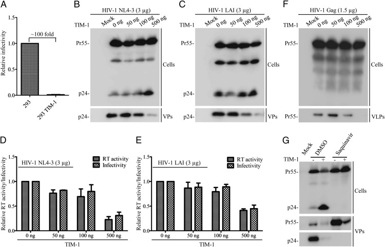
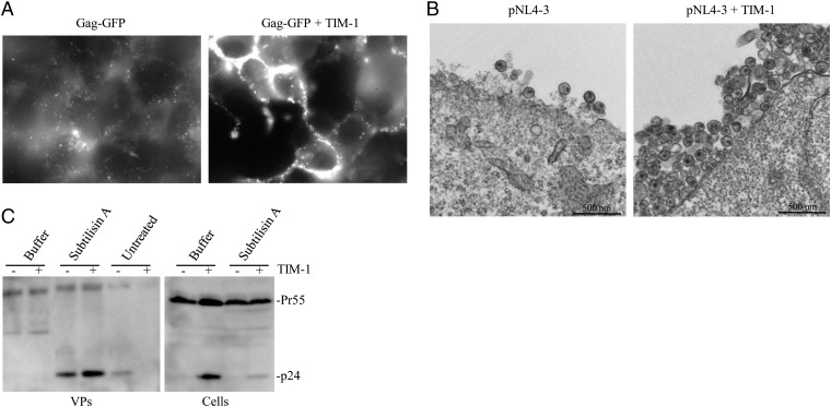
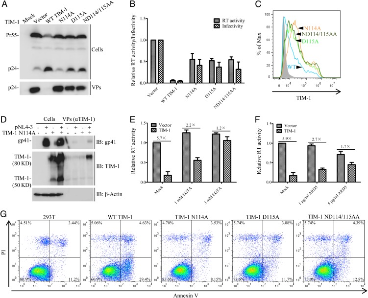
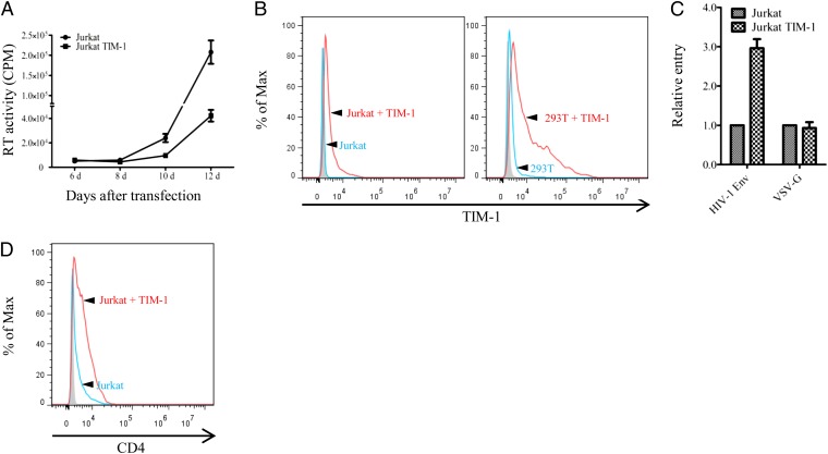
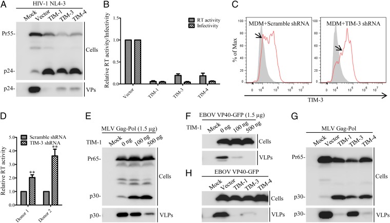
Accumulating evidence indicates that T-cell immunoglobulin (Ig) and mucin domain (TIM) proteins play critical roles in viral infections. Herein, we report that the TIM-family proteins strongly inhibit HIV-1 release, resulting in diminished viral production and replication. Expression of TIM-1 causes HIV-1 Gag and mature viral particles to accumulate on the plasma membrane. Mutation of the phosphatidylserine (PS) binding sites of TIM-1 abolishes its ability to block HIV-1 release. TIM-1, but to a much lesser extent PS-binding deficient mutants, induces PS flipping onto the cell surface; TIM-1 is also found to be incorporated into HIV-1 virions. Importantly, TIM-1 inhibits HIV-1 replication in CD4-positive Jurkat cells, despite its capability of up-regulating CD4 and promoting HIV-1 entry. In addition to TIM-1, TIM-3 and TIM-4 also block the release of HIV-1, as well as that of murine leukemia virus (MLV) and Ebola virus (EBOV); knockdown of TIM-3 in differentiated monocyte-derived macrophages (MDMs) enhances HIV-1 production. The inhibitory effects of TIM-family proteins on virus release are extended to other PS receptors, such as Axl and RAGE. Overall, our study uncovers a novel ability of TIM-family proteins to block the release of HIV-1 and other viruses by interaction with virion- and cell-associated PS. Our work provides new insights into a virus-cell interaction that is mediated by TIMs and PS receptors.


Conflict of interest statement

The authors declare no conflict of interest.

Figures

Fig. 1.
Fig. 1.
TIM-1 inhibits HIV-1 production and Gag release. (A) HEK293 or 293 cells stably expressing TIM-1 were transfected with pNL4-3 DNA, and viral infectivity was measured in HeLa-TZM cells. (BE) HEK293T cells were transfected with NL4-3 or LAI HIV-1 proviral clones plus indicated amounts of plasmids encoding TIM-1. Western blotting was performed to examine cell-associated Gag (Cells) and cell-free viral particles (VPs) or VLPs by using an anti-p24 antibody (B and C). “Mock” indicates untransfected 293T cells. The infectivity of cell-free virus was measured in HeLa-TZM indicator cells (D and E). The data shown in D and E represent three independent experiments. (F) HEK293T cells were transfected with plasmids expressing codon-optimized HIV-1 Gag and increasing amounts of TIM-1. Western blotting was performed to determine cell-associated Gag and VLPs. (G) HEK293T cells were transfected with HIV-1 NL4-3, and cells were treated either with DMSO or 5 µM Saquinavir. Western blotting was performed using an anti-p24 antibody. Positions of the Gag precursor Pr55Gag (Pr55) and the mature CA protein (p24) are indicated.
Fig. 2.
Fig. 2.
TIM-1 retains HIV-1 particles on the cell surface. (A) HEK293T cells were cotransfected with plasmids encoding HIV-1 Gag-GFP, with or without TIM-1 expression vector; cells were reseeded onto bottom-top dishes, and live cell images were acquired using a fluorescent microscope (Olympus, 100×). Note that numerous VLPs diffusely are present in “GFP-Gag” control cells compared with cells expressing “GFP-Gag + TIM-1,” where Gag-GFP accumulated at cell–cell contacts. (B) HEK293T cells were transfected pNL4-3 or pNL4-3 plus TIM-1 expression vector. Transfected cells were harvested at 24 h posttransfection, fixed in glutaraldehyde, and subjected to TEM. Representative images of thin-sectioned cells are shown. (C) HEK293T cells were transfected with pNL4-3 DNA with or without TIM-1 expression plasmid; viral particles released into supernatants were harvested 24 h after transfection (“untreated”). The cells were then treated with stripping buffer alone (“buffer”) or buffer containing 1 mg/mL subtilisin A for 15 min at 37 °C, followed by adding PMSF to stop the reaction. Cells were washed with PBS, lysed, and subjected to Western blotting (“Cell”). The stripped supernatants (“VPs”) were concentrated and analyzed by Western blotting. Positions of the Gag precursor Pr55Gag (Pr55) and the mature CA protein (p24) are indicated.
Fig. 3.
Fig. 3.
Mutation of the PS-binding sites of TIM-1 diminishes its ability to block HIV-1 release. (A) HEK293T cells were transfected with HIV-1 NL4-3 proviral DNA, along with plasmids encoding WT TIM-1 or its PS-binding mutants. Western blotting was performed to determine HIV-1 Gag expression in transfected 293T cells and purified viral particles. Positions of the Gag precursor Pr55Gag (Pr55) and the mature CA protein (p24) are indicated. (B) The RT activity and infectivity of HIV-1 harvested in A was determined by infection of HeLa-TZM cells. (C) Flow cytometric analysis of TIM-1 expression on the surface of transfected 293T cells using an anti–hTIM-1 antibody. (D) Incorporation of TIM-1 into HIV-1 virions. HEK293T cells were transfected with pNL4-3 proviral DNA in the presence or absence of the TIM-1 N114A plasmid. Released virions were purified and coimmunoprecipitated with an anti–TIM-1 antibody at 4 °C overnight. The bound virions, along with cell lysates were resolved by SDS/PAGE, followed by Western blotting using anti–HIV-1 gp41 or anti–TIM-1 antibodies. (E) Effect of EGTA on the TIM-1–mediated inhibition of HIV-1 release. The fold inhibitions between mock (“untreated”) and EGTA-treated cells were indicated. We set cells not expressing TIM-1 and untreated with EGTA to 1.0 for easy comparison. (F) Effect of anti–TIM-1 antibody, ARD5 (against IgV domain) on HIV-1 release. The fold differences in HIV-1 RT activity between mock (“untreated”) and antibody-treated cells are indicated. We set cells not expressing TIM-1 and untreated with anti–TIM-1 antibody to 1.0 for easy comparison. (G) HEK293T cells were transfected with plasmids encoding wild-type TIM-1 or its PS-binding mutants, and their ability to induce PS flipping to the outer leaflets of the plasma membrane was assessed by flow cytometry using Annexin V and Propidium iodide (PI) binding kit (Roche).
Fig. 4.
Fig. 4.
Effect of TIM-1 on HIV-1 replication and entry. (A) Jurkat or Jurkat cells expressing TIM-1 were transfected with pNL4-3 provial DNA, and viral replication kinetics were determined by measuring RT activities. (B) Comparison of TIM-1 expression in Jurkat/TIM-1 and 293T/TIM-1 cells by flow cytometry. (C) Expression of TIM-1 increases HIV-1 Env (NL4-3)–mediated but not VSV-G–mediated entry into Jurkat cells. Jurkat or Jurkat/TIM-1 cells were transduced by lentiviral vector (pLenti-GFP-puro) bearing NL4-3 Env or VSV-G and GFP-positive cells were scored by flow cytometry. (D) TIM-1 up-regulates CD4 expression in Jurkat cells. The levels of CD4 expression on the cell surface were determined by flow cytometry using an anti-CD4 antibody.
Fig. 5.
Fig. 5.
Effects of TIM-3 and TIM-4 on release of HIV-1, MLV, and EBOV. (A and B) HEK293T cells were cotransfected with HIV-1 NL4-3 proviral DNA, along with plasmids expressing TIM-1, TIM-3 or TIM-4. Western blotting was performed to monitor cell-associated Gag expression and cell-free virion release by anti–HIV-1 p24 antibody. HIV-1 production was measured by RT activity, and viral infectivity was determined by infecting HeLa-TZM cells. (C) Expression of TIM-3 in MDMs. MDMs were transiently transduced by lentiviral vectors expressing TIM-1 shRNA or scrambled shRNA, and TIM-3 expression was examined by flow cytometry using an anti–TIM-3 antibody. Arrows indicate TIM-3–positive signals. (D) Effect of TIM-3 knockdown on HIV-1 release. MDMs transiently transduced by lentiviral vectors expressing TIM-3 shRNA or scrambled shRNA were infected with NL4-3/KFS pseudovirions bearing VSV-G for 6 h. After 3 washes, cells were maintained for an additional 18 h, and the RT activity of the produced virions determined. Results were averages of three independent experiments from two healthy donors; the RT activity of MDMs transduced by scramble shRNA was set to 1. **P < 0.01. (E and F) HEK293T cells were transfected with plasmids expressing MoMLV Gag-Pol or EBOV VP40-GFP, along with different amounts of plasmids encoding TIM-1. Forty-eight hours posttransfection, Western blotting was performed to examine MoMLV Gag and EBOV VP40-GFP expression in the transfected cells and purified virus-like particles (VLPs). (G and H) Similar experimental procedures were performed as described for E and F except that TIM-3 and TIM-4 were tested. Positions of the HIV-1 Gag precursor Pr55Gag (Pr55), the MLV Gag precursor Pr65Gag (Pr65), the mature HIV-1 CA protein (p24) and mature MLV CA protein (p30) are indicated.

References

    1. Kuchroo VK, Umetsu DT, DeKruyff RH, Freeman GJ. The TIM gene family: Emerging roles in immunity and disease. Nat Rev Immunol. 2003;3(6):454–462. - PubMed
    1. Freeman GJ, Casasnovas JM, Umetsu DT, DeKruyff RH. TIM genes: A family of cell surface phosphatidylserine receptors that regulate innate and adaptive immunity. Immunol Rev. 2010;235(1):172–189. - PMC - PubMed
    1. Chae SC, et al. Molecular variations in the promoter and coding regions of human Tim-1 gene and their association in Koreans with asthma. Hum Immunol. 2003;64(12):1177–1182. - PubMed
    1. Umetsu DT, Umetsu SE, Freeman GJ, DeKruyff RH. TIM gene family and their role in atopic diseases. Curr Top Microbiol Immunol. 2008;321:201–215. - PubMed
    1. McIntire JJ, et al. Immunology: Hepatitis A virus link to atopic disease. Nature. 2003;425(6958):576. - PubMed

Publication types

MeSH terms