. 2014 Sep 2;111(35):12667-72.
doi: 10.1073/pnas.1406015111. Epub 2014 Aug 18.

Gyrification from constrained cortical expansion

Affiliations

Gyrification from constrained cortical expansion

Tuomas Tallinen et al. Proc Natl Acad Sci U S A. .

Abstract

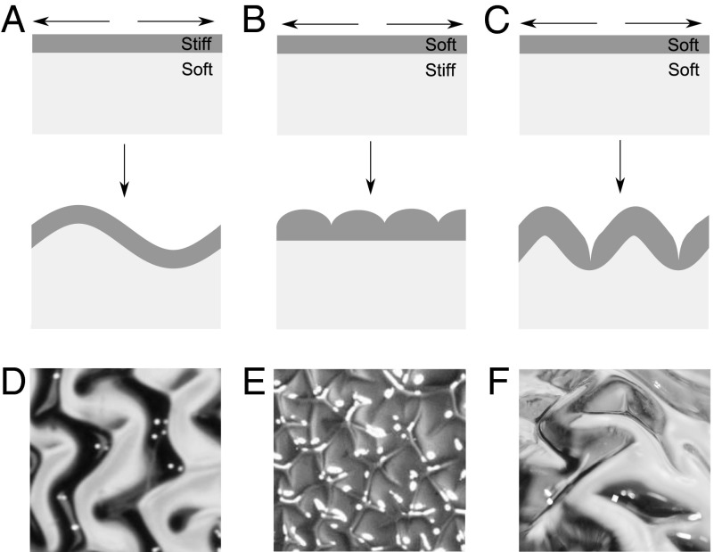
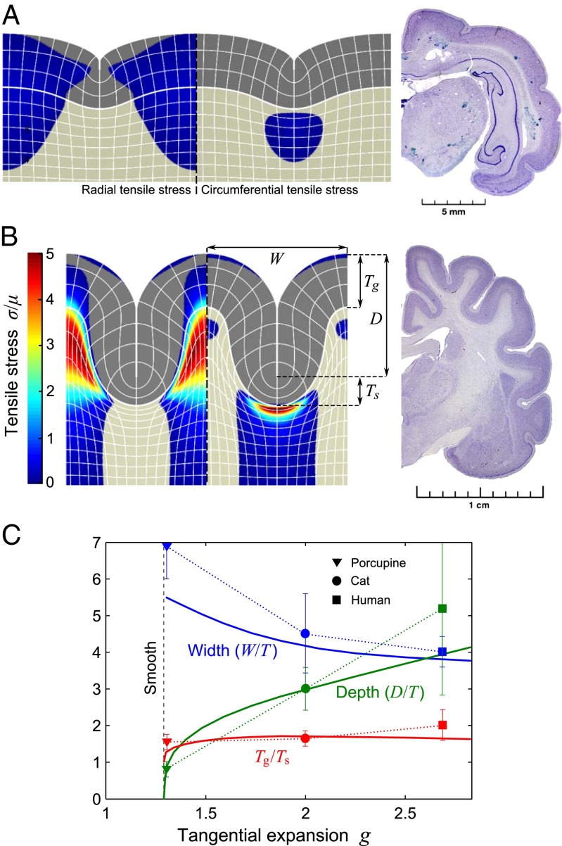
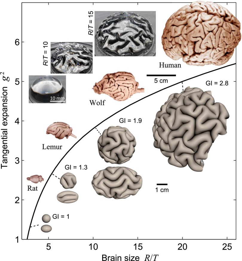
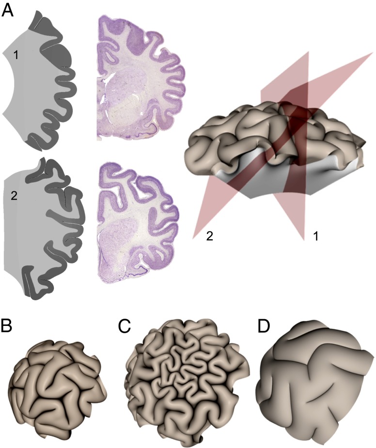
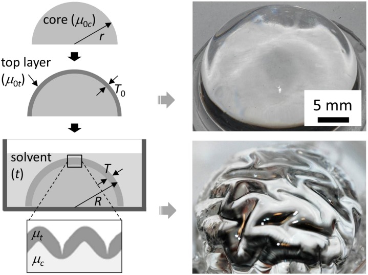
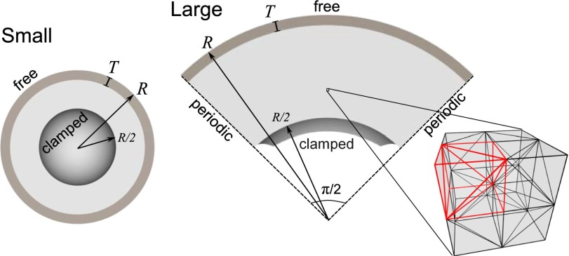
The exterior of the mammalian brain--the cerebral cortex--has a conserved layered structure whose thickness varies little across species. However, selection pressures over evolutionary time scales have led to cortices that have a large surface area to volume ratio in some organisms, with the result that the brain is strongly convoluted into sulci and gyri. Here we show that the gyrification can arise as a nonlinear consequence of a simple mechanical instability driven by tangential expansion of the gray matter constrained by the white matter. A physical mimic of the process using a layered swelling gel captures the essence of the mechanism, and numerical simulations of the brain treated as a soft solid lead to the formation of cusped sulci and smooth gyri similar to those in the brain. The resulting gyrification patterns are a function of relative cortical expansion and relative thickness (compared with brain size), and are consistent with observations of a wide range of brains, ranging from smooth to highly convoluted. Furthermore, this dependence on two simple geometric parameters that characterize the brain also allows us to qualitatively explain how variations in these parameters lead to anatomical anomalies in such situations as polymicrogyria, pachygyria, and lissencephalia.

Keywords: brain morphogenesis; elastic instability.


Conflict of interest statement

The authors declare no conflict of interest.

Figures

Fig. 1.
Fig. 1.
Wrinkling and sulcification in a layered material subject to differential growth. (A) If the growing gray matter is much stiffer than the white matter it will wrinkle in a smooth sinusoidal way. (B) If the gray matter is much softer than the white matter its surface will invaginate to form cusped folds. (C) If the two layers have similar moduli the gray matter will both wrinkle and cusp giving gyri and sulci. Physical realizations of A, B, and C, based on differential swelling of a bilayer gel (Materials and Methods), confirm this picture and are shown in D, E, and F, respectively.
Fig. 2.
Fig. 2.
Formation of a minimal sulcus. The 2D sulci with tangential expansion ratio of (A) g = 1.30 and (B) g = 2.25 of the gray matter (Eq. 2 and Fig. S1). Coloring shows radial and circumferential tensile stress in the left and right sulci, respectively. The stress is compressive in the noncolored areas. Grid lines correspond to every 20 rows or columns of the numerical discretization with nodes. The width W, depth D, and thickness of the gray matter in the sulcus (Ts) and gyrus (Tg) are indicated in B. For comparison with observations of brains, we also show sections of porcupine and cat brains, taken from www.brainmuseum.org. (C) Scaled dimensions of the simulated sulcus (solid lines) as a function of g compared with those in porcupine (triangles), cat (dots), and human (squares) show that our model can capture the basic observed geometry. Width and depth are given relative to the undeformed thickness T of the gray matter (for details of the measurements and error bars, see Fig. S2).
Fig. 3.
Fig. 3.
Known empirical scaling laws for gray-matter volume and thickness are mapped on a g2 vs. R/T diagram. Corresponding simulations for spherical brain configurations, with images shown at a few points, show that the surface remains smooth for the smallest brains, but becomes increasingly folded as the brain size increases. We also show patterns for ellipsoidal configurations (major axis = 1.5 × minor axes) that lead to anisotropic gyrification. Images of rat, lemur, wolf, and human brains illustrate the increasingly prominent folding with increasing size in real brains. Also shown are images of our physical mimic of the brain using a swelling bilayer gel of PDMS immersed in hexanes. The smooth initial state gives rise to gyrified states for different relative sizes of the brain R/T = 10, 15 (see also Fig. 5). All of the brain images are from www.brainmuseum.org.
Fig. 4.
Fig. 4.
(A) Sections of a simulated brain (section planes indicated at right) are compared with coronal sections of a raccoon brain (from www.brainmuseum.org). Cuts through the center of the brain (Upper) and the off the center (Lower) show that we can capture the hierarchical folds but emphasize how misleading sections can be in characterizing the sulcal architecture. (B) Confining our simulations with a uniform pressure of 0.7μ to mimic the meninges and skull leads to a familiar flattened sulcal morphology. (C) Changing the gray matter thickness in a small patch of the growing cortex leads to morphologies similar to polymicrogyria in our simulations. Here g2 = 5 and R/T = 20 except in the densely folded region where R/T = 40. (D) A simulated brain of same physical size as that in C but with a thickened cortex (R/T = 12) and reduced tangential expansion (g2 = 2) displays wide gyri and shallow sulci reminiscent of pachygyric brains.
Fig. 5.
Fig. 5.
A physical model of brainlike instability. To mimic the growth of the gray matter in the brain, a hemispherical elastomer (radius r, shear modulus μ0c) is coated with a top elastomer layer (thickness T0, shear modulus μ0t) that swells by absorbing solvent over time t. Representative images of a bilayer specimen in the initial (dried) state and swollen state (modulus ratio μt/μc ≈ 1) are shown at right.
Fig. 6.
Fig. 6.
Cross-section views of 3D simulation geometries for small and large brains in their initial undeformed states. The gray-matter thickness T, brain radius R, and boundary conditions are indicated. A detailed image of the regular mesh structure of the large brain domain shows the reflection symmetry between every pair of elementary cubes that share a face.

References

    1. Striedter GF. Principles of Brain Evolution. Sunderland, MA: Sinauer Associates; 2005.
    1. Lui JH, Hansen DV, Kriegstein AR. Development and evolution of the human neocortex. Cell. 2011;146(1):18–36. - PMC - PubMed
    1. Gabi M, et al. Cellular scaling rules for the brains of an extended number of primate species. Brain Behav Evol. 2010;76(1):32–44. - PMC - PubMed
    1. Neal J, et al. Insights into the gyrification of developing ferret brain by magnetic resonance imaging. J Anat. 2007;210(1):66–77. - PMC - PubMed
    1. Lancaster MA, et al. Cerebral organoids model human brain development and microcephaly. Nature. 2013;501(7467):373–379. - PMC - PubMed

Publication types