. 2014 Sep 2;111(35):E3641-9.
doi: 10.1073/pnas.1409781111. Epub 2014 Aug 18.

IL-6/STAT3 promotes regeneration of airway ciliated cells from basal stem cells

Affiliations

IL-6/STAT3 promotes regeneration of airway ciliated cells from basal stem cells

Tomomi Tadokoro et al. Proc Natl Acad Sci U S A. .

Abstract

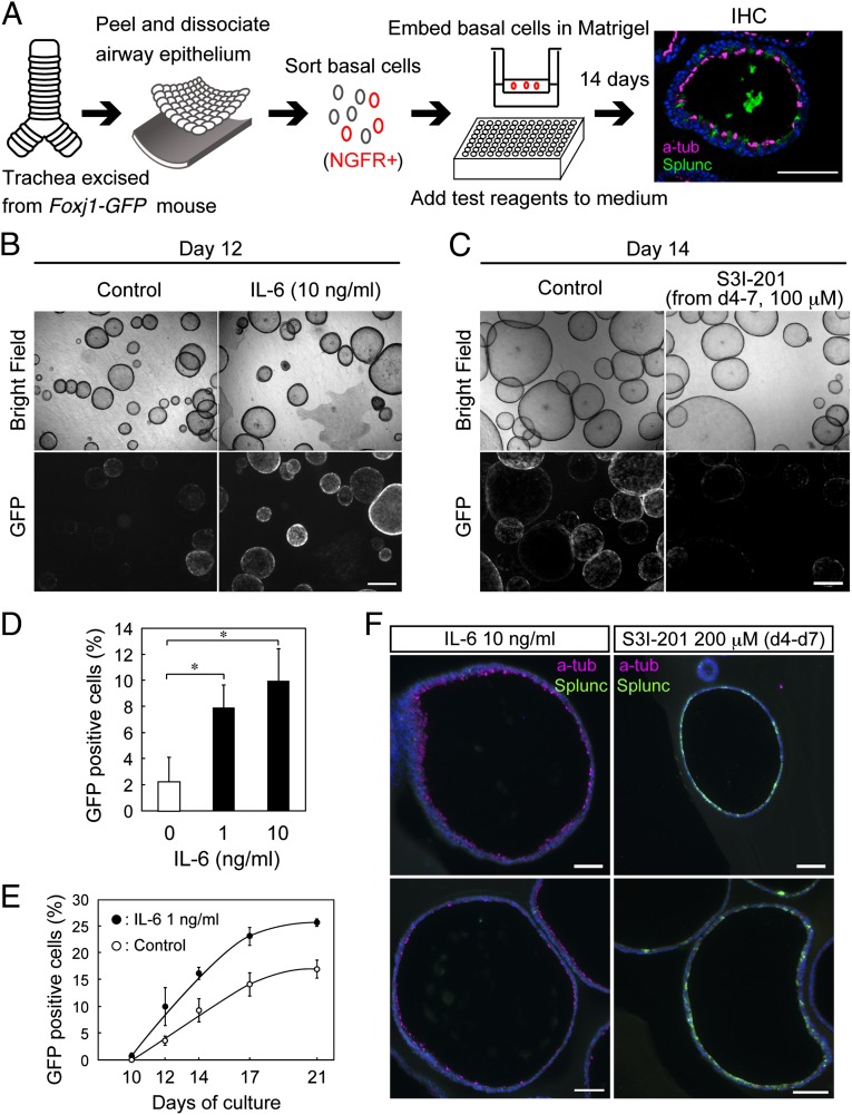
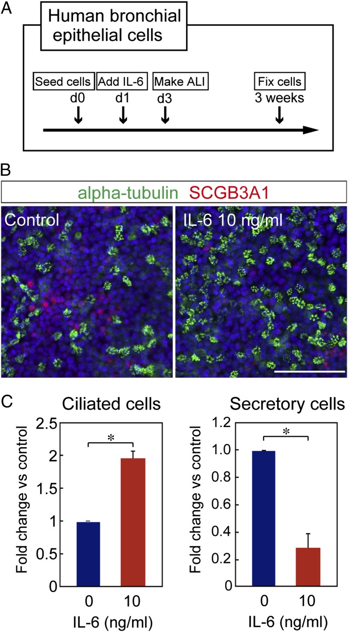
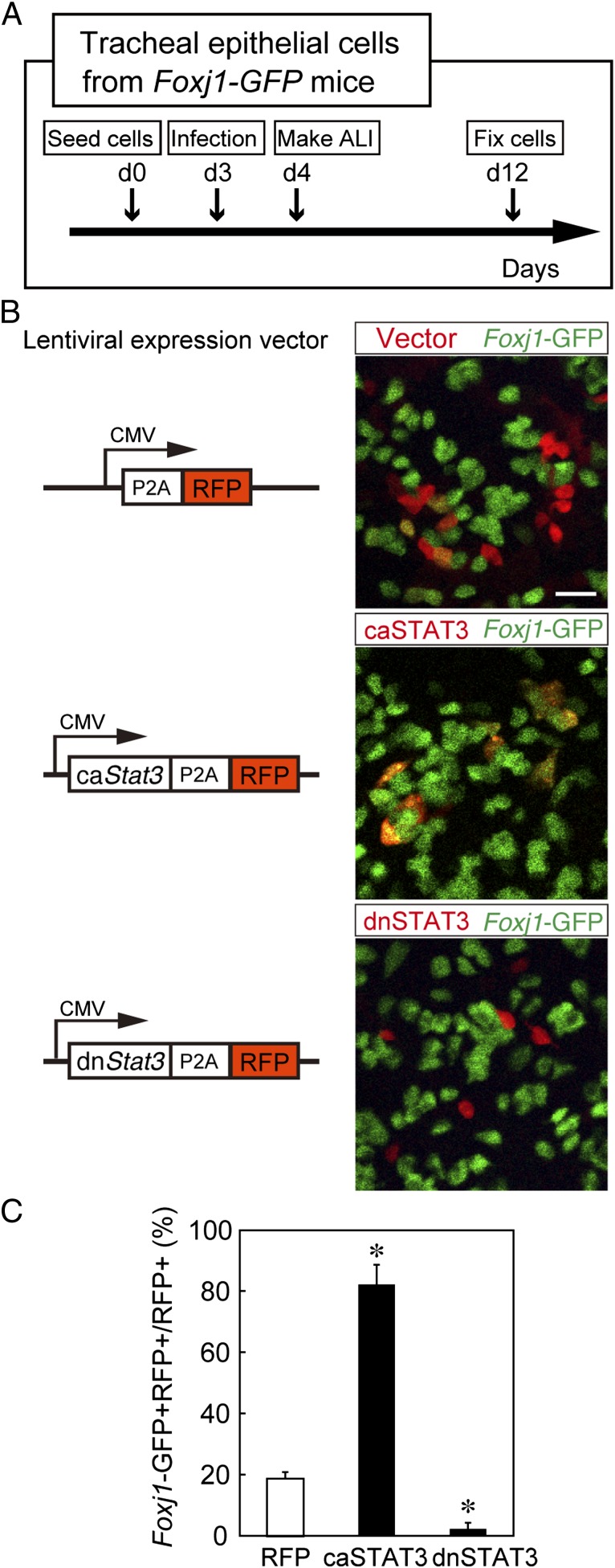
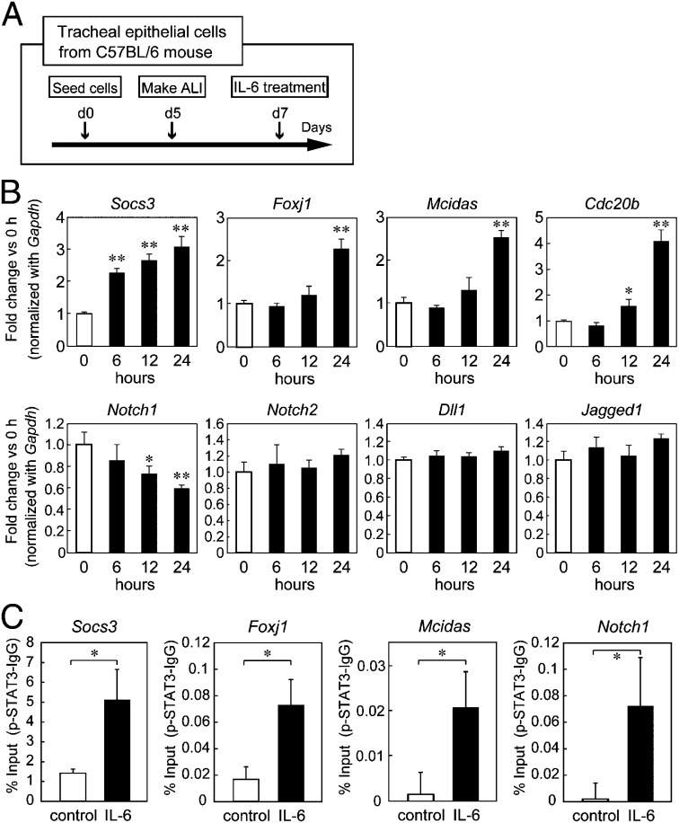
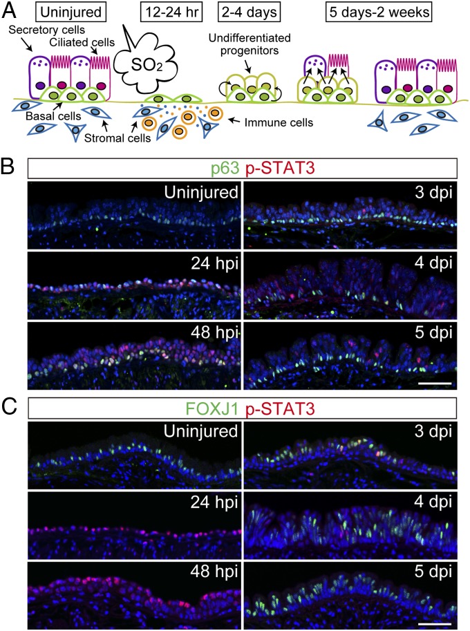
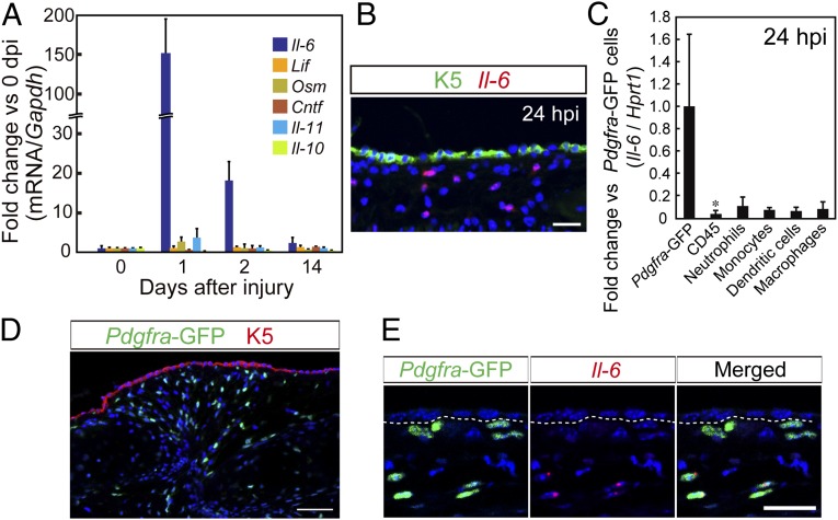
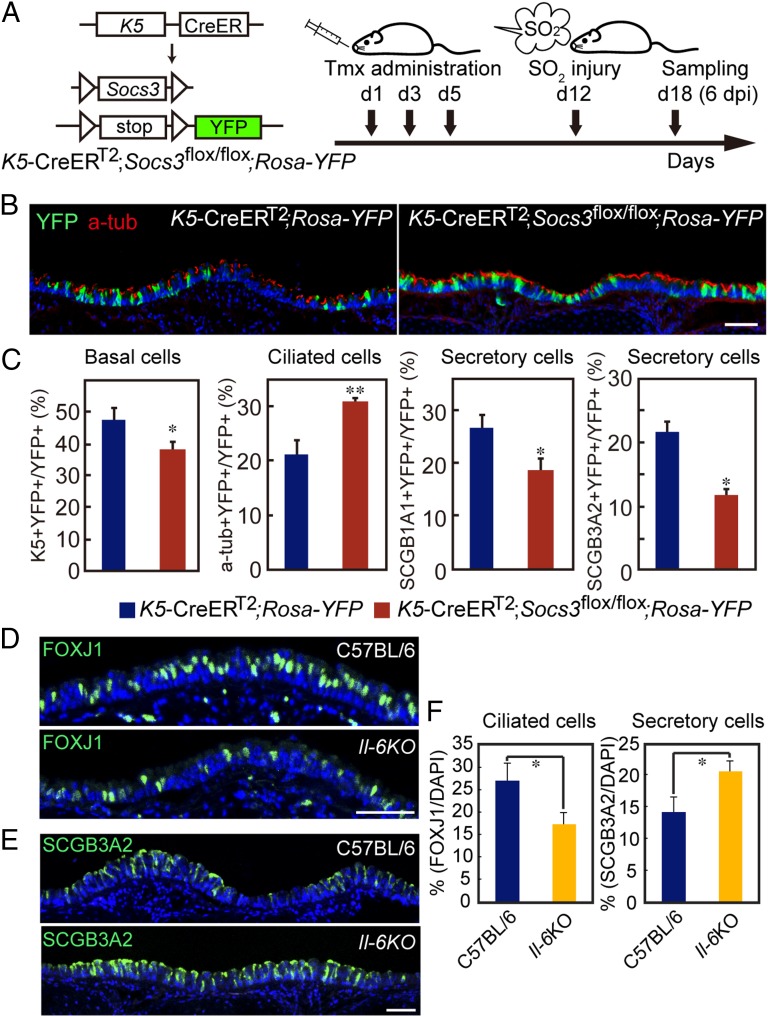
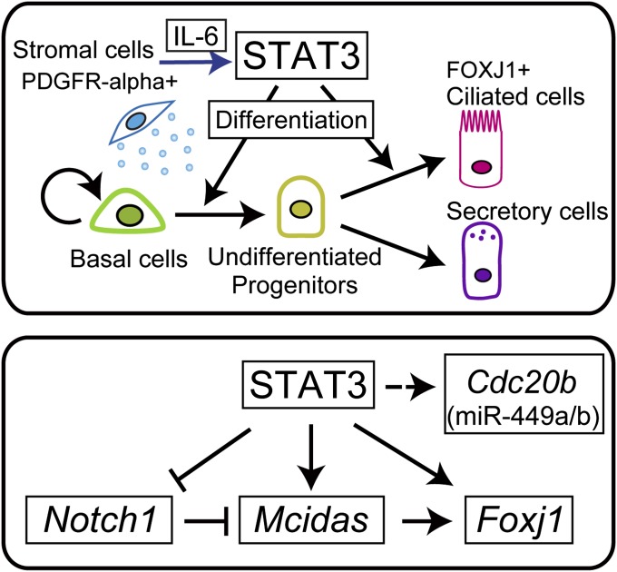
The pseudostratified airway epithelium of the lung contains a balanced proportion of multiciliated and secretory luminal cells that are maintained and regenerated by a population of basal stem cells. However, little is known about how these processes are modulated in vivo, and about the potential role of cytokine signaling between stem and progenitor cells and their niche. Using a clonal 3D organoid assay, we found that IL-6 stimulated, and Stat3 inhibitors reduced, the generation of ciliated vs. secretory cells from basal cells. Gain-of-function and loss-of-function studies with cultured mouse and human basal cells suggest that IL-6/Stat3 signaling promotes ciliogenesis at multiple levels, including increases in multicilin gene and forkhead box protein J1 expression and inhibition of the Notch pathway. To test the role of IL-6 in vivo genetically, we followed the regeneration of mouse tracheal epithelium after ablation of luminal cells by inhaled SO2. Stat3 is activated in basal cells and their daughters early in the repair process, correlating with an increase in Il-6 expression in platelet-derived growth factor receptor alpha(+) mesenchymal cells in the stroma. Conditional deletion in basal cells of suppressor of cytokine signaling 3, encoding a negative regulator of the Stat3 pathway, results in an increase in multiciliated cells at the expense of secretory and basal cells. By contrast, Il-6 null mice regenerate fewer ciliated cells and an increased number of secretory cells after injury. The results support a model in which IL-6, produced in the reparative niche, functions to enhance the differentiation of basal cells, and thereby acts as a "friend" to promote airway repair rather than a "foe."

Keywords: cell fate; epithelial repair; mucociliary epithelium.


Conflict of interest statement

The authors declare no conflict of interest.

Figures

Fig. 1.
Fig. 1.
IL-6 enhances Foxj1-GFP expression in the mouse tracheosphere culture assay. (A) Schematic of the assay. NGFR+ basal cells from Foxj1-GFP tracheas were cultured in 50% Matrigel in 96-well inserts. (Right) Section of a typical sphere with acetylated tubulin+ (a-tub) ciliated (magenta) and Splunc+ secretory cells (green). IHC, immunohistochemistry. The effect of IL-6 (B) and STAT3 inhibitor (C) on Foxj1-GFP expression is shown. Differential interference contrast images (Upper) and fluorescent images (Lower) of the same spheres are shown. (D) Quantification by FACS at day 11 of the percentage of GFP+ cells in dissociated spheres treated with IL-6 (0, 1, and 10 ng/mL). (E) Quantification at different times of GFP+ cells in spheres cultured with or without IL-6 (1 ng/mL). (F) Representative sections of spheres at day 14 treated with IL-6 (Left, 10 ng/mL) or S3I-201 (Right, 200 μM, days 4–7). Both sections were stained with antibodies to a-tub+ (magenta) and Splunc+ (green). *P < 0.02 against control (n = 3). Error bars indicate SD (n = 3). (Scale bars: AC, 500 μm; F, 100 μm.) (Also see Fig. S1.)
Fig. 2.
Fig. 2.
Effect of IL-6 on regeneration of human epithelium in ALI culture. (A) Schematic of ALI culture of primary HBE cells. (B) Whole-mount staining of day 21 cultures for ciliated (α-tubulin, green) and secretory (SCGB3A1, red) cells. Nuclei are blue (DAPI). (Scale bar: 100 μm.) (C) Quantification of whole-mount staining, shown as a fold change over untreated culture. The α-tubulin+ or SCGB3A1+ cells were counted in four randomly chosen areas (0.18 mm2) per filter. Values are mean ± SD for cultures from three different donors. *P < 0.001 against control (n = 3). Error bars indicate SD (n = 3). (Also see Fig. S2.)
Fig. 3.
Fig. 3.
STAT3 pathway regulates ciliogenesis in mouse epithelium in ALI culture. (A) Schematic of ALI culture of mouse tracheal epithelial cells. Subconfluent cultures are infected with lentivirus at day 3 when cells are undifferentiated. (B) Virus-infected cells are RFP+ (red), and Foxj1-expressing cells are GFP+ (green). The caSTAT3 promotes ciliogenesis (Middle), but the dnSTAT3 inhibits ciliogenesis (Bottom) compared with control (Top). (Scale bar: 20 μm.) (C) Quantification of results in B. *P < 0.001 against control (n = 3). Error bars indicate SD (n = 3).
Fig. 4.
Fig. 4.
IL-6 enhances expression of cilia-related genes and inhibits Notch1 expression in mouse ALI culture. (A) Schematic of ALI culture of mouse tracheal epithelial cells. At day 7, IL-6 (10 ng/mL) was added to culture medium in the lower chamber. Cells were harvested after 6, 12, and 24 h, and total RNA was extracted. (B) Quantitative RT-PCR shows that IL-6 treatment promotes the expression of the known target gene Socs3 and ciliogenesis-related genes, such as Multicilin (Mcidas) and Foxj1. IL-6 treatment also inhibits Notch1 and promotes expression of Cdc20b, the host gene for miR-449a/b. No significant changes were observed in the expression of Notch2, Dll1, or Jagged1. (C) ChIP assay shows that p-STAT3 binding to promoter regions of Socs3, Foxj1, Mcidas, and Notch1 is increased after IL-6 stimulation. *P < 0.05 against control; **P < 0.001 against control (n = 3). Error bars indicate SD (n = 3).
Fig. 5.
Fig. 5.
IL-6/STAT3 signaling is activated in tracheal epithelium during repair. (A) Schematic of the SO2 injury model. After exposure to SO2, luminal cells die. Basal cells spread, proliferate, and generate early progenitors. These progenitors differentiate into ciliated and secretory cells, and repair is complete in 2 wk. (B) Longitudinal midline sections stained with antibodies to p-STAT3 (red) and p63 (green), a marker for basal cells. (C) Expression of p-STAT3 (red) and FOXJ1 (green) during epithelial repair. Note the coexpression of p-STAT3 and FOXJ1 at 3 dpi. (Scale bars: B and C, 50 μm.) (Also see Fig. S3.)
Fig. 6.
Fig. 6.
IL-6 is up-regulated in PDGFRα+ stromal cells after SO2 injury. (A) RNAs were extracted from whole trachea at 0, 1, 2, and 14 d after injury and subjected to quantitative RT-PCR analysis. The mRNA expression level of cytokines was normalized to Gapdh. (B) In situ hybridization combined with immunohistochemistry shows that Il-6 mRNA (red) is expressed in cells in the stroma beneath basal cells (K5+, green) after SO2 injury. (C) Quantitative PCR analysis of Il-6 expression in sorted stromal cells [Pdgfrα (Pdgfra)-GFP+] and immune cell subpopulations from the trachea at 24 hpi. (D) Immunohistochemistry of a trachea section at 24 hpi shows Pdgfra-GFP+ cells (GFP+, green) in the stroma beneath the epithelium with basal cells (K5+, red). (E) In situ hybridization and immunohistochemistry show that Pdgfra-GFP+ cells (GFP+, green) express Il-6 mRNA (red) at 24 hpi. (Scale bars: B and E, 20 μm; D, 50 μm.) *P < 0.05 against control (n = 3). Error bars indicate SD (n = 3).
Fig. 7.
Fig. 7.
Effect of IL-6/STAT3 on tracheal epithelial repair in vivo. (A) Schematic of gain-of-function (K5-CreERT2; Socs3flox/flox; Rosa-YFP) model. Floxed alleles are deleted, and the YFP reporter is activated in basal cells with three doses of Tmx. One week later, mice are exposed to SO2 and tracheas are harvested at 6 dpi. (B) Representative midline sections of tracheas (ventral) stained with YFP (lineage label, green) and a-tub (ciliated cells, red) in control (K5-CreERT2; Rosa-YFP) and gain-of-function (K5-CreERT2; Socs3flox/flox; Rosa-YFP) mice. A similar analysis was carried out using antibodies to K5 for basal cells and SCGB1A1 and SCGB3A2 for secretory cells, respectively. (C) Percentage of total lineage-labeled cells (YFP+) throughout the trachea that are ciliated, secretory, or basal cells. Blue and red bars show K5-CreERT2; Rosa-YFP and K5-CreERT2; Socs3flox/flox; Rosa-YFP, respectively. (D) FOXJ1 staining (green) of airway epithelium at 4 dpi in WT and Il-6 null mice. (E) SCGB3A2 staining (green) of airway epithelium at 4 dpi in WT and Il-6 null mice. (F) In Il-6 null mice, there is a reduction of ciliated cells (FOXJ1+) and an increase of secretory cells (SCGB3A2+) after SO2 injury (4 dpi). *P < 0.05 against control; **P < 0.001 against control (n = 3). Error bars indicate SD (n = 3). (Scale bars: 50 μm.) (Also see Fig. S4.)
Fig. 8.
Fig. 8.
Model for regulation of ciliogenesis in airway epithelium by STAT3. (Upper) After injury, STAT3 in both basal cells and progenitors is activated by IL-6 secreted from PDGFRα+ stromal cells. Ciliogenesis is likely promoted both at the level of cell fate determination and at the level of differentiation/maturation of the progenitors of multiciliated cells. (Lower) Schematic model for how STAT3 may directly regulate ciliogenesis-related genes during repair of the tracheal epithelium.

Similar articles

Cited by

References

    1. Borthwick DW, Shahbazian M, Krantz QT, Dorin JR, Randell SH. Evidence for stem-cell niches in the tracheal epithelium. Am J Respir Cell Mol Biol. 2001;24(6):662–670. - PubMed
    1. Rawlins EL, Ostrowski LE, Randell SH, Hogan BLM. Lung development and repair: Contribution of the ciliated lineage. Proc Natl Acad Sci USA. 2007;104(2):410–417. - PMC - PubMed
    1. Rock JR, et al. Notch-dependent differentiation of adult airway basal stem cells. Cell Stem Cell. 2011;8(6):639–648. - PMC - PubMed
    1. Rock JR, et al. Basal cells as stem cells of the mouse trachea and human airway epithelium. Proc Natl Acad Sci USA. 2009;106(31):12771–12775. - PMC - PubMed
    1. Lai H, Rogers DF. New pharmacotherapy for airway mucus hypersecretion in asthma and COPD: Targeting intracellular signaling pathways. J Aerosol Med Pulm Drug Deliv. 2010;23(4):219–231. - PubMed

Publication types

MeSH terms