. 2014 Sep 2;111(35):E3679-88.
doi: 10.1073/pnas.1413726111. Epub 2014 Aug 18.

A sharp T-cell antigen receptor signaling threshold for T-cell proliferation

Affiliations

A sharp T-cell antigen receptor signaling threshold for T-cell proliferation

Byron B Au-Yeung et al. Proc Natl Acad Sci U S A. .

Abstract

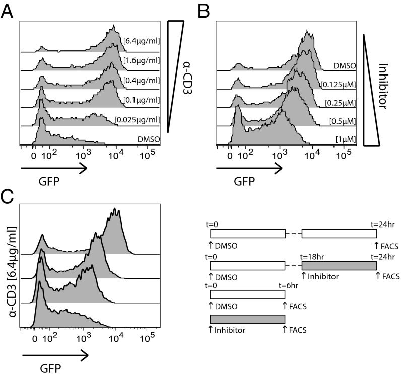
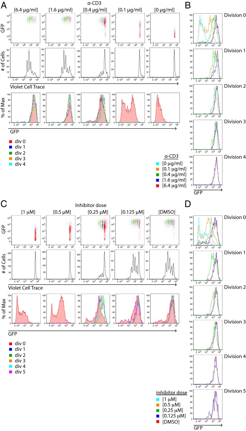
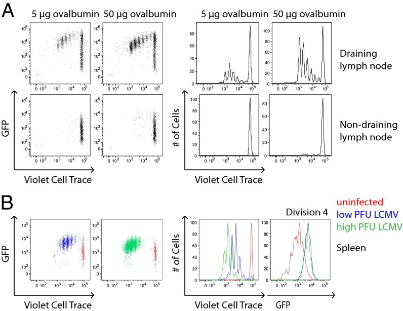
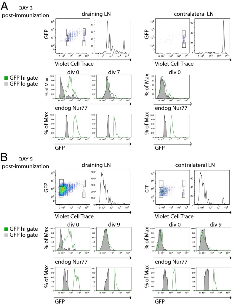
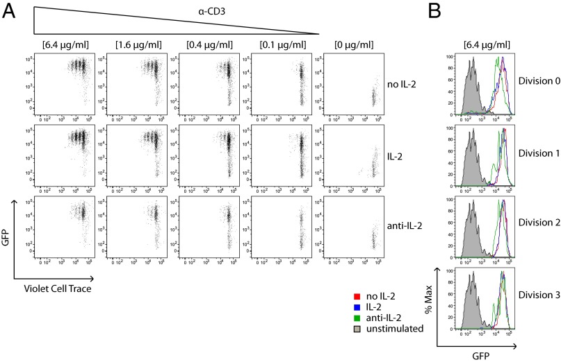
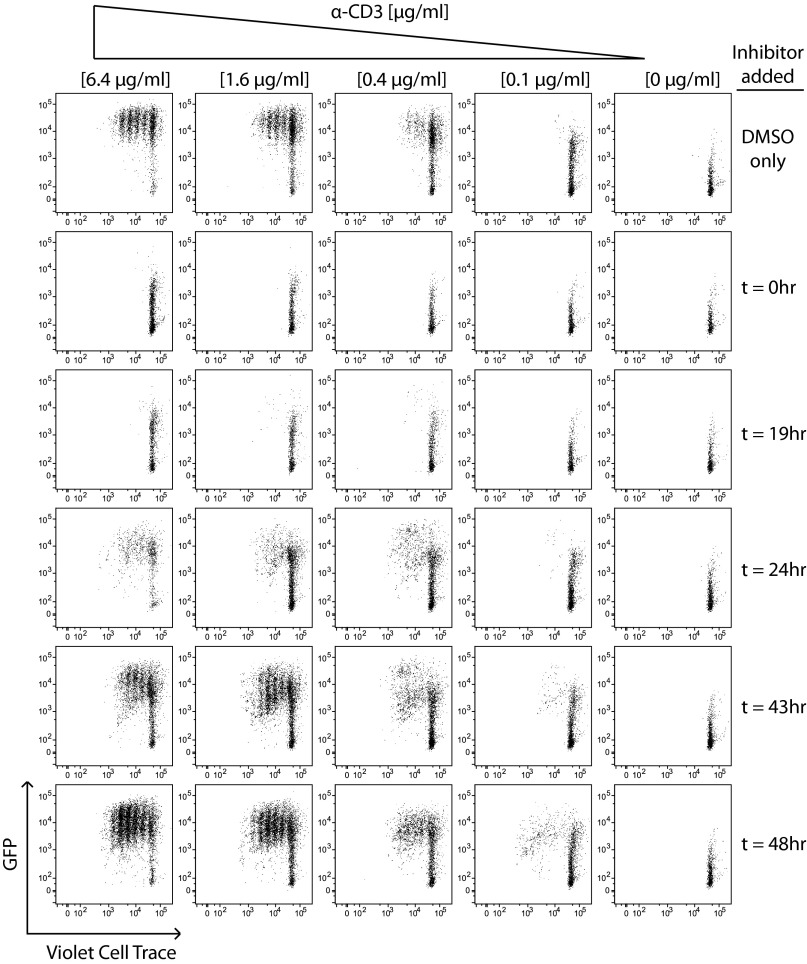
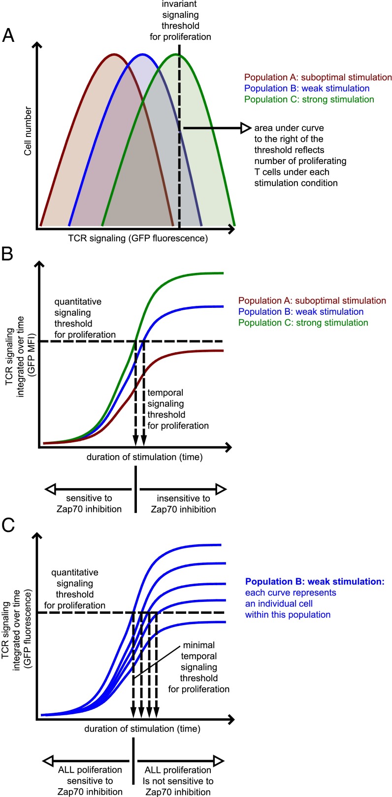
T-cell antigen receptor (TCR) signaling is essential for activation, proliferation, and effector function of T cells. Modulation of both intensity and duration of TCR signaling can regulate these events. However, it remains unclear how individual T cells integrate such signals over time to make critical cell-fate decisions. We have previously developed an engineered mutant allele of the critical T-cell kinase zeta-chain-associated protein kinase 70 kDa (Zap70) that is catalytically inhibited by a small molecule inhibitor, thereby blocking TCR signaling specifically and efficiently. We have also characterized a fluorescent reporter Nur77-eGFP transgenic mouse line in which T cells up-regulate GFP uniquely in response to TCR stimulation. The combination of these technologies unmasked a sharp TCR signaling threshold for commitment to cell division both in vitro and in vivo. Further, we demonstrate that this threshold is independent of both the magnitude of the TCR stimulus and Interleukin 2. Similarly, we identify a temporal threshold of TCR signaling that is required for commitment to proliferation, after which T cells are able to proliferate in a Zap70 kinase-independent manner. Taken together, our studies reveal a sharp threshold for the magnitude and duration of TCR signaling required for commitment of T cells to proliferation. These results have important implications for understanding T-cell responses to infection and optimizing strategies for immunomodulatory drug delivery.


Conflict of interest statement

The authors declare no conflict of interest.

Figures

Fig. 1.
Fig. 1.
A system for titration and detection of TCR signaling. CD4+ T cells from Zap-deficient mice harboring the Zap70(AS) allele and the Nur77–GFP reporter (Zap70(AS)–GFP mice) were stimulated overnight with (A) the indicated concentrations of plate-bound anti-CD3 or with (B) a fixed dose of anti-CD3 (6.4 μg/mL) and the indicated concentrations of analog inhibitor, HXJ42. (C) Zap70(AS) CD4+ cells were stimulated with 6.4 μg/mL anti-CD3 and a high concentration (1 μM) of HXJ42 or vehicle alone was added at 0 h for flow cytometric analysis of GFP expression at 6 h, or cells were stimulated for 18 h, and vehicle or HXJ42 was added for 6 h before analysis of GFP expression at 24 h. Data are representative of at least three independent experiments.
Fig. 2.
Fig. 2.
Nur77–GFP reporter reveals a dose-independent TCR signaling threshold for T-cell proliferation in vitro. (A) CD4+ T cells from Nur77–GFP reporter mice were loaded with violet cell trace dye and stimulated with the indicated concentrations of plate-bound anti-CD3 for 4 d. Cells were subsequently stained for T-cell markers and assessed for dye dilution and GFP fluorescence by flow cytometry. Individual cell divisions are color coded (Top Row). (B) Histograms show GFP expression of cells within each cell division that were stimulated with varying concentrations of anti-CD3. (C) CD4+ T cells from Zap70(AS)–GFP mice were loaded with violet cell trace dye and stimulated with a fixed dose of anti-CD3 (6.4 μg/mL) and the indicated concentrations of HXJ42 for 4 d. Cells were subsequently stained for T-cell markers and assessed for dye dilution and GFP fluorescence by flow cytometry. (D) Histograms show GFP expression of cells within each cell division that were treated with varying concentrations of HXJ42. Data are representative of at least three independent experiments.
Fig. 3.
Fig. 3.
Nur77–GFP reporter reveals a dose-independent TCR signaling threshold for T-cell proliferation in vivo. (A) CD4+ T cells from OT2 transgenic mice harboring the Nur77–GFP reporter (OT2–GFP mice) were loaded with violet cell trace dye and adoptively transferred into CD45.1+ congenic hosts. The following day, host mice were immunized via s.c. footpad injection with 5 μg or 50 μg ovalbumin protein in complete Freund's adjuvant. At day 3 postimmunization, draining and nondraining contralateral popliteal lymph node cells were harvested, stained for T-cell markers, and assessed for dye dilution and GFP fluorescence by flow cytometry. (B) CD4+ T cells from SMARTA transgenic mice harboring the Nur77–GFP reporter (SMARTA–GFP mice) were loaded with violet cell trace dye and adoptively transferred into congenic CD45.1+ hosts. The following day, host mice were infected with low (2 × 10⁴ cfu) or high (2 × 10⁶ cfu) infectious doses of LCMV. On day 3 of infection, splenocytes were harvested, treated with collagenase, stained for T-cell markers, and assessed for dye dilution and GFP fluorescence by flow cytometry. Data are representative of at least three biological replicates.
Fig. 4.
Fig. 4.
Endogenous Nur77 expression reveals loss of active TCR signaling in proliferating T cells in vivo. CD4+ T cells from OT2–GFP mice were loaded with violet cell trace dye, transferred into congenic CD45.1+ hosts, and immunized as described in Fig. 3A. On (A) day 3 and (B) day 5 postimmunization, draining and nondraining contralateral popliteal lymph node cells were harvested, fixed, permeabilized, and stained for both endogenous Nur77 and T-cell markers. Cells were subsequently assessed for GFP fluorescence, dye dilution, and endogenous Nur77 expression by flow cytometry. Top panels represent gating scheme to identify cells that have undergone no divisions or many divisions, with either low or high GFP expression. Middle panels represent endogenous Nur77 and Bottom panels represent GFP fluorescence in these gates. Data are representative of at least three biological replicates.
Fig. 5.
Fig. 5.
TCR signaling threshold is not enforced by IL-2. (A) CD4+ T cells from Nur77–GFP reporter mice were loaded with violet cell trace dye and stimulated with varying doses of plate-bound anti-CD3 without additional reagents (Top), in the presence of exogenous IL-2 (Middle), or presence of neutralizing IL-2 mAb (Bottom) for 4 d. Cells were subsequently stained for T-cell markers and assessed for dye dilution and GFP fluorescence by flow cytometry. (B) Histograms show GFP expression within each cell division for the highest concentration of anti-CD3 used (6.4 μg/mL). Data are representative of at least three independent experiments.
Fig. 6.
Fig. 6.
Continuous TCR signaling is dispensable after 24 h for commitment to proliferation. CD4+ T cells from Zap70(AS)–GFP mice were loaded with violet cell trace dye and stimulated with varying doses of plate-bound anti-CD3 along with a fixed dose of anti-CD28 (2 μg/mL). Samples were then treated with a fixed concentration of HXJ42 (1 μM) or vehicle alone at various time points after initial stimulation, and all samples were then harvested at 96 h. Cells were stained for T-cell markers and assessed for GFP fluorescence. Data are representative of at least three independent experiments.
Fig. 7.
Fig. 7.
A quantitative and temporal threshold model for T-cell proliferation. (A) Integrated TCR signaling among a population of T cells exposed to a uniform stimulus nevertheless exhibits a distribution of GFP expression. This distribution reflects the strength of TCR stimulation, titrated by Zap70 kinase inhibition. The cells within a population in which their integrated TCR signaling (as read out by Nur77–eGFP fluorescence) cross an invariant threshold enter the proliferating pool. This model is compatible with both an invariant signaling threshold at the single-cell level and with a titration of the proliferating cell population by varying TCR signal strength. Importantly, a weak stimulus that barely drives cells over this threshold is predicted to result in slightly lower GFP MFI among the population of proliferating T cells. (B) Each curve here represents the mean perceived TCR signal strength (GFP MFI) of an entire population, including both proliferating and nonproliferating cells. TCR signaling is integrated over time and GFP MFI therefore increases with time. Once cells within a population cross an invariant signaling threshold, further TCR signaling to commit to proliferation is dispensable, as demonstrated by insensitivity of proliferating population to Zap70 inhibition beyond this time point. Cells in population “A” receive subthreshold/suboptimal stimulation and these cells never cross the invariant signaling threshold and never proliferate. (C) As depicted in A, a population of stimulated T cells displays a distribution of GFP fluorescence at any given time point. In this model, each curve represents the perceived TCR signal strength of individual T cells. Only a fraction of cells within this population crosses the invariant signaling threshold at any given time point. There exists a minimum time required for any cells to do so, beyond which those cells committed to proliferation are not sensitive to Zap70 inhibition. However, prolonged duration of stimulation is capable of driving increasing numbers of cells across the threshold. In this manner, prolonged signal duration can recruit more and more cells into the proliferating pool, consistent with prior observations of a temporal threshold and a titration effect on clonal burst size.

References

    1. Iezzi G, Karjalainen K, Lanzavecchia A. The duration of antigenic stimulation determines the fate of naive and effector T cells. Immunity. 1998;8(1):89–95. - PubMed
    1. Irvine DJ, Purbhoo MA, Krogsgaard M, Davis MM. Direct observation of ligand recognition by T cells. Nature. 2002;419(6909):845–849. - PubMed
    1. Sojka DK, Bruniquel D, Schwartz RH, Singh NJ. IL-2 secretion by CD4+ T cells in vivo is rapid, transient, and influenced by TCR-specific competition. J Immunol. 2004;172(10):6136–6143. - PubMed
    1. Huang J, et al. A single peptide-major histocompatibility complex ligand triggers digital cytokine secretion in CD4(+) T cells. Immunity. 2013;39(5):846–857. - PMC - PubMed
    1. Kimachi K, Croft M, Grey HM. The minimal number of antigen-major histocompatibility complex class II complexes required for activation of naive and primed T cells. Eur J Immunol. 1997;27(12):3310–3317. - PubMed

Publication types

MeSH terms

Substances