. 2014 Sep 9;111(36):13057-62.
doi: 10.1073/pnas.1408710111. Epub 2014 Aug 18.

Local anesthetic and antiepileptic drug access and binding to a bacterial voltage-gated sodium channel

Affiliations

Local anesthetic and antiepileptic drug access and binding to a bacterial voltage-gated sodium channel

Céline Boiteux et al. Proc Natl Acad Sci U S A. .

Abstract

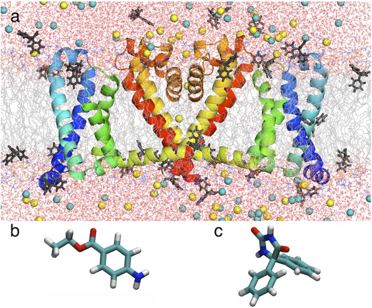
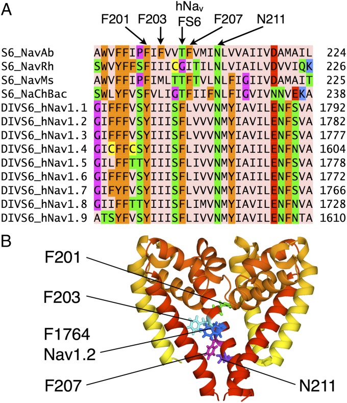
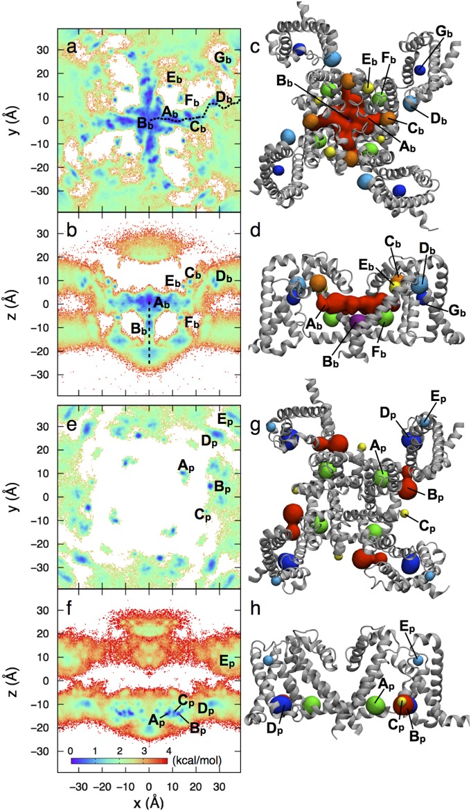
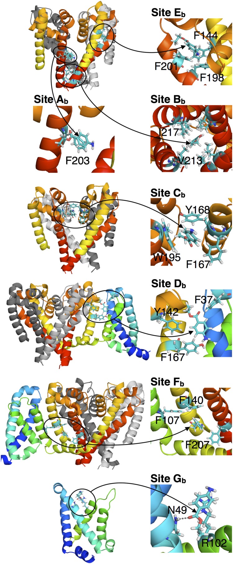
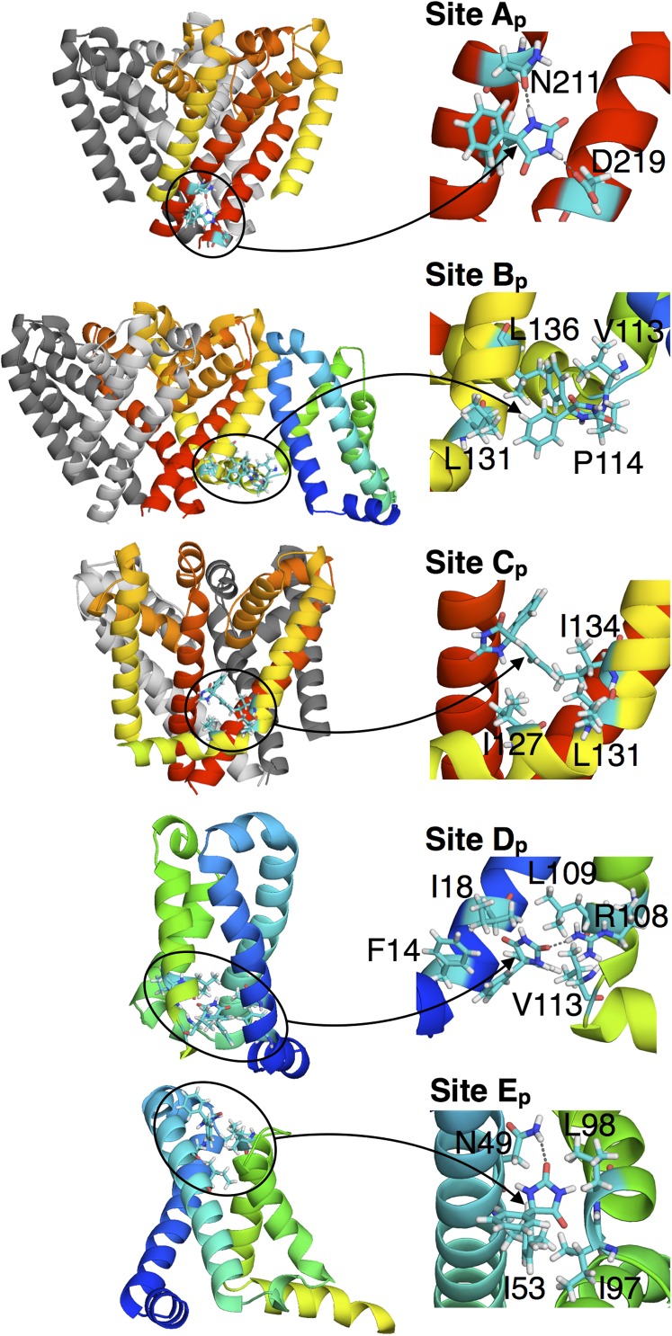
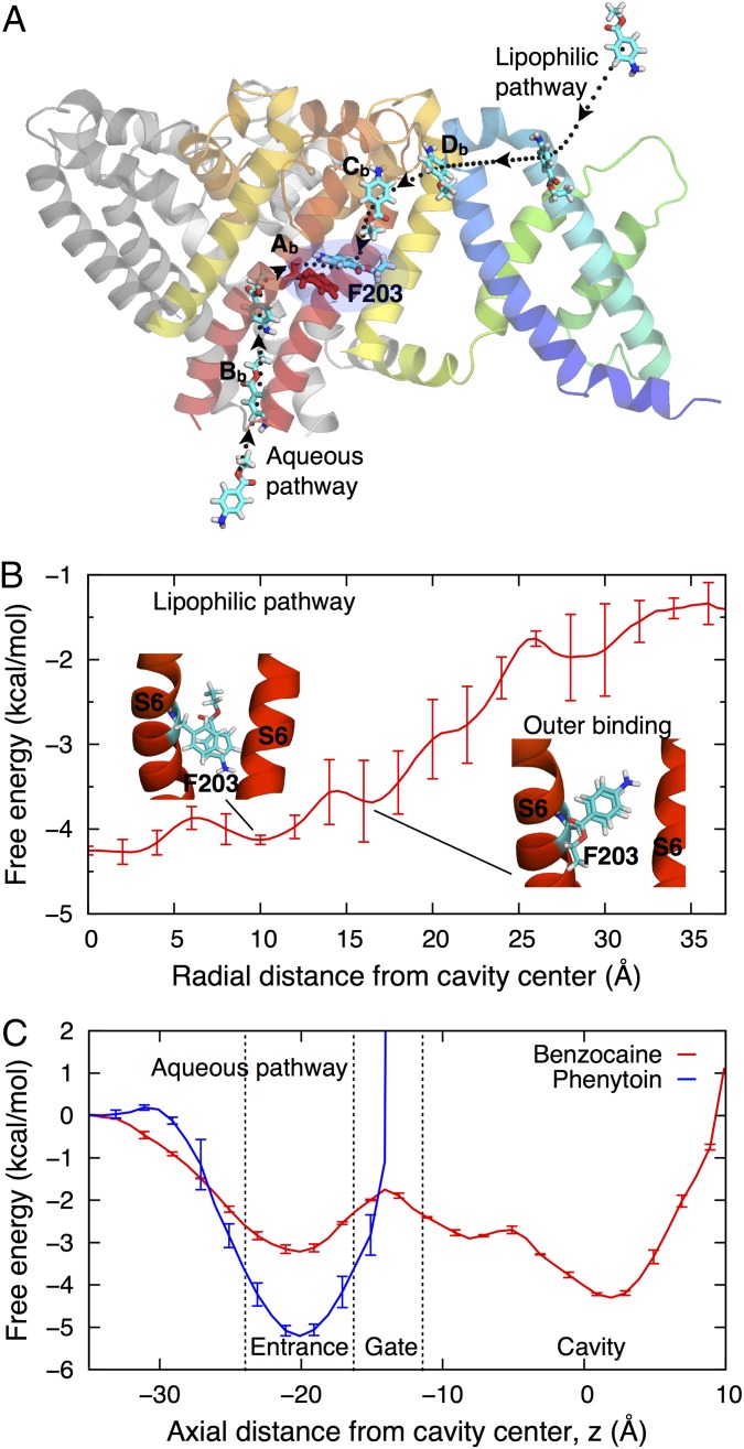
Voltage-gated sodium (Nav) channels are important targets in the treatment of a range of pathologies. Bacterial channels, for which crystal structures have been solved, exhibit modulation by local anesthetic and anti-epileptic agents, allowing molecular-level investigations into sodium channel-drug interactions. These structures reveal no basis for the "hinged lid"-based fast inactivation, seen in eukaryotic Nav channels. Thus, they enable examination of potential mechanisms of use- or state-dependent drug action based on activation gating, or slower pore-based inactivation processes. Multimicrosecond simulations of NavAb reveal high-affinity binding of benzocaine to F203 that is a surrogate for FS6, conserved in helix S6 of Domain IV of mammalian sodium channels, as well as low-affinity sites suggested to stabilize different states of the channel. Phenytoin exhibits a different binding distribution owing to preferential interactions at the membrane and water-protein interfaces. Two drug-access pathways into the pore are observed: via lateral fenestrations connecting to the membrane lipid phase, as well as via an aqueous pathway through the intracellular activation gate, despite being closed. These observations provide insight into drug modulation that will guide further developments of Nav inhibitors.

Keywords: bacterial sodium channel; drug binding.


Conflict of interest statement

The authors declare no conflict of interest.

Figures

Fig. 1.
Fig. 1.
(A) Simulation system showing NavAb (two of four subunits as ribbons, with voltage sensor domain (VSD) S1 and S2 in blue and S3 and S4 in green, pore domain (PD) S5 in yellow and S6 in red, P-loop P1, P2, and selectivity filter (SF; orange) in a hydrated lipid bilayer (chains as gray lines; water as red/white sticks; NaCl as yellow/cyan balls) and PHT (dark gray sticks). (B and C) Stick representations of BZC (B) and PHT (C).
Fig. 2.
Fig. 2.
(A) Sequence alignment of segment S6 of NavAb to other Nav channels (showing only FS6-containing domain IV; see SI Appendix, Fig. S1, for full comparison). F201, F203, F207, and N211 in NavAb and F1764 in Nav1.2 are indicated. Amino acids were colored with Jalview (46, 47) using the Zappo scheme. (B) Aligned structures (showing three subunits) of NavAb and model Nav1.2 (based on NavAb) comparing NavAb F201, F203, and F207 to Nav1.2 F1764. Conserved N211 (NavAb numbering) is also indicated.
Fig. 3.
Fig. 3.
Free energy surfaces (Left) and binding sites (Right) for BZC (AD) and PHT (EH), with color scale as inset. For clarity, B for BZC includes only −10 < y < 10 Å (full data in SI Appendix, Fig. S5). Binding sites for BZC (C and D) and PHT (G and H), identified from free energy minima, are shown as spheres (colored for clarity, not by free energy).
Fig. 4.
Fig. 4.
Interactions involved in binding sites Ab to Gb of BZC, with segments colored as in Fig. 1. (Insets) Arrows point directly at the BZC molecule.
Fig. 5.
Fig. 5.
Interactions involved in the binding sites Ap to Fp of PHT. (Insets) Arrows point directly at the PHT molecule.
Fig. 6.
Fig. 6.
Drug-binding pathways. (A) Side view of the channel showing movement of BZC from water to the central cavity along lipophilic and aqueous pathways (dotted curves). The full PD and only one VSD subunit are shown. (B and C) The free energy profile for BZC movement through a fenestration (B) and those for BZC (red) and PHT (blue) movements through the activation gate (C). The free energy is set to zero in water, using SI Appendix, Fig. S2A to set the membrane reference for B.

References

    1. England S, de Groot MJ. Subtype-selective targeting of voltage-gated sodium channels. Br J Pharmacol. 2009;158(6):1413–1425. - PMC - PubMed
    1. Mike A, Lukacs P. The enigmatic drug binding site for sodium channel inhibitors. Curr Mol Pharmacol. 2010;3(3):129–144. - PubMed
    1. Payandeh J, Scheuer T, Zheng N, Catterall WA. The crystal structure of a voltage-gated sodium channel. Nature. 2011;475(7356):353–358. - PMC - PubMed
    1. McCusker EC, et al. Structure of a bacterial voltage-gated sodium channel pore reveals mechanisms of opening and closing. Nat Commun. 2012;3:1102. - PMC - PubMed
    1. Zhang X, et al. Crystal structure of an orthologue of the NaChBac voltage-gated sodium channel. Nature. 2012;486(7401):130–134. - PMC - PubMed

Publication types

MeSH terms