. 2014 Jul 31;9(1):373.
doi: 10.1186/1556-276X-9-373. eCollection 2014.

Enhanced antibacterial and anti-biofilm activities of silver nanoparticles against Gram-negative and Gram-positive bacteria

Affiliations

Enhanced antibacterial and anti-biofilm activities of silver nanoparticles against Gram-negative and Gram-positive bacteria

Sangiliyandi Gurunathan et al. Nanoscale Res Lett. .

Abstract

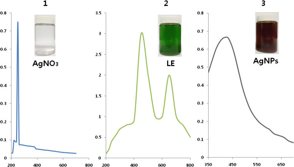
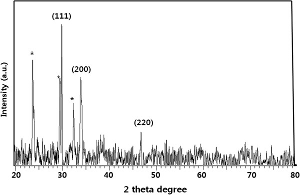
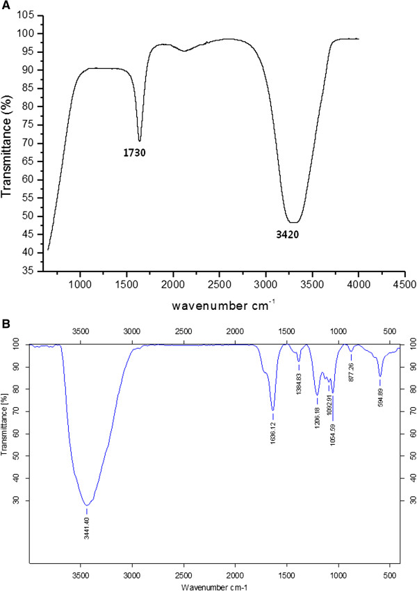
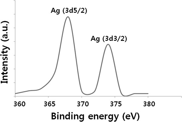
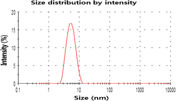
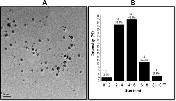
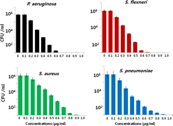
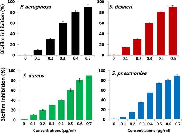
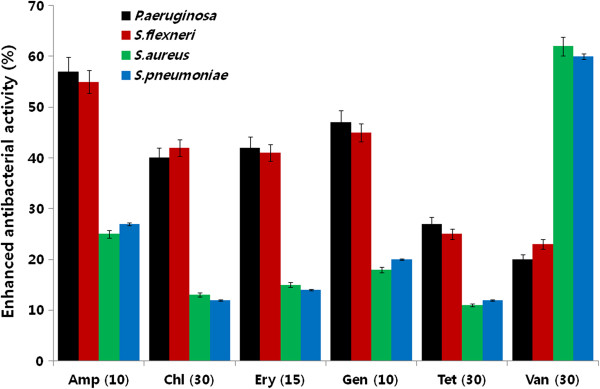
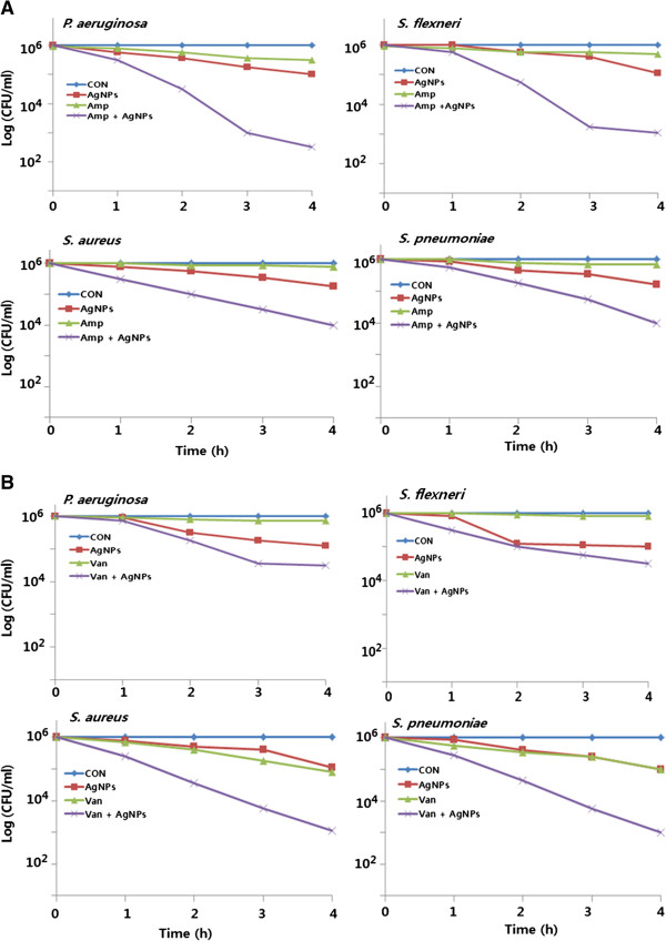
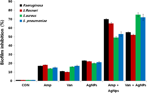
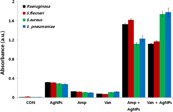
Silver nanoparticles (AgNPs) have been used as antibacterial, antifungal, antiviral, anti-inflammtory, and antiangiogenic due to its unique properties such as physical, chemical, and biological properties. The present study was aimed to investigate antibacterial and anti-biofilm activities of silver nanoparticles alone and in combination with conventional antibiotics against various human pathogenic bacteria. Here, we show that a simple, reliable, cost effective and green method for the synthesis of AgNPs by treating silver ions with leaf extract of Allophylus cobbe. The A. cobbe-mediated synthesis of AgNPs (AgNPs) was characterized by ultraviolet-visible absorption spectroscopy, X-ray diffraction (XRD), Fourier transform infrared spectroscopy (FTIR), X-ray photoelectron spectroscopy (XPS), dynamic light scattering (DLS), and transmission electron microscopy (TEM). Furthermore, the antibacterial and anti-biofilm activity of antibiotics or AgNPs, or combinations of AgNPs with an antibiotic was evaluated using a series of assays: such as in vitro killing assay, disc diffusion assay, biofilm inhibition, and reactive oxygen species generation in Pseudomonas aeruginosa, Shigella flexneri, Staphylococcus aureus, and Streptococcus pneumonia. The results suggest that, in combination with antibiotics, there were significant antimicrobial and anti-biofilm effects at lowest concentration of AgNPs using a novel plant extract of A. cobbe, otherwise sublethal concentrations of the antibiotics. The significant enhancing effects were observed for ampicillin and vancomycin against Gram-negative and Gram-positive bacteria, respectively. These data suggest that combining antibiotics and biogenic AgNPs can be used therapeutically for the treatment of infectious diseases caused by bacteria. This study presented evidence of antibacterial and anti-biofilm effects of A. cobbe-mediated synthesis of AgNPs and their enhanced capacity against various human pathogenic bacteria. These results suggest that AgNPs could be used as an adjuvant for the treatment of infectious diseases.

Keywords: Allophylus cobbe; Anti-biofilm activity; Antibacterial activity; Antibiotics; Silver nanoparticles; Sublethal concentrations.


Figures

Figure 1
Figure 1
Characterization of AgNPs synthesized using A. cobbe leaf extracts. The absorption spectra of AgNPs exhibited a strong, broad peak at 420 nm. This band was attributed to the surface plasmon resonance of the AgNPs. The images show the spectrum of AgNO3(1), leaf extract (2), and mixture of AgNO3 and leaf extract (3) at 6 h exposure. After exposure for 6 h, the color of the colloidal solution of AgNPs turned from green to dark brown, indicating the formation of AgNPs.
Figure 2
Figure 2
XRD pattern of silver nanoparticles synthesized using A. cobbe leaf broth.
Figure 3
Figure 3
FTIR spectra of A. cobbe leaf broth (A), silver nanoparticles synthesized by A. cobbe leaf broth (B).
Figure 4
Figure 4
XPS analysis of AgNPs.
Figure 5
Figure 5
Size distribution analysis of AgNPs was determined by dynamic light scattering. The particle size distribution analysis revealed that the average particle size was approximately 5 nm.
Figure 6
Figure 6
Determination of size and shape of AgNPs. The size and morphology of AgNPs were determined using transmission electron microscopy. TEM micrograph of AgNPs prepared from A. cobbe(A). The average particle size was found to be 5 nm. Particle size distributions from TEM images (B).
Figure 7
Figure 7
Effect of AgNPs on cell survival. Dose-dependent effects of AgNPs on bacterial survival. All test strains were incubated in the presence of different concentrations of AgNPs. Bacterial survival was determined at 4 h by a CFU assay. The results are expressed as the means ± SD of three separate experiments each of which contained three replicates. Treated groups showed statistically significant differences from the control group by the Student's t test (p < 0.05).
Figure 8
Figure 8
Effect of AgNPs on biofilm inhibition. The anti-biofilm activity of AgNPs was assessed by incubating all test strains with different concentrations of AgNPs for 4 h in a 96-well plate. The results are expressed as the means ± SD of three separate experiments each of which contained three replicates. Treated groups showed statistically significant differences from the control group by the Student's t test (p < 0.05).
Figure 9
Figure 9
Enhancement of antibacterial activity of antibiotic in the presence of AgNPs. Antibacterial activities were determined by the agar diffusion method. The MICs of AgNPs for each test strain were loaded into the wells formed on plates containing a bacterial lawn. Growth inhibition was determined by measuring the zone of inhibition after 24 h. Experiments were performed in triplicate. The percentage of enhanced antibacterial activity was calculated using the formula (B - A/A) × 100. The results are expressed as the means ± SD of three separate experiments. Treated groups showed statistically significant differences from the control group by the Student's t test (p < 0.05).
Figure 10
Figure 10
Enhanced antibacterial effect of antibiotics in the presence of AgNPs. All test strains were treated for 4 h with sublethal concentrations of ampicillin or AgNPs, or combinations of AgNPs and ampicillin (A) or combinations of AgNPs and vancomycin (B). All test strains were treated for 4 h with sublethal concentrations of vancomycin or AgNPs, or combinations of AgNPs and vancomycin. Bacterial survival was determined at 4 h by the CFU assay. The results are expressed as the means ± SD of three separate experiments, each of which contained three replicates. Treated groups showed statistically significant differences from the control group by the Student's t test (p < 0.05).
Figure 11
Figure 11
Enhanced biofilm inhibitory activitity of antibiotics and AgNPs. The anti-biofilm activity of AgNPs was assessed by incubating all test strains with sublethal concentrations of ampicillin or AgNPs, or combinations of AgNPs with the ampicillin antibiotic for 4 h. The results are expressed as the means ± SD of three separate experiments, each of which contained three replicates. Treated groups showed statistically significant differences from the control group by the Student's t test (p < 0.05).
Figure 12
Figure 12
Enhanced effect of antibiotics and AgNPs on ROS generation. All test strains were treated with sublethal concentrations of antibiotics or AgNPs, or combinations of AgNPs with antibiotics for 12 h. ROS generation was measured by the XTT assay. The results are expressed as the means ± SD of three separate experiments, each of which contained three replicates. Treated groups showed statistically significant differences from the control group by the Student's t test (p < 0.05).

References

    1. Chen X, Schluesener HJ. Nanosilver: a nanoproduct in medical application. Toxicol Lett. 2008;9:1–12. - PubMed
    1. Lok CN, Ho CM, Chen R, He QY, Yu WY, Sun H, Tam PK, Chiu JF, Che CM. Silver nanoparticles: partial oxidation and antibacterial activities. J Biol Inorg Chem. 2007;9:527–534. - PubMed
    1. Malik MA, O'Brien P, Revaprasadu N. A simple route to the synthesis of core/shell nanoparticles of chalcogenides. Chem Mater. 2002;9:2004–2010.
    1. Gurunathan S, Kalishwaralal K, Vaidyanathan R, Deepak V, Pandian SRK, Muniyandi J, Hariharan N, Eom SH. Biosynthesis, purification and characterization of silver nanoparticles using Escherichia coli. Colloid Surf B. 2009;9:328–335. - PubMed
    1. Gurunathan S, Han JW, Eppakayala V, Jeyaraj M, Kim JH. Cytotoxicity of biologically synthesized silver nanoparticles in MDA-MB-231 human breast cancer cells. Biomed Res Int. 2013;9:535796. - PMC - PubMed