Review
. 2014 Sep;8(3):187-94.
doi: 10.1007/s11693-014-9142-x. Epub 2014 Apr 2.

Microtubule networks for plant cell division

Affiliations
Review

Microtubule networks for plant cell division

Jeroen de Keijzer et al. Syst Synth Biol. 2014 Sep.

Abstract

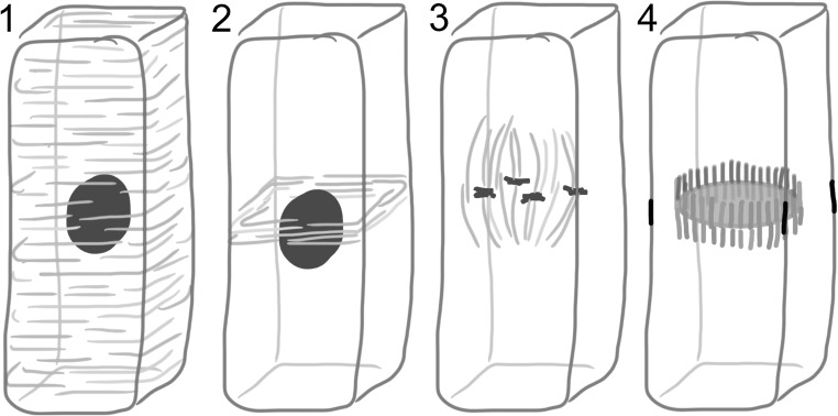
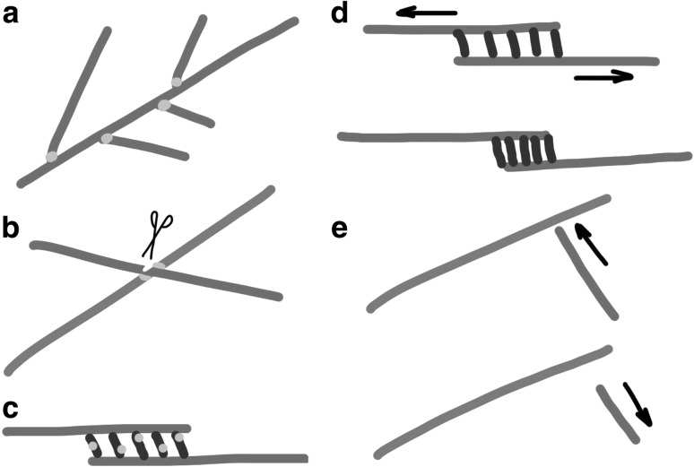
During cytokinesis the cytoplasm of a cell is divided to form two daughter cells. In animal cells, the existing plasma membrane is first constricted and then abscised to generate two individual plasma membranes. Plant cells on the other hand divide by forming an interior dividing wall, the so-called cell plate, which is constructed by localized deposition of membrane and cell wall material. Construction starts in the centre of the cell at the locus of the mitotic spindle and continues radially towards the existing plasma membrane. Finally the membrane of the cell plate and plasma membrane fuse to form two individual plasma membranes. Two microtubule-based cytoskeletal networks, the phragmoplast and the pre-prophase band (PPB), jointly control cytokinesis in plants. The bipolar microtubule array of the phragmoplast regulates cell plate deposition towards a cortical position that is templated by the ring-shaped microtubule array of the PPB. In contrast to most animal cells, plants do not use centrosomes as foci of microtubule growth initiation. Instead, plant microtubule networks are striking examples of self-organizing systems that emerge from physically constrained interactions of dispersed microtubules. Here we will discuss how microtubule-based activities including growth, shrinkage, severing, sliding, nucleation and bundling interrelate to jointly generate the required ordered structures. Evidence mounts that adapter proteins sense the local geometry of microtubules to locally modulate the activity of proteins involved in microtubule growth regulation and severing. Many of the proteins and mechanisms involved have roles in other microtubule assemblies as well, bestowing broader relevance to insights gained from plants.

Keywords: Cortical array; Cytokinesis; Cytoskeleton; Microtubule; Phragmoplast; Plant.


Figures

Fig. 1
Fig. 1
Spatial relations between microtubule networks in plant cell division. The cortical microtubule array 1 develops into the dense ring-like pre-prophase band 2 whose location coincides with the cell nucleus blue. Other microtubules that are initially formed around the nuclear envelope develop into a mitotic spindle that segregates sister chromatids 3. The phragmoplast 4 forms out of the spindle remnant and expands radially towards the location of the erstwhile pre-prophase band black leaving a finished cell plate in its wake yellow. (Color figure online)
Fig. 2
Fig. 2
Cytokinesis visualized by live cell imaging in the moss Physcomitrella patens. Microtubules are shown in green and chromatin and cell-plate material in red. Chromatids are segregated towards spindle poles and the remaining microtubules of the mitotic spindle (top) are used as template for the initial phragmoplast (middle; 6 min later). The reforming daughter nuclei are seen at the phragmoplast poles while accumulation of cell plate material is visible in the central plane of the phragmoplast where microtubules of opposite poles form antiparallel overlaps (arrowheads). Fifteen minutes later the central region of the cell plate has matured and the phragmoplast has expanded laterally (bottom). Microtubules were visualized using GFP fused to α-tubulin (Hiwatashi et al. 2008), chromatin using mRFP fused to histone2B (Nakaoka et al. 2012) and the membranous material delivered to the cell plate with the lipophilic styryl dye FM4-64. The scale bar indicates 10 µm
Fig. 3
Fig. 3
Feedback between microtubule organization and microtubule activities a Augmin-mediated branching of new microtubules from the sides of existing microtubules. b Points of microtubule crossover in plants are sites of microtubule severing by katanin, possibly regulated by SPIRAL2. c Crosslinking proteins in between overlapping microtubules recruit proteins that locally regulate dynamic instability or drive microtubule sliding. d Crosslinkers confined in between sliding microtubules are driven together by convening microtubule ends. e Existing microtubules obstruct the growth of incoming microtubules triggering shrinkage in the near two-dimensional geometry of the plant cortex

References

    1. Al-Bassam J, Kim H, Brouhard G, et al. CLASP promotes microtubule rescue by recruiting tubulin dimers to the microtubule. Dev Cell. 2010;19:245–258. doi: 10.1016/j.devcel.2010.07.016. - DOI - PMC - PubMed
    1. Allard JF, Ambrose JC, Wasteneys GO, Cytrynbaum EN. A mechanochemical model explains interactions between cortical microtubules in plants. Biophys J. 2010;99:1082–1090. doi: 10.1016/j.bpj.2010.05.037. - DOI - PMC - PubMed
    1. Allard JF, Wasteneys GO, Cytrynbaum EN. Mechanisms of self-organization of cortical microtubules in plants revealed by computational simulations. Mol Biol Cell. 2010;21:278–286. doi: 10.1091/mbc.E09-07-0579. - DOI - PMC - PubMed
    1. Ambrose C, Wasteneys GO. Nanoscale and geometric influences on the microtubule cytoskeleton in plants: thinking inside and outside the box. Protoplasma. 2012;249(Suppl):S69–S76. doi: 10.1007/s00709-011-0334-x. - DOI - PubMed
    1. Ambrose C, Allard JF, Cytrynbaum EN, Wasteneys GO. A CLASP-modulated cell edge barrier mechanism drives cell-wide cortical microtubule organization in Arabidopsis. Nat Commun. 2011;2:430. doi: 10.1038/ncomms1444. - DOI - PMC - PubMed

LinkOut - more resources