Review
. 2014 Sep;8(3):249-69.
doi: 10.1007/s11693-014-9145-7. Epub 2014 May 27.

Divided we stand: splitting synthetic cells for their proliferation

Affiliations
Review

Divided we stand: splitting synthetic cells for their proliferation

Yaron Caspi et al. Syst Synth Biol. 2014 Sep.

Abstract

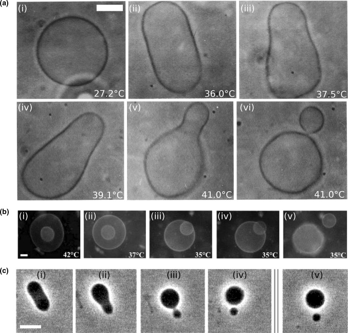
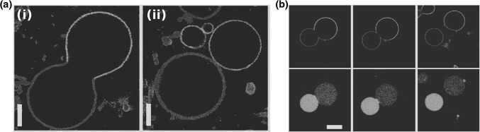
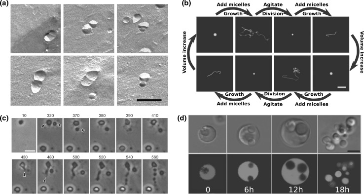
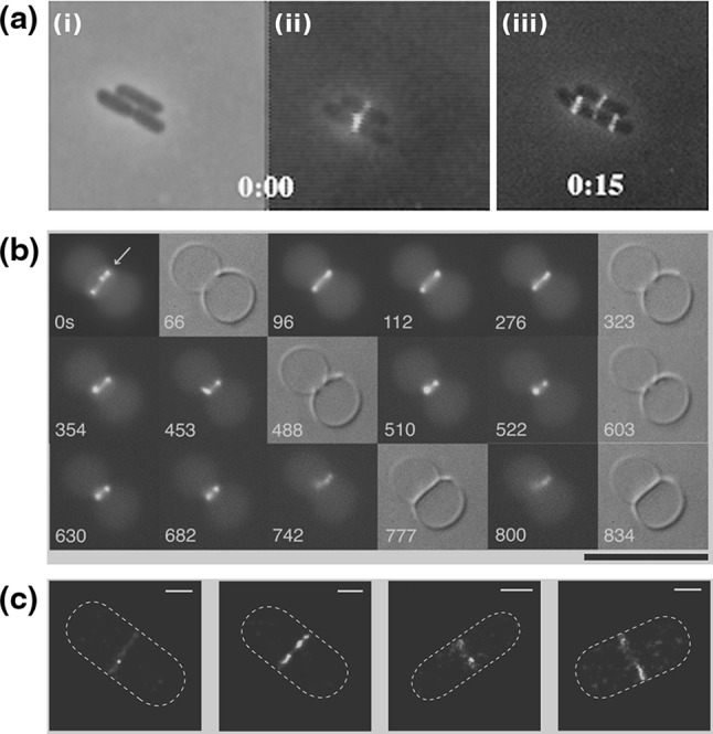
With the recent dawn of synthetic biology, the old idea of man-made artificial life has gained renewed interest. In the context of a bottom-up approach, this entails the de novo construction of synthetic cells that can autonomously sustain themselves and proliferate. Reproduction of a synthetic cell involves the synthesis of its inner content, replication of its information module, and growth and division of its shell. Theoretical and experimental analysis of natural cells shows that, whereas the core synthesis machinery of the information module is highly conserved, a wide range of solutions have been realized in order to accomplish division. It is therefore to be expected that there are multiple ways to engineer division of synthetic cells. Here we survey the field and review potential routes that can be explored to accomplish the division of bottom-up designed synthetic cells. We cover a range of complexities from simple abiotic mechanisms involving splitting of lipid-membrane-encapsulated vesicles due to physical or chemical principles, to potential division mechanisms of synthetic cells that are based on prokaryotic division machineries.

Keywords: Cell division; Minimal cells; Synthetic cells; Vesicle splitting.


Figures

Fig. 1
Fig. 1
Shape trajectories for vesicles that are physically manipulated. a A DPMC vesicle is heated from formula image (i) to formula image (v). A further increase of the temperature by formula image transferred the shape of the vesicle from a pear to the limiting shape of two spheres that stay connected by a narrow neck (without abscission of the neck). Area increased from 2,570 to 2,820 formula image, while volume changed only slightly from 12,200 to 12,000 formula image. Note that before the heating cycle the vesicle was stiff and did not have any excess surface area. Scale bar 10 formula image. b Time series of the birthing of an inner vesicle through the membrane of a mother vesicle. A DLPE:DPPC 3:7 vesicle was cooled down from formula image (i) to formula image (iiiv). The cooling process created an inner pressure that caused an inner bud (that had formed previously due to a heating process) to be ejaculated from the lumen of the mother vesicle. Scale barformula image. c Fission of a DPPC:cholesterol vesicle due to the incorporation of lyso-PC. Budding and fission were induced by the local injection of a solution of 1 formula image palmitoyl-lyso-PC. Time after injection are 0, 17, 18, 40, and 240 s for (i)–(v), respectively. Scale bar 10 formula image. a Modified with permissions from reference Käs and Sackmann (1991) formula image (1991) Elsevier Limited. b Modified with permissions from reference Sakuma and Imai (2011) formula image (2011) American Physical Society. c Modified with permission from reference Tanaka et al. (2004) formula image (2004) American Chemical Society
Fig. 2
Fig. 2
Fission of buds in liposomes with liquid-disordered phase membranes. a Fission of a bud at the liquid-ordered/liquid-disordered separation line upon heating from formula image (i) to formula image (ii). Vesicle composition 0.615:0.135:0.25 SM:DOPC:cholesterol. formula image phase was imaged in blue by incorporating small amount of a Dil dye, formula image phase was imaged in red by incorporating a small amount of N-lissamine rhodamine dipalmitoylphosphoethanolamine. Scale barformula image. b Fission of vesicle due to an encapsulation of an ATPS system. 0.35:0.35:0.3 POPC:DPPC:cholesterol vesicle encapsulating a PEG:dextran ATPS. Osmotic pressure increases form left to right. Blue indicates lectin SBA Alexa 647 (dextran phase); red indicates formula image domain lipid (DOPE-rhodamine); and green indicates formula image domain through streptavidin-Alexa488 (bound to the lipid DSPE-PEG-2K-biotin). Scale bar 10 microns. a Modified with permissions from reference Baumgart et al. (2003) formula image (2003) Nature Publishing Group. b Modified with permission from reference Andes-Koback and Keating (2011) formula image (2011) American Chemical Society. (Color figure online)
Fig. 3
Fig. 3
Division due to membrane growth of liposomes and cells. a Freeze-fracture electron microscopy images of twin-vesicles that appear to have a septum between them, formed from the addition of oleate micelles to pre-formed oleic acid/oleate vesicles. Scale bar 200 nm. b Coupled growth and division cycle of a multilamellar oleate vesicle, induced by the incorporation of oleate micelles followed by division of the resulted tubes into small spherical vesicles by gentle hydrodynamic forces. Scale bar 20 formula image. c Division of L-form formula image B. subtilis cells by an extrusion–resolution mechanism. The cell marked with an arrow at formula image minutes start to form protrusions after 430 min which resolved into a string of six cells. The cell marked with an asterisk did not divide. Scale bar 500 nm. d Phase-contrast (upper row) and fluorescence microscopy (lower row) of different phases of the L-form L. monocytogenes growth and multiplication. Intra-cellular bodies are formed within the mother cell. After they grow, the mother cell ruptures. Upon release, most of the intra-cellular bodies gain metabolic activity. Metabolic function was followed by synthesis and/or subsequent maturation of intracellular GFP (green). Time is in hours after incubation on soft agar. a Modified with permission of IOP Publishing, from reference Stano et al. (2006) formula image (2006) IOP Publishing. doi:10.1088/0953-8984/18/33/S37. All rights reserved. b Modified with permission from Zhu and Szostak (2009) Copyright (2009) American Chemical Society. c Modified with permission from reference Leaver et al. (2009) formula image (2009) Nature Publishing Group. d Modified with permission from reference Dell’Era et al. (2009) formula image (2009) John Wiley and Sons, Inc. (Color figure online)
Fig. 4
Fig. 4
FtsZ in the cytokinesis of cells and liposomes. a Localization of the Z-ring during the cell cycle of E. coli. i Phase-contrast image of three cells. ii Overlay of a phase-contrast image and a conventional fluorescence microscopy image of FtsZ–GFP. iii Overlay of a phase-contrast image and a conventional fluorescence microscopy image of FtsZ–GFP after 15 min. Note that initially one FtsZ ring appears in the lower cell, while two new rings appears in the two daughter cells after the mother cell has divided. b Time lapse recording of the formation of a septum between two liposomes containing equimolar FtsZ–YFP and a mutant formula image proteins, together with GTP and ATP. Time represents seconds after the start of the recording. Black background images are epi-fluorescence images while gray background images are DIC ones. c Four high-resolution images of the Z-ring in E. coli cells (PALM images of FtsZ–Emos2). Note the inhomogeneity of the fluorescence intensity, indicating that the Z-ring is not a perfect continuous filament. Scale bar 500 nm. Initially, an FtsZ arc is seen localized at a constriction site. After several spatial reorganization steps, the system seems to end up in an intra-vesicular septum. Scale bar 10 formula image. a Modified with permission from reference Sun and Margolin (1998) formula image (1998) American Society of Microbiology. b Modified with permission from reference Osawa and Erickson (2013) formula image (2013) National Academy of Sciences, USA. c Modified from reference Fu et al. (2010) under the Creative Commons Attribution (CC BY) license http://creativecommons.org/licenses/by/3.0/. (Color figure online)
Fig. 5
Fig. 5
Binary fission of cells driven by ESCRT/Cdv or mechanical forces. a Three-dimensional reconstruction of ESCRT-III treated giant unilaminar vesicles (GUV). i Membrane staining of the intra-vesicular bodies that were formed after reconstitution of the eukaryote ESCRT-III complex on the outside of a GUV. ii Z-stack confocal image of the same GUV showing that the intra-vesicular bodies were filled with the extra-vesicular content as a result of ESCRT-III-induced inward budding. Scale barformula image. b In situ immunofluorescence localization of CdvA (red middle column) and CdvB (green right column) during the constriction of four S. acidocaldarius cells. Note that both CdvA and CdvB are localized between the two segregated chromosomes (blue stained with DAPI). Left column phase-contrast illumination. c Stages in the division of formula image M. genitalium. Newborn cells possess a single terminal organelle. Division starts when the terminal organelle duplicates, leading to second organelle at the other cell pole. Cytoskeleton filaments then elongate the cell, and create a constricted tube at the cell middle. After the formation of a chain of filaments, some cells are torn off the chain and can start a new reproduction cycle. Scale bar 500 nm. a Modified with permission from reference Wollert et al. (2009) formula image (2009) Nature Publishing Group. b Modified with permission from reference Samson et al. (2008) formula image (2008) National Academy of Sciences, USA. c Modified with permission from reference Lluch-Senar et al. (2010) formula image (2010) John Wiley and Sons, Inc. (Color figure online)
Fig. 6
Fig. 6
Mechanisms for division apparatus localization. a i Assay for the reconstitution of MinDE in microchambers (side view). Microchambers are fabricated in PDMS, coated with a supported lipid bilayer, and buffer with MinD, MinE and ATP is placed on top. ii Top-view images of MinDE dynamic localization in one of the microchambers of a. Time is in seconds; 10 % of the MinE proteins are labeled with Alexa-488. Scale bar 10 formula image. b Localization of the nucleoid and FtsZ-GFP in rod shaped (iiii left column) and anomalously shaped E. coli cells (ivvi right column). Nucleoid is labeled with the nucleoid-associated protein HupA conjugated to RFP. Red dashed line represents the outline of the cells. i, iv Chromosomes profile. ii, v FtsZ-GFP. iii, vi Overlay of the HupA-RPF (orange) and the FtsZ-GFP (green) images. Scale bar in both cases is 1 formula image. a Modified with permission from reference Zieske and Schwille (2013) formula image (2013) John Wiley and Sons, Inc. b Modified with permission from reference Männik et al. (2012) formula image (2012) National Academy of Sciences, USA. (Color figure online)

References

    1. Aarsman MEG, Piette A, Fraipont C, Vinkenvleugel TMF, Nguyen-Distèche M, den Blaauwen T. Maturation of the Escherichia coli divisome occurs in two steps. Mol Microbiol. 2005;55:1631–1645. - PubMed
    1. Adams DW, Errington J. Bacterial cell division: assembly, maintenance and disassembly of the Z ring. Nat Rev Microbiol. 2009;7:642–653. - PubMed
    1. Akerlund T, Gullbrand B, Nordström K. Effects of the Min system on nucleoid segregation in Escherichia coli. Microbiology. 2002;148:3213–3222. - PubMed
    1. Albers SV, Meyer BH. The archaeal cell envelope. Nat Rev Microbiol. 2011;9:414–426. - PubMed
    1. Anderson DE, Gueiros-Filho FJ, Erickson HP. Assembly dynamics of FtsZ rings in Bacillus subtilis and Escherichia coli and effects of FtsZ-regulating proteins. J Bacteriol. 2004;186:5775–5781. - PMC - PubMed

LinkOut - more resources