Review
. 2014:2014:591374.
doi: 10.1155/2014/591374. Epub 2014 Jul 21.

Hox transcription factors: modulators of cell-cell and cell-extracellular matrix adhesion

Affiliations
Review

Hox transcription factors: modulators of cell-cell and cell-extracellular matrix adhesion

Yasushi Taniguchi. Biomed Res Int. 2014.

Abstract

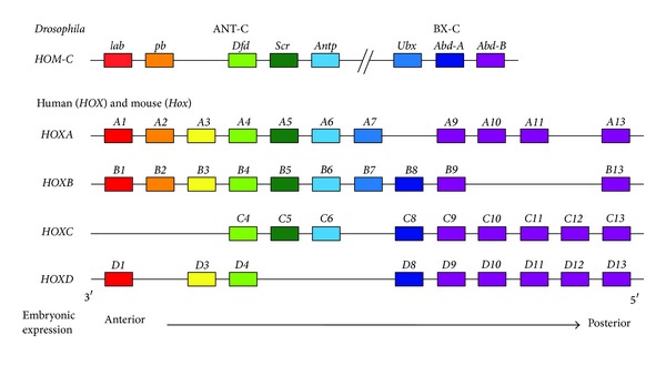
Hox genes encode homeodomain-containing transcription factors that determine cell and tissue identities in the embryo during development. Hox genes are also expressed in various adult tissues and cancer cells. In Drosophila, expression of cell adhesion molecules, cadherins and integrins, is regulated by Hox proteins operating in hierarchical molecular pathways and plays a crucial role in segment-specific organogenesis. A number of studies using mammalian cultured cells have revealed that cell adhesion molecules responsible for cell-cell and cell-extracellular matrix interactions are downstream targets of Hox proteins. However, whether Hox transcription factors regulate expression of cell adhesion molecules during vertebrate development is still not fully understood. In this review, the potential roles Hox proteins play in cell adhesion and migration during vertebrate body patterning are discussed.


Figures

Figure 1
Figure 1
Arrangement of Hox genes in the Drosophila and mammalian genomes. In Drosophila, eight Hox genes clustered on a single chromosome, the homeotic complex (HOM-C), are divided into two groups: the Antennapedia complex (ANT-C) and Bithorax complex (BX-C). ANT-C comprises five Hox genes: labial (lab), proboscipedia (pb), Deformed (Dfd), Sex combs reduced (Scr), and Antennapedia (Antp). The BX-C consists of three Hox genes: Ultrabithorax (Ubx), Abdominal-A (Abd-A), and Abdominal-B (Abd-B). In mammals, 39 Hox genes are divided into four separate clusters (HoxA, HoxB, HoxC, and HoxD) on four different chromosomes. In each cluster, Hox genes are tandem arranged in sequence from 3′ to 5′. Hox genes with the same number are referred to as paralogs. In the embryo, expression of the 3′ paralogs occurs earlier and more anteriorly along the anterior-posterior axis, whereas the 5′ paralogs are expressed later and more posteriorly.
Figure 2
Figure 2
Transition from epithelial to mesenchymal morphology caused by HOXD3 expression in lung cancer A549 cells. A549 cells stably transfected with empty vector (A549-vec) or HOXD3 expression vector (A549-HOXD3) [33] were fixed and stained for nuclei and F-actin by using DAPI and phalloidin-rhodamine, respectively. A549-vec cells have epithelial morphology (a, b), while A549-HOXD3 cells have spindle-shape mesenchymal morphology (c, d). A reduction in E-cadherin expression and an increase in α3 and β3 integrin expression were observed in A549-HOXD3 cells, as compared to A549-vec cells [33].
Figure 3
Figure 3
Reduced N-cadherin expression induced by HOXD3 overexpression in the roof plate of the early mouse embryo. (a, b) Expression of lacZ and HOXD3 genes in transverse neural tube sections at the thoracic level of 12.5-day transgenic embryos. Transgenic embryos were generated, in which lacZ and HOXD3 are expressed in the roof plate cells under the control of the Wnt1 regulatory element [34]. These embryos were sectioned and analyzed using in situ hybridization. Expression of lacZ (control) is restricted to roof plate cells within the neural tube, while HOXD3 expression is localized not only in the dorsal neural tube, but also within the ventricular zone and in ventral regions of the neural tube. (c, d) N-Cadherin expression in the thoracic neural tubes of 12.5-day lacZ- and HOXD3-expressing transgenic embryos. Transverse sections were stained using anti-human N-cadherin antibodies [34]. N-Cadherin is strongly expressed in the ventricular zone of lacZ-expressing embryos, whereas the ventricular zone in HOXD3-expressing embryos is composed of a number of progenitor cells that do not express N-cadherin. The ventricular zone is surrounded by dotted lines. Insets show that N-cadherin expression levels in the sympathetic ganglia of lacZ-expressing embryos are similar to those of HOXD3-expressing embryos. (e, f) Summary of the neural tube phenotype in transgenic embryos expressing lacZ and HOXD3. In embryos expressing lacZ, N-cadherin expression (green) is distributed throughout the neural tube. In HOXD3-expressing embryos, roof plate cells expressing HOXD3 (red circles) expand ventrally into the ventricular zone, where almost all N-cadherin-expressing cells are lost. R, roof plate; IZ, intermediate zone; VZ, ventricular zone. Scale bar: 100 μm.

References

    1. Lewis EB. A gene complex controlling segmentation in Drosophila . Nature. 1978;276(5688):565–570. - PubMed
    1. Qian YQ, Billeter M, Otting G, Müller M, Gehring WJ, Wüthrich K. The structure of the Antennapedia homeodomain determined by NMR spectroscopy in solution: comparison with prokaryotic repressors. Cell. 1989;59(3):573–580. - PubMed
    1. Levine M, Hoey T. Homeobox proteins as sequence-specific transcription factors. Cell. 1988;55(4):537–540. - PubMed
    1. McGinnis W, Krumlauf R. Homeobox genes and axial patterning. Cell. 1992;68(2):283–302. - PubMed
    1. Abramovich C, Pineault N, Ohta H, Humphries RK. Hox genes: from leukemia to hematopoietic stem cell expansion. Annals of the New York Academy of Sciences. 2005;1044:109–116. - PubMed

MeSH terms

LinkOut - more resources