. 2014 Sep 23;8(9):8765-75.
doi: 10.1021/nn503513p. Epub 2014 Aug 22.

Addressing the instability of DNA nanostructures in tissue culture

Affiliations

Addressing the instability of DNA nanostructures in tissue culture

Jaeseung Hahn et al. ACS Nano. .

Abstract

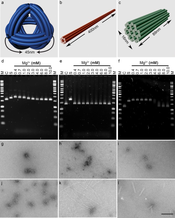
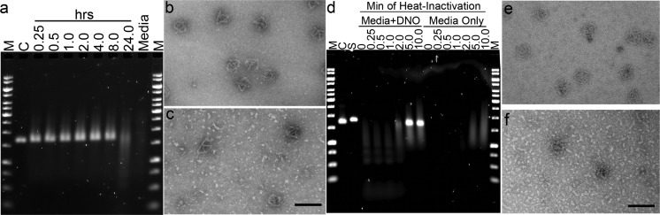
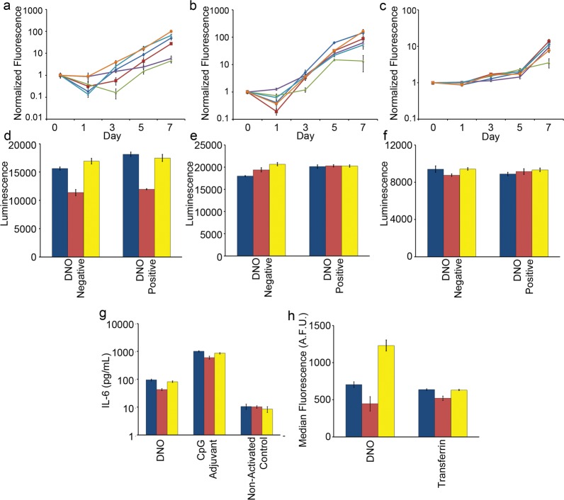
DNA nanotechnology is an advanced technique that could contribute diagnostic, therapeutic, and biomedical research devices to nanomedicine. Although such devices are often developed and demonstrated using in vitro tissue culture models, these conditions may not be compatible with DNA nanostructure integrity and function. The purpose of this study was to characterize the sensitivity of 3D DNA nanostructures produced via the origami method to the in vitro tissue culture environment and identify solutions to prevent loss of nanostructure integrity. We examined whether the physiological cation concentrations of cell culture medium and the nucleases present in fetal bovine serum (FBS) used as a medium supplement result in denaturation and digestion, respectively. DNA nanostructure denaturation due to cation depletion was design- and time-dependent, with one of four tested designs remaining intact after 24 h at 37 °C. Adjustment of medium by addition of MgSO4 prevented denaturation. Digestion of nanostructures by FBS nucleases in Mg(2+)-adjusted medium did not appear design-dependent and became significant within 24 h and when medium was supplemented with greater than 5% FBS. We estimated that medium supplemented with 10% FBS contains greater than 256 U/L equivalent of DNase I activity in digestion of DNA nanostructures. Heat inactivation at 75 °C and inclusion of actin protein in medium inactivated and inhibited nuclease activity, respectively. We examined the impact of medium adjustments on cell growth, viability, and phenotype. Adjustment of Mg(2+) to 6 mM did not appear to have a detrimental impact on cells. Heat inactivation was found to be incompatible with in vitro tissue culture, whereas inclusion of actin had no observable effect on growth and viability. In two in vitro assays, immune cell activation and nanoparticle endocytosis, we show that using conditions compatible with cell phenotype and nanostructure integrity is critical for obtaining reliable experimental data. Our study thus describes considerations that are vital for researchers undertaking in vitro tissue culture studies with DNA nanostructures and some potential solutions for ensuring that nanostructure integrity and functions are maintained during experiments.

Keywords: DNA nanotechnology; DNA origami; cation; cells; in vitro; nanorobot; nuclease; stability; structural integrity; tissue culture.


Figures

Figure 1
Figure 1
DNA nanostructure sensitivity to cation depletion in tissue culture medium. The three test nanostructures are (a) DNA nano-octahedron (DNO), (b) six-helix bundle nanotube (NT), and (c) 24-helix nanorod (NR). (d–f) Three nanostructures were incubated for 24 h at 37 °C in unmodified RPMI tissue culture medium containing 0.4 mM Mg2+ or adjusted to 0.7–10 mM Mg2+, and the products were analyzed by agarose gel electrophoresis (AGE). In comparison to the control lanes of stable sample, migration of the DNO and NR is retarded after low Mg2+ incubation, indicative of denaturation, whereas no obvious difference in migration is observed with the NT. (g–i) Transmission electron microscopy images of nanostructures incubated in unmodified medium, showing varying levels of denaturation. (j–l) With RPMI medium adjusted to 6 mM Mg2+, structural integrity is maintained in all three designs. M = molecular weight ladder, C = nanostructure in TE + 10 mM Mg2+, S = M13 scaffold. Scale bar = 100 nm.
Figure 2
Figure 2
Kinetics of nanostructure digestion by nucleases present in serum and heat inactivation. (a) DNO nanostructure (5 nM) was incubated at 37 °C for 0.25–24 h in RPMI + 6 mM Mg2+ + 10% FBS. Analysis by AGE shows smearing and decreased intensity of the product band as digestion progresses. (b) TEM image of DNO control sample showing intact nanostructures. (c) Partially digested DNO sample after 2 h incubation in medium + 10% FBS. Top left: example of a partially digested DNO. (d) Aliquots of FBS were heat-treated for 0.25–10.0 min at 75 °C prior to medium preparation. DNO was incubated for 24 h at 37 °C and analyzed by AGE. Medium without nanostructures was analyzed on the right half of the gel, showing a change in appearance from 5 and 10 min heat treatment. (e) TEM of a control and (f) DNO incubated in RPMI medium prepared with FBS heat-inactivated for 5 min. M = molecular weight ladder, C = nanostructure in TE + 10 mM Mg2+, S = M13 scaffold. Scale bars = 100 nm.
Figure 3
Figure 3
Measuring the impact of medium modifications on cell growth, viability and phenotype. (a) Mouse 3T3 fibroblasts, (b) human HEK-293 embryonic kidney, and (c) human H441 adenocarcinoma cell lines were seeded into standard medium (dark blue line), Mg2+-adjusted (red line), heat-inactivated FBS (green line), Mg2+ + heat-inactivated FBS (purple line), 200 nM actin (light blue line), or combination Mg2+ + 200 nM actin medium (orange line). Change in cell number was estimated by fluorescence of CyQuant stain over time and was normalized to signal on day 0. Cell viability and metabolism of (d) 3T3, (e) HEK-293, and (f) H441 cells were profiled after 24 h incubation with and without 5 nM DNO. (g) Primary mouse immune cells were isolated from spleens and incubated with 1 nM DNO, an equivalent mass of CpG phosphorothioate oligonucleotide adjuvant, or medium only, and the concentration of IL-6 cytokine released into the supernatant was determined by ELISA. (h) 3T3 fibroblast cells were incubated for 16 h with 1 nM fluorescently labeled DNO or transferrin, and endocytosis of the two agents was measured by flow cytometry after extensive washing to remove surface-bound particles. (d–h) Culture conditions are standard medium (blue), medium + 6 mM Mg2+ + heat-inactivated FBS (red), or medium + 6 mM Mg2+ + FBS + 200 nM actin (yellow). (a–h) Error bars represent standard error of the mean of n = 5 replicates.

References

    1. Seeman N. C. Nanomaterials Based on DNA. Annu. Rev. Biochem. 2010, 79, 65–87. - PMC - PubMed
    1. Rothemund P. W. K. Folding DNA To Create Nanoscale Shapes and Patterns. Nature 2006, 440, 297–302. - PubMed
    1. Douglas S. M.; Dietz H.; Liedl T.; Högberg B.; Graf F.; Shih W. M. Self-Assembly of DNA into Nanoscale Three-Dimensional Shapes. Nature 2009, 459, 414–418. - PMC - PubMed
    1. Dietz H.; Douglas S. M.; Shih W. M. Folding DNA into Twisted and Curved Nanoscale Shapes. Science 2009, 325, 725–730. - PMC - PubMed
    1. Liedl T.; Högberg B.; Tytell J.; Ingber D. E.; Shih W. M. Self-Assembly of Three-Dimensional Prestressed Tensegrity Structures from DNA. Nat. Nanotechnol. 2010, 5, 520–524. - PMC - PubMed

Publication types