. 2014 Aug 19;9(8):e102684.
doi: 10.1371/journal.pone.0102684. eCollection 2014.

Selective HDAC inhibition for the disruption of latent HIV-1 infection

Affiliations

Selective HDAC inhibition for the disruption of latent HIV-1 infection

Kirston M Barton et al. PLoS One. .

Abstract

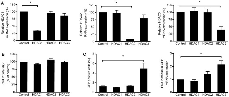
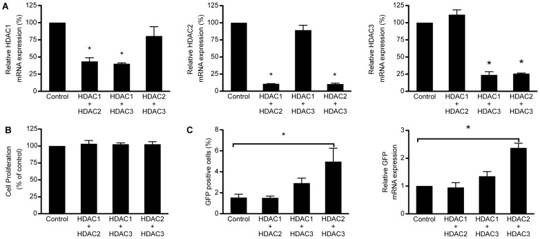
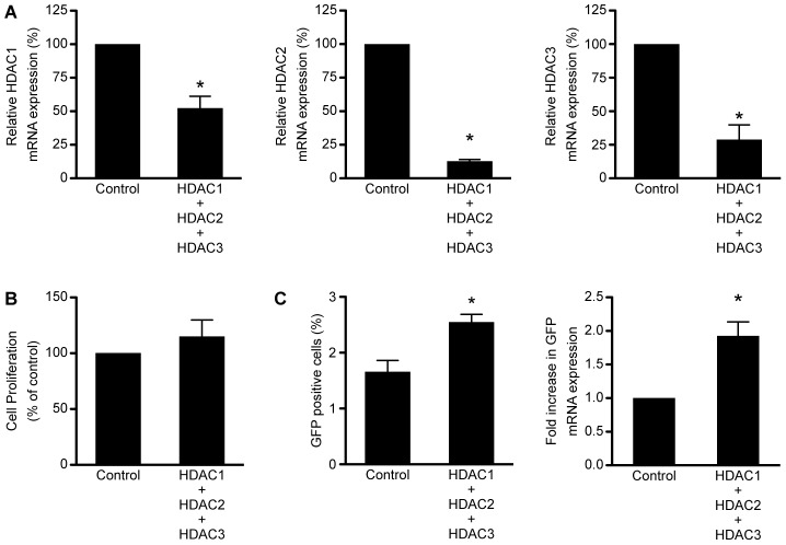
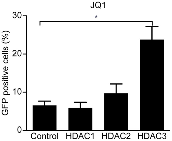
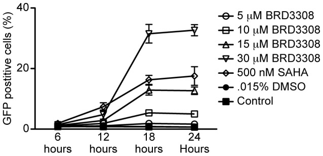
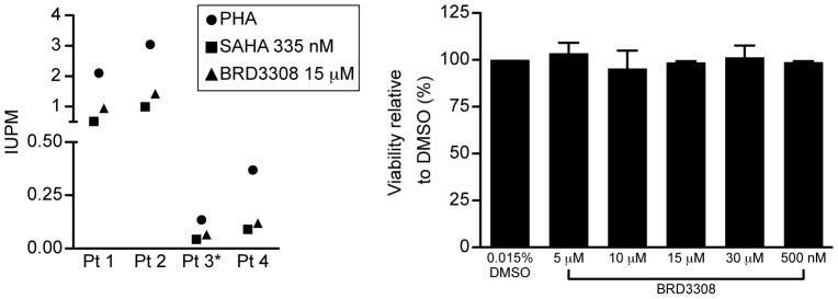
Selective histone deacetylase (HDAC) inhibitors have emerged as a potential anti-latency therapy for persistent human immunodeficiency virus type 1 (HIV-1) infection. We utilized a combination of small molecule inhibitors and short hairpin (sh)RNA-mediated gene knockdown strategies to delineate the key HDAC(s) to be targeted for selective induction of latent HIV-1 expression. Individual depletion of HDAC3 significantly induced expression from the HIV-1 promoter in the 2D10 latency cell line model. However, depletion of HDAC1 or -2 alone or in combination did not significantly induce HIV-1 expression. Co-depletion of HDAC2 and -3 resulted in a significant increase in expression from the HIV-1 promoter. Furthermore, concurrent knockdown of HDAC1, -2, and -3 resulted in a significant increase in expression from the HIV-1 promoter. Using small molecule HDAC inhibitors of differing selectivity to ablate the residual HDAC activity that remained after (sh)RNA depletion, the effect of depletion of HDAC3 was further enhanced. Enzymatic inhibition of HDAC3 with the selective small-molecule inhibitor BRD3308 activated HIV-1 transcription in the 2D10 cell line. Furthermore, ex vivo exposure to BRD3308 induced outgrowth of HIV-1 from resting CD4+ T cells isolated from antiretroviral-treated, aviremic HIV+ patients. Taken together these findings suggest that HDAC3 is an essential target to disrupt HIV-1 latency, and inhibition of HDAC2 may also contribute to the effort to purge and eradicate latent HIV-1 infection.


Conflict of interest statement

Competing Interests: D.M.M. is a consultant for and has received grants from Merck Research Laboratories. A.S.E. is an employee of Merck Research Laboratories. E.B.H. is named on patents for HDAC inhibitors held by the Broad Institute and is an equity holder at KDAc Therapeutics. E.B.H. and F.F.W. are both consultants to KDAc Therapeutics, which has licensed the patent estate around HDAC3 selective compounds and HDAC inhibitors. This does not alter the authors' adherence to PLOS ONE policies on sharing data and materials. All remaining authors declare no conflicts of interest.

Figures

Figure 1
Figure 1. Depletion of HDAC3 significantly increases expression from the HIV-1 promoter.
A. Expression of the indicated HDAC mRNAs following shRNA mediated depletion of HDAC1, −2, or −3 in 2D10 cells. Depletion of HDAC1, −2, or −3 was significant and specific for the targeted HDAC. B. Cell proliferation was not significantly affected following depletion of HDAC1, −2, or −3 in 2D10 cells. C. The percentage of 2D10 cells expressing GFP protein from the HIV-1 promoter following depletion significantly increased following depletion of HDAC3 in comparison to control cells transduced with the scrambled shRNA, but not following depletion of HDAC1 or −2. The same effect was seen when GFP mRNA expression was directly measured. Expression was not significantly altered from baseline following transduction with non-specific shRNA. (*p<0.05).
Figure 2
Figure 2. Co-depletion of HDAC2 and HDAC3 significantly increases expression from the HIV-1 promoter.
A. Expression of the indicated HDAC mRNAs following shRNA mediated depletion of HDAC1 and −2, HDAC1 and −3, or HDAC2 and −3. The depletion was significant and specific for the targeted HDACs. B. Cell proliferation and viability of 2D10 cells that were transduced with shRNAs targeting HDAC1 and −2, HDAC1 and −3, or HDAC2 and −3. No significant changes in cell proliferation and viability were observed following co- depletion of these HDACs as compared to cells that were transduced with the scrambled shRNA control. C. Expression of GFP from the HIV-1 promoter following depletion of HDAC2 and −3 increased compared to the control cells transduced with a scrambled shRNA. However, depletion of HDAC1 and −2 or HDAC1 and −3 did not have a significant effect on GFP expression. This same result was seen when GFP mRNA expression was directly measured. Expression was not significantly altered from baseline following transduction with non-specific shRNA. (*p<0.05).
Figure 3
Figure 3. Depletion of HDAC1, −2, and −3 significantly increases expression from the HIV-1 promoter.
A. Fold change of HDAC1, −2, and −3 mRNA expression as compared to 2D10 cells transduced with the scrambled shRNA control. A significant reduction in the mRNA expression of all three HDACs was observed. B. Cell viability and proliferation as a percentage of the 2D10 cells transduced with the scrambled shRNA control. Depletion of HDAC1, −2, and −3 did not significantly affect cell viability and proliferation. C. A significant increase in the percentage of cells expressing GFP protein from the HIV-1 promoter was observed following depletion of HDAC1, −2, and −3. Furthermore, a significant increase in expression of GFP mRNA from the HIV-1 promoter was observed following depletion of HDAC1, −2, and −3. (*p<0.05).
Figure 4
Figure 4. Chemical inhibition of HDACs following depletion of HDAC3 significantly increases expression from the HIV-1 promoter.
A. 2D10 cell that had been depleted of HDAC1, −2, or −3 or had been infected with the scrambled control lentivirus were exposed to 0.015% DMSO as the vehicle control for 24 hours. B. Chemical inhibition of HDAC1 and −2 using Mrk 12 (20 µM) does not result in a significant increase in the percentage of 2D10 cells expressing GFP following depletion of HDAC1 or −2. However, similar to depletion of HDAC3 alone, a significant increase in the percent of cells expressing GFP was observed when Mrk 12 was added to cells depleted of HDAC3. C. Chemical inhibition of HDAC1, −2 and −3 with Mrk 13 (200 nM) resulted in a significant increase in the percent of GFP positive 2D10 cells in cells depleted of HDAC2 or −3, but not HDAC1. D. A submaximal (250 nM) concentration of SAHA resulted in a significant increase in the percent of cells expressing GFP following depletion of HDAC2 and −3. E. A maximal concentration of SAHA (500 nM) resulted in a significant increase in the percent of cells expressing GFP in cells depleted of HDAC3. However, in D. or E. depletion of HDAC1 did not increase the percent of cells expressing GFP. F. Chemical inhibition of HDACs with the non-selective inhibitor TSA (25 nM) resulted in a significant increase in the percent of GFP expressing 2D10 cells in cells depleted of HDAC3, but not HDAC1 or HDAC2. G. Chemical inhibition of HDAC3, −6, and −8 with the selective HDAC inhibitor Droxinostat (2 µM) resulted in a significant increase in the percent of 2D10 cells expressing GFP in cells that had been depleted of HDAC3 but not HDAC1 or −2. (*p<0.05).
Figure 5
Figure 5. JQ1 enhances the effects of HDAC3 depletion.
A significant increase in GFP protein expression from the HIV-1 promoter was observed in 2D10 cells that had been depleted of HDAC3 and had been treated with JQ1 (50 nM) for 24 hours over cells that were transduced with the scrambled shRNA control and treated with JQ1. However, no effect was observed in cells depleted of HDAC1 or HDAC2. (*p<0.05).
Figure 6
Figure 6. An HDAC3 selective inhibitor induces HIV-1 expression from 2D10 cells.
2D10 cells were treated with BRD3308 at the indicated concentration for 6 hours, 12 hours, 18 hours, or 24 hours and collected for analysis at 24 hours. The percent of 2D10 cells expressing GFP was measured using flow cytometry. HIV-1 expression was observed with 10 µM of BRD3308 and higher after 12 hours of treatment.
Figure 7
Figure 7. Inhibition of HDAC3 induces outgrowth of HIV-1 from latently infected patient cells ex vivo.
A. HIV-1 outgrowth from latently infected CD4+ T cells following overnight exposure to 15 µM BRD3308 or 335 nM SAHA. The frequency of viral outgrowth following mitogen stimulation of HDAC inhibitor exposure is expressed as infected cells per million resting CD4+ T cells (IUPM). In four patients (Pt 1, Pt 2, Pt 3, and Pt 4) exposure to BRD3308 induced a comparable frequency of outgrowth to that observed following exposure to SAHA. *IUPM for PHA for Pt.3 is obtained from three prior assays; all other assays are contemporaneous. B. PBMCs were exposed to 5, 10, 15, or 30 µM of BRD3308, and viability was measured. The results are expressed as the viability relative to the control DMSO condition. BRD3308 does not significantly affect viability of PBMCs.

Similar articles

Cited by

References

    1. Finzi D, Hermankova M, Pierson T, Carruth LM, Buck C, et al. (1997) Identification of a reservoir for HIV-1 in patients on highly active antiretroviral therapy. Science 278: 1295–1300. - PubMed
    1. Finzi D, Blankson J, Siliciano JD, Margolick JB, Chadwick K, et al. (1999) Latent infection of CD4+ T cells provides a mechanism for lifelong persistence of HIV-1, even in patients on effective combination therapy. Nat Med 5: 512–517. - PubMed
    1. Chun TW, Engel D, Berrey MM, Shea T, Corey L, et al. (1998) Early establishment of a pool of latently infected, resting CD4(+) T cells during primary HIV-1 infection. Proc Natl Acad Sci U S A 95: 8869–8873. - PMC - PubMed
    1. UNAIDS (2008) 2008 Report on the global AIDS epidemic.
    1. Ganchi PA, Sun SC, Greene WC, Ballard DW (1992) I kappa B/MAD-3 masks the nuclear localization signal of NF-kappa B p65 and requires the transactivation domain to inhibit NF-kappa B p65 DNA binding. Mol Biol Cell 3: 1339–1352. - PMC - PubMed

Publication types

MeSH terms