

High IL-17E and low IL-17C dermal expression identifies a fibrosis-specific motif common to morphea and systemic sclerosis
- PMID: 25136988
- PMCID: PMC4138152
- DOI: 10.1371/journal.pone.0105008
High IL-17E and low IL-17C dermal expression identifies a fibrosis-specific motif common to morphea and systemic sclerosis
Abstract
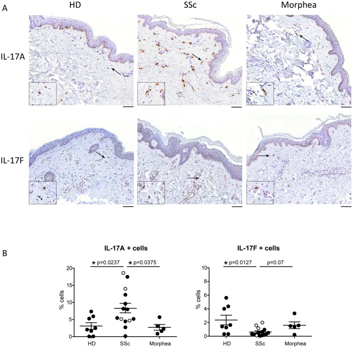
Background: High interleukin (IL)-17A levels are characteristically found in the skin of systemic sclerosis (SSc) individuals. Our aim was to investigate whether the dermal expression of IL-17A and related IL-17 family members (i.e. IL-17C, IL-17E and IL-17F) could distinguish fibrotic from healthy skin and could show similarities in SSc and morphea, two disorders with presumed distinct pathogenesis, but characterized by skin fibrosis.
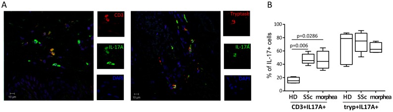
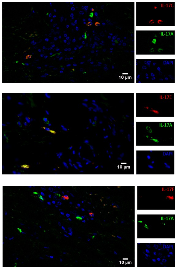
Methods: Biopsies were obtained from the involved skin of 14 SSc, 5 morphea and 8 healthy donors (HD) undergoing plastic surgery. Immunohistochemistry/immunofluorescence techniques were coupled to a semi-automated imaging quantification approach to determine the presence of the IL-17 family members in the skin. The in vitro effects induced by the IL-17 family members on fibroblasts from normal and SSc individuals were assessed by ELISA and RIA.
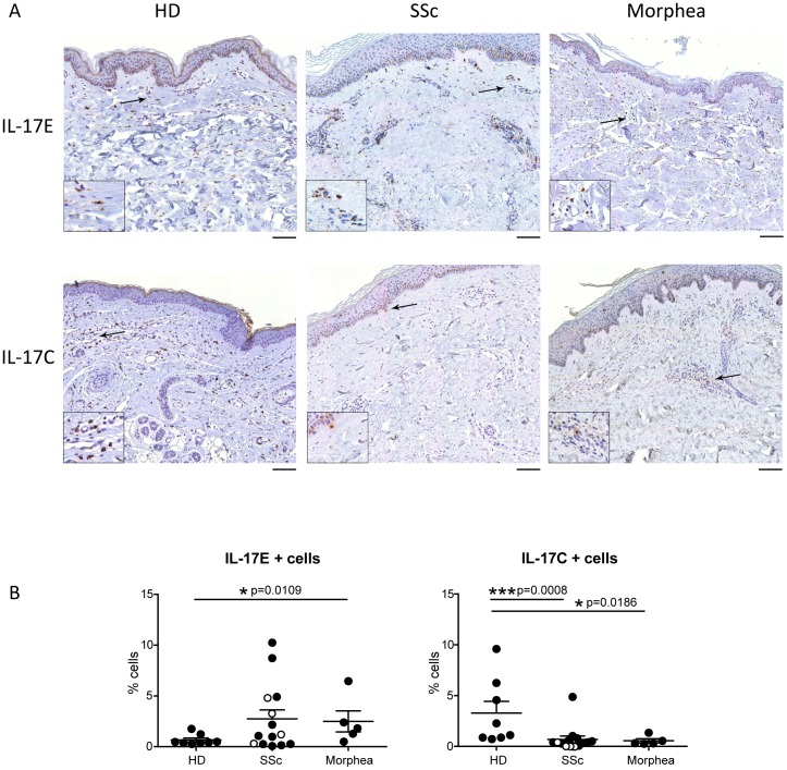
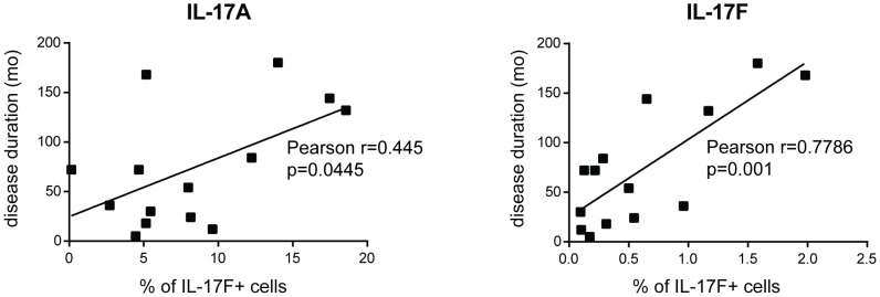
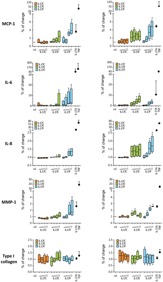
Results: Positive cells for each of the IL-17 isoforms investigated were present in the dermis of all the individuals tested, though with variable frequencies. SSc individuals had increased frequency of IL-17A+ (p = 0.0237) and decreased frequency of IL-17F+ (p = 0.0127) and IL-17C+ cells (p = 0.0008) when compared to HD. Similarly, morphea individuals had less frequent IL-17C+ cells (p = 0.0186) in their skin but showed similar number of IL-17A+ and IL-17F+ cells when compared to HD. Finally, IL-17E+ cells were more numerous in morphea (p = 0.0109) and tended to be more frequent in SSc than in HD. Fibroblast production of IL-6, MMP-1 and MCP-1 was enhanced in a dose-dependent manner in the presence of IL-17E and IL-17F, but not in the presence of IL-17C. None of the cytokine tested had significant effect on type I collagen production. Of interest, in SSc the frequency of both IL-17A and IL-17F positive cells increased with disease duration.
Conclusions: The frequency of IL-17A and IL-17F distinguish SSc to morphea individuals while dermal expression of IL-17C (low) and IL-17E (high) identifies a fibrosis-specific motif. The specific IL-17C/IL-17E cytokine combination may thus play a role in the development of fibrosis.
Conflict of interest statement
Figures
Similar articles
-
Interleukin-17A+ cell counts are increased in systemic sclerosis skin and their number is inversely correlated with the extent of skin involvement.Arthritis Rheum. 2013 May;65(5):1347-56. doi: 10.1002/art.37860. Arthritis Rheum. 2013. PMID: 23335253
-
IL-22 capacitates dermal fibroblast responses to TNF in scleroderma.Ann Rheum Dis. 2016 Sep;75(9):1697-705. doi: 10.1136/annrheumdis-2015-207477. Epub 2015 Oct 9. Ann Rheum Dis. 2016. PMID: 26452537
-
Th17 cells favor inflammatory responses while inhibiting type I collagen deposition by dermal fibroblasts: differential effects in healthy and systemic sclerosis fibroblasts.Arthritis Res Ther. 2013 Oct 10;15(5):R151. doi: 10.1186/ar4334. Arthritis Res Ther. 2013. PMID: 24289089 Free PMC article.
-
Abnormal collagen V deposition in dermis correlates with skin thickening and disease activity in systemic sclerosis.Autoimmun Rev. 2012 Sep;11(11):827-35. doi: 10.1016/j.autrev.2012.02.017. Epub 2012 Mar 2. Autoimmun Rev. 2012. PMID: 22406224 Review.
-
Recent Advances in Treatment of Systemic Sclerosis and Morphea.Am J Clin Dermatol. 2024 Mar;25(2):213-226. doi: 10.1007/s40257-023-00831-2. Epub 2023 Dec 12. Am J Clin Dermatol. 2024. PMID: 38087156 Review.
Cited by
-
Interleukin-17 Family Cytokines in Metabolic Disorders and Cancer.Genes (Basel). 2022 Sep 13;13(9):1643. doi: 10.3390/genes13091643. Genes (Basel). 2022. PMID: 36140808 Free PMC article. Review.
-
Therapeutic Options for Systemic Sclerosis: Current and Future Perspectives in Tackling Immune-Mediated Fibrosis.Biomedicines. 2022 Jan 29;10(2):316. doi: 10.3390/biomedicines10020316. Biomedicines. 2022. PMID: 35203525 Free PMC article. Review.
-
The IL-17 Family of Cytokines in Psoriasis: IL-17A and Beyond.Front Immunol. 2018 Aug 2;9:1682. doi: 10.3389/fimmu.2018.01682. eCollection 2018. Front Immunol. 2018. PMID: 30127781 Free PMC article. Review.
-
The role of the acquired immune response in systemic sclerosis.Semin Immunopathol. 2015 Sep;37(5):519-28. doi: 10.1007/s00281-015-0509-1. Epub 2015 Jul 8. Semin Immunopathol. 2015. PMID: 26152639 Review.
-
The role of interleukin-17 in the pathogenesis of systemic sclerosis: Pro-fibrotic or anti-fibrotic?J Scleroderma Relat Disord. 2021 Oct;6(3):227-235. doi: 10.1177/23971983211039421. Epub 2021 Aug 14. J Scleroderma Relat Disord. 2021. PMID: 35387209 Free PMC article. Review.
References
-
- Gabrielli A, Avvedimento EV, Krieg T (2009) Scleroderma. N Engl J Med 360: 1989–2003. - PubMed
-
- Chizzolini C, Brembilla NC, Montanari E, Truchetet ME (2011) Fibrosis and immune dysregulation in systemic sclerosis. Autoimmun Rev 10: 276–281. - PubMed
-
- Meier FM, Frommer KW, Dinser R, Walker UA, Czirjak L, et al. (2012) Update on the profile of the EUSTAR cohort: an analysis of the EULAR Scleroderma Trials and Research group database. Ann Rheum Dis 71: 1355–1360. - PubMed
-
- Yaqub A, Chung L, Rieger KE, Fiorentino DF (2013) Localized cutaneous fibrosing disorders. Rheum Dis Clin North Am 39: 347–364. - PubMed
-
- Chung L, Lin J, Furst DE, Fiorentino D (2006) Systemic and localized scleroderma. Clin Dermatol 24: 374–392. - PubMed
Publication types
MeSH terms
Substances
LinkOut - more resources
Full Text Sources
Other Literature Sources
Medical
Miscellaneous
