

In vivo fluorescence imaging and urinary monoamines as surrogate biomarkers of disease progression in a mouse model of pheochromocytoma
- PMID: 25137029
- PMCID: PMC4256828
- DOI: 10.1210/en.2014-1431
In vivo fluorescence imaging and urinary monoamines as surrogate biomarkers of disease progression in a mouse model of pheochromocytoma
Abstract
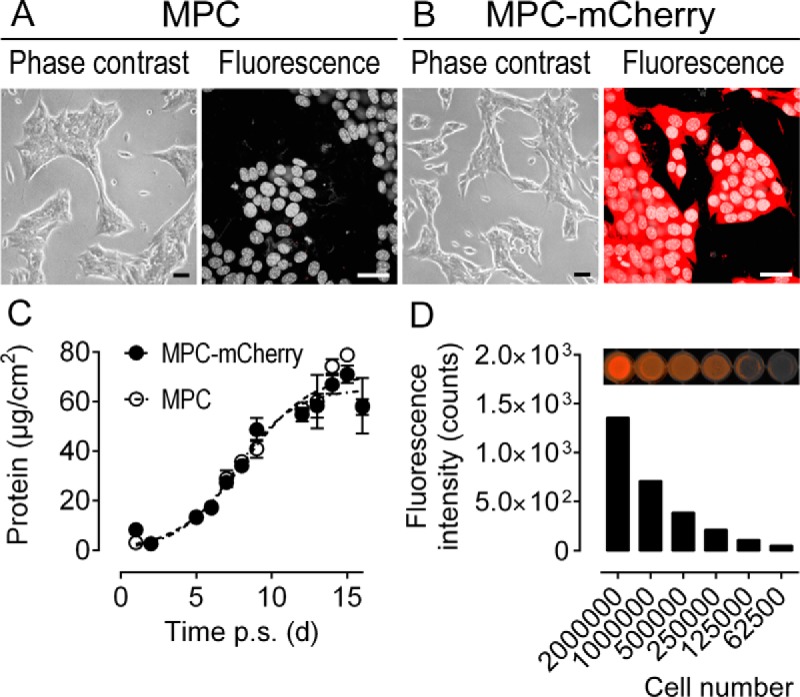
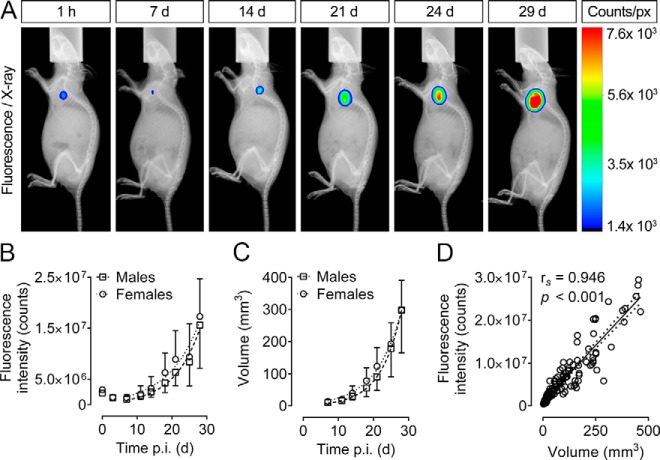
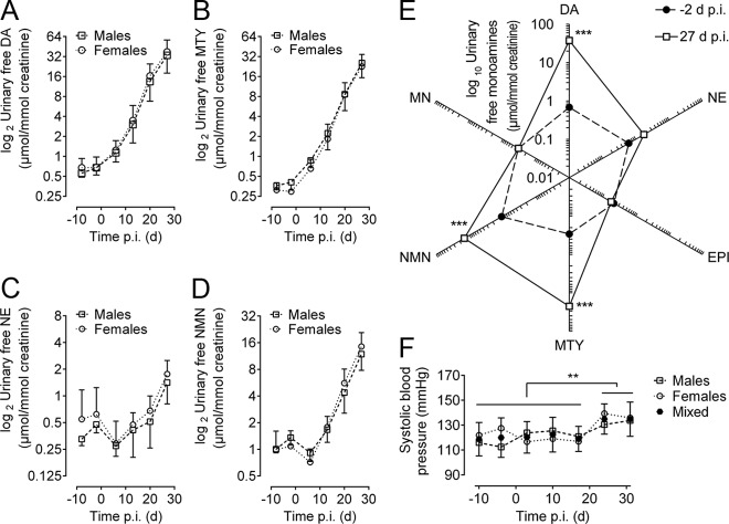
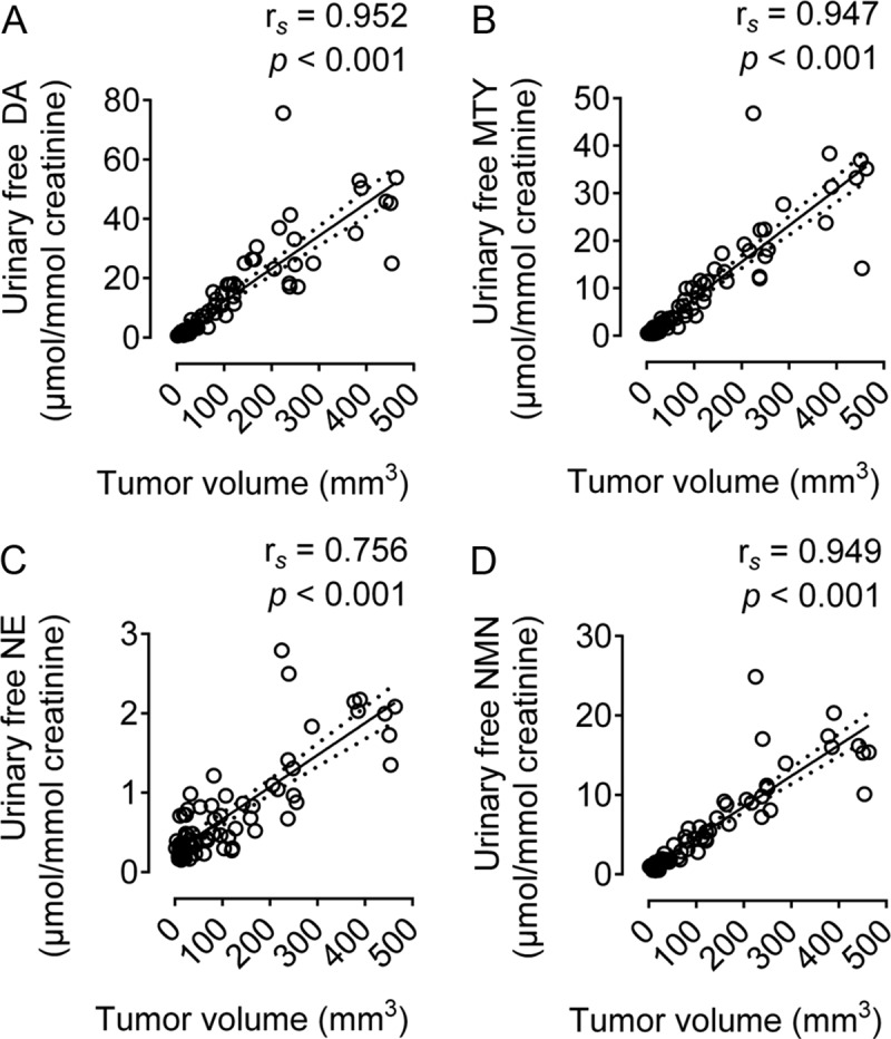
Pheochromocytoma (PHEO) is a rare but potentially lethal neuroendocrine tumor arising from catecholamine-producing chromaffin cells. Especially for metastatic PHEO, the availability of animal models is essential for developing novel therapies. For evaluating therapeutic outcome in rodent PHEO models, reliable quantification of multiple organ lesions depends on dedicated small-animal in vivo imaging, which is still challenging and only available at specialized research facilities. Here, we investigated whether whole-body fluorescence imaging and monitoring of urinary free monoamines provide suitable parameters for measuring tumor progression in a murine allograft model of PHEO. We generated an mCherry-expressing mouse PHEO cell line by lentiviral gene transfer. These cells were injected subcutaneously into nude mice to perform whole-body fluorescence imaging of tumor development. Urinary free monoamines were measured by liquid chromatography with tandem mass spectrometry. Tumor fluorescence intensity and urinary outputs of monoamines showed tumor growth-dependent increases (P < .001) over the 30 days of monitoring post-tumor engraftment. Concomitantly, systolic blood pressure was increased significantly during tumor growth. Tumor volume correlated significantly (P < .001) and strongly with tumor fluorescence intensity (rs = 0.946), and urinary outputs of dopamine (rs = 0.952), methoxytyramine (rs = 0.947), norepinephrine (rs = 0.756), and normetanephrine (rs = 0.949). Dopamine and methoxytyramine outputs allowed for detection of lesions at diameters below 2.3 mm. Our results demonstrate that mouse pheochromocytoma (MPC)-mCherry cell tumors are functionally similar to human PHEO. Both tumor fluorescence intensity and urinary outputs of free monoamines provide precise parameters of tumor progression in this sc mouse model of PHEO. This animal model will allow for testing new treatment strategies for chromaffin cell tumors.
Figures
Similar articles
-
Deconjugated urinary metanephrine, normetanephrine and 3-methoxytyramine in laboratory diagnosis of pheochromocytoma and paraganglioma.Physiol Res. 2015;64(Suppl 2):S313-22. doi: 10.33549/physiolres.933109. Physiol Res. 2015. PMID: 26680494
-
Strain-specific metastatic phenotypes in pheochromocytoma allograft mice.Endocr Relat Cancer. 2018 Oct 5;25(12):993-1004. doi: 10.1530/ERC-18-0136. Endocr Relat Cancer. 2018. PMID: 30288966 Free PMC article.
-
Urinary catecholamine and metanephrine to creatinine ratios in dogs with hyperadrenocorticism or pheochromocytoma, and in healthy dogs.J Vet Intern Med. 2010 Sep-Oct;24(5):1093-7. doi: 10.1111/j.1939-1676.2010.0578.x. Epub 2010 Aug 12. J Vet Intern Med. 2010. PMID: 20707840
-
Genetics, diagnosis, and management of medullary thyroid carcinoma and pheochromocytoma/paraganglioma.Endocr Pract. 2014 Feb;20(2):176-87. doi: 10.4158/EP13268.RA. Endocr Pract. 2014. PMID: 24449662 Review.
-
[Pheochromocytoma: update on diagnosis and therapy].Dtsch Med Wochenschr. 2014 Mar;139(10):486-90. doi: 10.1055/s-0033-1360082. Epub 2014 Feb 25. Dtsch Med Wochenschr. 2014. PMID: 24570195 Review. German.
Cited by
-
Primary fibroblast co-culture stimulates growth and metabolism in Sdhb-impaired mouse pheochromocytoma MTT cells.Cell Tissue Res. 2018 Dec;374(3):473-485. doi: 10.1007/s00441-018-2907-x. Epub 2018 Aug 29. Cell Tissue Res. 2018. PMID: 30159755 Free PMC article.
-
Instant kit preparation of 68Ga-radiopharmaceuticals via the hybrid chelator DATA: clinical translation of [68Ga]Ga-DATA-TOC.EJNMMI Res. 2019 May 23;9(1):48. doi: 10.1186/s13550-019-0516-7. EJNMMI Res. 2019. PMID: 31123943 Free PMC article.
-
Deciphering the Tumor Uptake of Heterobivalent (SST2/Albumin) [64Cu]Cu-NODAGA-cLAB-TATEs.J Med Chem. 2025 Jun 12;68(11):12029-12046. doi: 10.1021/acs.jmedchem.5c00890. Epub 2025 May 20. J Med Chem. 2025. PMID: 40393943 Free PMC article.
-
Imaging pheochromocytoma in small animals: preclinical models to improve diagnosis and treatment.EJNMMI Res. 2021 Dec 11;11(1):121. doi: 10.1186/s13550-021-00855-x. EJNMMI Res. 2021. PMID: 34894301 Free PMC article. Review.
-
Impact of Extrinsic and Intrinsic Hypoxia on Catecholamine Biosynthesis in Absence or Presence of Hif2α in Pheochromocytoma Cells.Cancers (Basel). 2019 Apr 28;11(5):594. doi: 10.3390/cancers11050594. Cancers (Basel). 2019. PMID: 31035382 Free PMC article.
References
-
- Harari A, Inabnet WB., 3rd Malignant pheochromocytoma: A review. Am J Surg. 2011; 201(5):700–708. - PubMed
-
- Lenders JW, Eisenhofer G, Mannelli M, Pacak K. Phaeochromocytoma. Lancet. 2005;366(9486):665–675. - PubMed
-
- Jemal A, Bray F, Center MM, Ferlay J, Ward E, Forman D. Global cancer statistics. CA Cancer J Clin. 2011;61(2):69–90. - PubMed
-
- Elder EE, Elder G, Larsson C. Pheochromocytoma and functional paraganglioma syndrome: No longer the 10% tumor. J Surg Oncol. 2005;89(3):193–201. - PubMed
-
- Harding JL, Yeh MW, Robinson BG, Delbridge LW, Sidhu SB. Potential pitfalls in the diagnosis of phaeochromocytoma. Med J Aust. 2005;182(12):637–640. - PubMed
Publication types
MeSH terms
Substances
LinkOut - more resources
Full Text Sources
Other Literature Sources
Medical
