. 2015 Mar;52(3):377-86.
doi: 10.1165/rcmb.2014-0142OC.

Wood smoke enhances cigarette smoke-induced inflammation by inducing the aryl hydrocarbon receptor repressor in airway epithelial cells

Affiliations

Wood smoke enhances cigarette smoke-induced inflammation by inducing the aryl hydrocarbon receptor repressor in airway epithelial cells

Elias G Awji et al. Am J Respir Cell Mol Biol. 2015 Mar.

Abstract

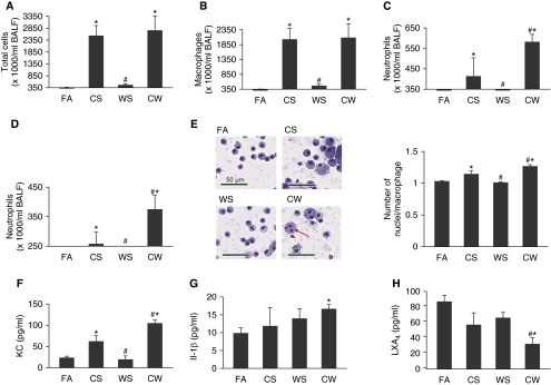
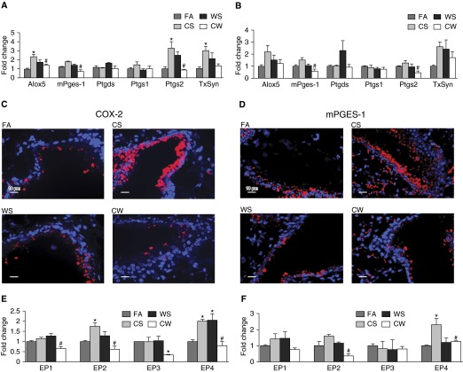
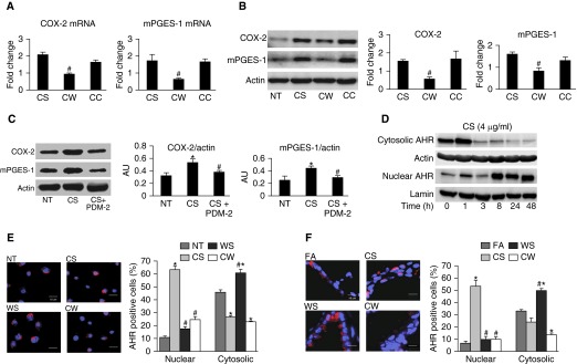
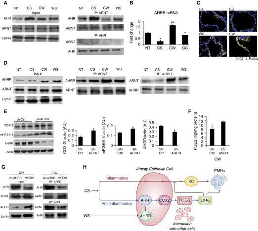
Our previous studies showed that cigarette smokers who are exposed to wood smoke (WS) are at an increased risk for chronic bronchitis and reduced lung function. The present study was undertaken to determine the mechanisms for WS-induced adverse effects. We studied the effect of WS exposure using four cohorts of mice. C57Bl/6 mice were exposed for 4 or 12 weeks to filtered air, to 10 mg/m(3) WS for 2 h/d, to 250 mg/m(3) cigarette smoke (CS) for 6 h/d, or to CS followed by WS (CW). Inflammation was absent in the filtered air and WS groups, but enhanced by twofold in the bronchoalveolar lavage of the CW compared with CS group as measured by neutrophil numbers and levels of the neutrophil chemoattractant, keratinocyte-derived chemokine. The levels of the anti-inflammatory lipoxin, lipoxin A4, were reduced by threefold along with cyclo-oxygenase (COX)-2 and microsomal prostaglandin E synthase (mPGES)-1 in airway epithelial cells and PGE2 levels in the bronchoalveolar lavage of CW compared with CS mice. We replicated, in primary human airway epithelial cells, the changes observed in mice. Immunoprecipitations showed that WS blocked the interaction of aryl hydrocarbon receptor (AHR) with AHR nuclear transporter to reduce expression of COX-2 and mPGES-1 by increasing expression of AHR repressor (AHRR). Collectively, these studies show that exposure to low concentrations of WS enhanced CS-induced inflammation by inducing AHRR expression to suppress AHR, COX-2, and mPGES-1 expression, and levels of PGE2 and lipoxin A4. Therefore, AHRR is a potential therapeutic target for WS-associated exacerbations of CS-induced inflammation.

Keywords: air pollution; arachidonic acid pathway; exacerbation; lipoxin; neutrophilic inflammation.


Figures

Figure 1.
Figure 1.
Wood smoke (WS) exacerbates cigarette smoke (CS)-induced inflammation in mice. Total (A) and differential cell counts of macrophages (B), neutrophils at 4 weeks (C), and 12 weeks (D) of exposure, in mice exposed to filtered air (FA), CS, WS, or CS followed by WS (CW). (E) Changes in macrophage phenotypes in the cytospins of bronchoalveolar lavage (BAL) cell pellets from mice exposed to FA, CS, WS, or CW at 4 weeks. Increased size and multinucleation of macrophages in CS and CW mice (left) and higher number of nuclei per macrophage in CW than CS mice are observed. Multinucleated cells are marked with a red arrow. Scale bars, 50 μm. BAL fluid (BALF) levels of the proinflammatory mediators, keratinocyte-derived chemokine (KC) after 12 weeks (F), IL-1β after 12 weeks (G), and the anti-inflammatory lipid mediator, lipoxin A4 (LXA4) after 4 weeks (H) of exposure of mice to FA, CS, WS, or CW and as measured by ELISA (n = 9 mice for each treatment). Data are presented as mean ± SEM. *P < 0.05 compared with FA; #P < 0.05 compared with CS.
Figure 2.
Figure 2.
WS modifies CS-induced expression of genes within the arachidonic acid (AA) pathway. mRNA levels of arachidonate 5-lipoxygenase (ALOX-5), prostaglandin (PG)–endoperoxide synthase 1 (PTGS-1; or cyclo-oxygenase [COX]-1), PTGS-2 (or COX-2), thromboxane synthase (TXsyn), PGD synthase (PTGDS), and microsomal PGE synthase (mPGES)-1 in whole lung tissues of mice exposed to FA, CS, WS, or CW for 4 (A) and 12 (B) weeks, and as determined by quantitative real-time PCR (qRT-PCR; n = 9 for each treatment). Immunofluorescence analysis of COX-2 (C) and mPGES-1 (D) proteins in lung tissue sections showing increased expression of both proteins predominantly in the bronchial epithelium of CS airways, and significantly reduced immunostaining in CW airways compared with CS airways. Scale bars, 10 μm. mRNAs for the four E-prostanoid (EP) receptors 1–4 in whole lung tissues of FA, CS, WS, and CW mice at 4 (E) and 12 weeks (F) of exposure, as determined by qRT-PCR. Data are presented as means ± SEM. *P < 0.05 compared with FA; #P < 0.05 compared with CS.
Figure 3.
Figure 3.
WS down-regulates CS-induced COX-2 and mPGES-1 levels in an aryl hydrocarbon receptor (AHR)–dependent manner. mRNA (A) and protein (B) levels in N3 cells treated with: (1) normal growth medium for 40 hours followed by a 3-hour treatment with fresh growth medium (nontreated (NT)); (2) CS (4 μg/ml) for 40 hours followed by a 3-hour treatment with normal growth medium (CS); (3) CS (4 μg/ml) for 40 hours followed by a 3-hour treatment with WS (10 ng/ml) (CW); or (4) CS (4 μg/ml) for 40 hours followed by a 3-hour treatment with CS (10 ng/ml) (CC) as an additional control. Means ± SEM (n ≥ 3 repeats for each treatment). #P < 0.05 compared with CS. (C) Inhibition of AHR with POU domain protein 2 (PDM-2) treatment reduced CS-induced COX-2 and mPGES-1 protein levels. Human airway epithelial cells (HAECs) were untreated (NT), treated with CS (4 μg/ml) for 40 hours, or pretreated with the selective AHR antagonist (PDM-2), followed by CS (4 μg/ml) for 40 hours (CS + PDM-2). AU, arbitrary units. (D) CS induces translocation of AHR from the cytoplasm to the nucleus in a time-dependent manner. Primary HAECs were treated with CS (4 μg/ml) for indicated times and AHR levels in the cytosolic and nuclear fractions were measured by Western blot (WB) analysis. Actin and lamin were used as loading controls. (E) WS down-regulates CS-induced AHR nuclear translocation in HAECs. Primary HAECs were treated as described previously here with NT, CS, CW, or WS (10 ng/ml) for 3 hours and immunofluorescence staining performed for AHR protein (red) and nuclei (blue). The percentage of cells showing positive staining for cytosolic and nuclear AHR is shown. Scale bars, 10 μm. (F) WS down-regulates CS-induced AHR nuclear translocation in airway cells of mice. Immunostaining and quantification for AHR protein expression was performed using lung tissues from mice exposed to FA, CS, CW, and WS, as described previously here for HAECs. Scale bars, 10 μm. Data presented are means ± SEM (n ≥ 3 repeats experiment for each treatment. *P < 0.05 compared with NT; #P < 0.05 compared with CS.
Figure 4.
Figure 4.
WS increases AHR repressor (AHRR) expression to suppress CS-induced AHR activity and suppress COX-2 and mPGES-1 expression, and AHRR deficiency mitigates the WS effect. (A) WS affects CS-induced AHR–AHR nuclear transporter (ARNT) interaction. HAECs were treated as described in Figure 3. Nuclear extracts were prepared and immunoprecipitation (IP)-immunoblot (IB) analyses were performed using antibodies to both AHR and ARNT. (B) CW induces higher AHRR mRNA expression than CS. The levels of AHRR mRNA were measured and normalized by cyclin-dependent kinase inhibitor 1 β (CDKN1β) levels (n ≥ 3). *P < 0.05 compared with NT; #P < 0.05 compared with CS. (C) WS increases AHRR protein expression in mice. Lung tissue sections from FA, CS, CW, and WS mice were stained with AHRR antibodies. Scale bars, 10 μm. (D) WS enhances AHRR–ARNT interaction. Nuclear extracts were prepared from HAECs treated as described in (A) with FA, CS, CW, and WS, and IP–IB analyses were performed using antibodies to both AHRR and ARNT. (E) Short hairpin–AHRR and sh control (sh-Ctrl) cells were treated with CW and COX-2, and mPGES-1 protein levels were measured by Western blotting. HAECs with reduced AHRR protein levels were generated by infecting parental cells with a retroviral vector expressing shRNA against AHRR (sh-AHRR). Control cells were infected with vector only (sh-Ctrl). (F) Supernatants from CW-treated cells showed a near-significant (P = 0.059) increase in PGE2 levels in sh-AHRR compared with sh-Ctrl cells. (G) Nuclear extracts were prepared from sh-AHRR and sh-Ctrl cells treated with CW, and IP followed by IB analyses were performed using antibodies to both ARNT and AHR. Nuclear extract was analyzed for AHR, ARNT, and AHRR by Western blotting (input). sh-AHRR cells show a greater AHR–ARNT interaction than sh-Ctrl cells. Data represent means ± SEM (n = 3). *P < 0.05 compared with sh-Ctrl. (H) Proposed model for WS-induced enhancement of CS-induced inflammation. CS induces KC expression and neutrophilic inflammation, but also induces AHR to increase COX-2 expression that activates other cell types to produce LXA4 to limit neutrophilic inflammation and KC production by airway epithelial cells. WS blocks this anti-inflammatory pathway of CS by up-regulating AHRR and suppressing AHR activity. PMNs, polymorphonuclear cells.

References

    1. Diette GB, Accinelli RA, Balmes JR, Buist AS, Checkley W, Garbe P, Hansel NN, Kapil V, Gordon S, Lagat DK, et al. Obstructive lung disease and exposure to burning biomass fuel in the indoor environment. Glob Heart. 2012;7:265–270. - PMC - PubMed
    1. Larson TV, Koenig JQ. Wood smoke: emissions and noncancer respiratory effects. Annu Rev Public Health. 1994;15:133–156. - PubMed
    1. Orozco-Levi M, Garcia-Aymerich J, Villar J, Ramírez-Sarmiento A, Antó JM, Gea J. Wood smoke exposure and risk of chronic obstructive pulmonary disease. Eur Respir J. 2006;27:542–546. - PubMed
    1. Gan WQ, FitzGerald JM, Carlsten C, Sadatsafavi M, Brauer M. Associations of ambient air pollution with chronic obstructive pulmonary disease hospitalization and mortality. Am J Respir Crit Care Med. 2013;187:721–727. - PubMed
    1. Sood A, Petersen H, Blanchette CM, Meek P, Picchi MA, Belinsky SA, Tesfaigzi Y. Wood smoke exposure and gene promoter methylation are associated with increased risk for COPD in smokers. Am J Respir Crit Care Med. 2010;182:1098–1104. - PMC - PubMed

Publication types

MeSH terms