Review
. 2014 Sep;15(9):815-23.
doi: 10.1038/ni.2938.

The self-obsession of T cells: how TCR signaling thresholds affect fate 'decisions' and effector function

Affiliations
Review

The self-obsession of T cells: how TCR signaling thresholds affect fate 'decisions' and effector function

Kristin A Hogquist et al. Nat Immunol. 2014 Sep.

Abstract

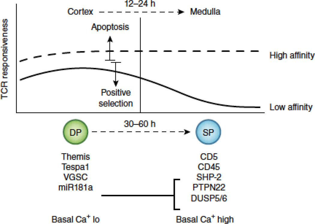
Self-reactivity was once seen as a potential characteristic of T cells that was eliminated by clonal selection to protect the host from autoimmune pathology. It is now understood that the T cell repertoire is in fact broadly self-reactive, even self-centered. The strength with which a T cell reacts to self ligands and the environmental context in which this reaction occurs influence almost every aspect of T cell biology, from development to differentiation to effector function. Here we highlight recent advances and discoveries that relate to T cell self-reactivity, with a particular emphasis on T cell antigen receptor (TCR) signaling thresholds.


Figures

Figure 1
Figure 1. TCR sensitivity changes as cells mature from cortical (DP) to medullary (SP) thymocytes
DP thymocytes reside in the cortex and respond efficiently to both low and high affinity TCR ligands. High affinity ligands can trigger apoptosis (clonal deletion), whereas low affinity ligands are more likely to induce survival and differentiation (positive selection). 12–24 hours after positive selection, DP thymocytes migrate to the medulla. At the same time they begin differentiating to the SP stage—a process that is not complete for another 1–2 days. In the medulla, SP thymocytes remain responsive to high affinity ligands, but their response to low affinity ligands decreases, a process sometimes referred to as “tuning”. DP thymocytes have a unique gene expression profile that serves two purposes. First, it allows them to interpret graded TCR signals in a digital fashion, to induce life or death in the cortex. The protein Themis is hypothesized to be crucial for this feature. Second, it allows them to respond more efficiently than SP thymocytes to low affinity ligands. Evidence suggests that the protein Tespa1, the voltage gated sodium channel (VGSC) and the microRNA mir181a all contribute to this property. Mir181a represses expression of several phosphatases (SHP-2, PTPN22, and DUSP5/6) that are known to negatively regulate TCR signaling. Another phosphatase (CD45) and a cell surface protein known to interact with phosphatases (CD5) are upregulated in SP thymocyte independently of mir181a. These gene expression changes, together with an increase in the basal level of intracelluar Ca++, are all thought to contribute to the developmental tuning of the TCR response.
Figure 2
Figure 2. The anatomic context of TCR signaling is crucial for thymocyte fate
Cortex: TCR signal strength is central in DP thymocyte fate outcome, but extrinsic factors provided by antigen presenting cells in the cortex are also key. Weak interactions between the nascent TCR and self-pMHC complexes on cortical thymic epithelial cells (cTEC) are crucial for positive selection. The pMHC repertoire expressed by cTEC is unique, due to the function of several genes involved in endogenous or endosomal proteolysis, such as β5T, TSSP, and cathepsin L. Although this unique repertoire is essential for efficient positive selection, we do not fully understand why. Strong interactions between the newly expressed TCR and self-pMHC complexes in the cortex classically induces apoptosis (clonal deletion) and co-stimulation by B7 family members enhances this outcome. Strong TCR signals can also induce cells to become IEL precursors (IELp), although it remains unclear how frequently this occurs (relative to deletion), if it requires unique extrinsic factors, and whether IELp express a transcription factor that acts as a “master-regulator” of this fate. In the case of iNKT cells, other DP thymocytes expressing CD1d are a crucial APC. The semi-invariant iNKT TCR must recognize a high affinity CD1d/lipid ligand, but SAP-dependent signals from homotypic interactions between SLAM family members on DP thymocytes is also required. These integrated signals result in the upregulation of PLZF. Medulla: The cells positively selected by weak TCR signals in the cortex will migrate to the medulla and, by downregulating the inappropriate co-receptor, will form the medullary SP thymocyte pool. SP thymocytes interact with distinct APC, including medullary TEC (mTEC), which express the nuclear factor AIRE. As AIRE promotes the expression of tissue-specific antigens, mTEC also display a unique pMHC repertoire. Strong interactions between the TCR and new pMHC complexes displayed by mTEC or DC classically induce apoptosis (clonal deletion), and both express B7 family members as a source of co-stimulation. Strong TCR signals can also induce CD4+ SP cells to become Treg cells, through the induction of the transcription factor FoxP3. Treg cell induction requires signals from TNFR family members, including OX40 and GITR, the ligands of which are expressed on mTEC. Treg cell induction also requires TGFβ, which is abundantly produced by mTEC. Like for IELp cells in the cortex, it remains unclear how frequently Treg cell induction occurs (relative to deletion). SP thymocytes that continue to weakly recognize self-pMHC ligands ultimately emigrate from the thymus and form the naïve T cell pool in peripheral lymphoid organs.
Figure 3
Figure 3. Self-reactivity establishes the activation potential of naïve T cells
A. Naïve T cells, as a population, display a heterogeneous level of CD5, and this heterogeneity reflects the strength with which the TCR of a particular clone recognizes self-pMHC. This was confirmed using Nur77GFP mice, where CD5 expression levels correlate with GFP expression. An additional means by which CD5 heterogeneity may arise is by recognition of public versus private self-pMHC. We refer to public self-pMHC as those that are displayed by both positive-selecting APC in the thymic cortex (cTEC) and dendritic cells in the periphery. Private self-pMHC are those displayed only by cTEC, due to their unique expression of proteolysis factors. A clone selected on private pMHC will fail to continue to recognize the same pMHC in the periphery, resulting in lower CD5 levels. CD5hi clones have a higher level of basal TCRζ phosphorylation and altered gene expression patterns compared to CD5lo clones, factors that contribute to their activation potential. B. CD5 expression levels on naïve T cells correlate with their ability to respond to foreign antigens during an immune response. CD5lo cells are less efficiently recruited into immune responses, produce IL-2 more slowly, and undergo less clonal expansion. Interestingly, naïve CD4 T cells have an overall higher level of CD5 (and GFP in Nur77GFP mice) than naïve CD8 T cells and are more effective at rapidly producing IL-2. Because CD4 T cells can undergo activation induced cell death (AICD) when over-stimulated, the CD4 clones with the very highest CD5 levels will not be as well represented in memory response.

References

    1. Hogquist KA, Baldwin TA, Jameson SC. Central tolerance: learning self-control in the thymus. Nature reviews Immunology. 2005;5(10):772–782. - PubMed
    1. Stritesky GL, Xing Y, Erickson JR, Kalekar LA, Wang X, Mueller DL, et al. Murine thymic selection quantified using a unique method to capture deleted T cells. Proceedings of the National Academy of Sciences of the United States of America. 2013;110(12):4679–4684. Using Bim deficient Nur77GFP reporter mice, this study reported that the extent of negative selection is far greater than previously appreciated.

    1. Daley SR, Hu DY, Goodnow CC. Helios marks strongly autoreactive CD4+ T cells in two major waves of thymic deletion distinguished by induction of PD-1 or NF-kappaB. The Journal of experimental medicine. 2013;210(2):269–285. This study reports that helios expression distinguishes cells undergoing positive and negative selection in the thymus. Analogous to the previous study, they analyzed helios expresion in Bim deficient mice to define the extent of clonal deletion.

    1. Sinclair C, Bains I, Yates AJ, Seddon B. Asymmetric thymocyte death underlies the CD4:CD8 T-cell ratio in the adaptive immune system. Proceedings of the National Academy of Sciences of the United States of America. 2013;110(31):E2905–E2914. Sinclair and colleagues estimated rates of death and differentiation using mathematical analysis of synchronized cohorts of thymocytes developing in an inducible ZAP70 model. Their results suggested an asymmetry in the death rates of Class I and Class II restricted thymocytes, and concurred remarkably well with the previous two studies that the majority of cells that start selection fail to complete it.

    1. Garcia KC, Gapin L, Adams JJ, Birnbaum ME, Scott-Browne JP, Kappler JW, et al. A closer look at TCR germline recognition. Immunity. 2012;36(6):887–888. author reply 889–890. - PMC - PubMed

Publication types

MeSH terms

Substances