Review
. 2014 Aug 18;24(16):R762-76.
doi: 10.1016/j.cub.2014.06.043.

Aberrant regulation and function of microRNAs in cancer

Affiliations
Review

Aberrant regulation and function of microRNAs in cancer

Brian D Adams et al. Curr Biol. .

Abstract

Malignant neoplasms are consistently among the top four leading causes of death in all age groups in the United States, despite a concerted effort toward developing novel therapeutic approaches. Our understanding of and therapeutic strategy for treating each of these neoplastic diseases have been improved through decades of research on the genetics, signaling pathways, and cellular biology that govern tumor cell initiation, progression and maintenance. Much of this work has concentrated on post-translational modifications and abnormalities at the DNA level, including point mutations, amplifications/deletions, and chromosomal translocations, and how these aberrant events affect the expression and function of protein-coding genes. Only recently has a novel class of conserved gene regulatory molecules been identified as a major contributor to malignant neoplastic disease. This review focuses on how these small non-coding RNA molecules, termed microRNAs (miRNAs), can function as oncogenes or tumor suppressors, and how the misexpression of miRNAs and dysregulation of factors that regulate miRNAs contribute to the tumorigenic process. Specific focus is given to more recently discovered regulatory mechanisms that go awry in cancer, and how these changes alter miRNA expression, processing, and function.


Conflict of interest statement

The authors have no conflicts of interest to disclose.

Figures
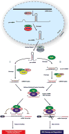
Figure 1
Figure 1. Biogenesis of miRNAs
In the canonical miRNA biogenesis pathway miRNAs are transcribed by RNA Polymerase II into long miRNA primary transcripts termed pri-miRNAs. These pri-miRNAs serve as substrates for Drosha and DGCR8, the former cleaves the flanking single stranded RNA to generate an ~85ntprecursor miRNA (pre-miRNA). After nuclear export to the cytoplasm via RanGTP/Exportin-5, the majority of pre-miRNAs are directly processed by Dicer and are subsequently cleaved to generate an ~22nt miRNA duplex (I). However, pre-miRNAs can be cleaved by Ago2 to generate an ac-pre-miRNA, which is then recognized by Dicer (II). Once the miRNA-duplex is unwound and the passenger strand degraded, the guide strand (highlighted in red), is incorporated into RISC, which contains Ago and other proteins. A miRNA loaded RISC mediates gene silencing via mRNA target cleavage and degradation, or translational repression and deadenylation depending on the complementarity between the miRNA and the targeted mRNA transcript. It should be appreciated that there are additional non-canonical ways in which miRNAs can be generated (i.e. miRtrons), and that miRNAs can function is non-classical ways.
Figure 2
Figure 2. Consequencesof Aberrant miRNA Biogenesis in Cancer
(A) Depicted is the Knudson’s model of haploinsufficiency for a tumor suppressor gene (adapted from Figure 1 [147]). The controlled expression of Dicer has important functions in both developmental and tumorigenic processes. Red dashed line indicates a haploid dose of Dicer expression is sufficientfor embryonic development, while haploidy is insufficient for tumor suppression. Given that tumors retain some Dicer expression and Dicer loss is embryonic lethal, complete loss (KO gene dosage) of Dicer is presumably detrimental to both normal and tumor cell viability. As noted in the text, tumor suppressor genes such as Rb are haplosufficient for tumor suppression (see red-dashed line), and usually require KO gene dosage to impair tumor suppressive activity. (B)In general, reduced miRNA levels are associated with tumor development, in part through decreased miRNA biogenesis proteins such as Drosha and Dicer. This suggests a majority of miRNAs harbor tumor suppressive functions by controlling the expression of genes with oncogenic activity when aberrantly expressed. Whether loss of Drosha/Dicer is an initiating event that allows for a permissive tumorigenic environment, or if established tumors actively suppress these proteins to maintain a tumorigenic state is still unclear. (C) Ago2 can function either as a tumor suppressor (left panel), or as an oncogene (right panel) depending on cell context. As a tumor suppressor, loss of Ago2 would result in reduced formation of Ago2-miRNP complexes and P-body formation, ultimately reducing miRNA activity. As a result, normal gene regulation and dampening of transcriptional noise would decrease and potentially lead to enhanced abundance of aberrantly expressed transcripts that harbor oncogenic activity. In some cancers, AGO2 expression is elevated suggesting an oncogenic role. While the mechanism is unclear, it is known that EGF/MAPK signaling pathways promote the phosphorylation of Ago2 and its localization to P-bodies. Presumably the resulting enhancement in miRNA activity reduces certain tumor suppressive transcripts highly abundant in those tumor cells, or through other Ago2-mediated mechanisms such as chromatin modification. Since proteasome inhibitors reduce AGO2 levels[26], other mechanisms for gain of Ago2 expression in cancer could be through loss of certain E3 ligases such as BRCA1, which is know to be mutated or lost in certain breast cancers.
Figure 3
Figure 3. Mechanisms Disrupting or Promoting miRNA Biogenesis in Cancer
(A) LIN28 mediated regulation of let-7. Left panel depicts LIN28 binding to the stem-loop region of either pri-let-7 or pre-let-7, blocking the miRNA biogenesis machinery from recognizing their respective substrates, and ultimately reducing mature let-7 levels. Right panel depicts a separate but not mutually exclusive mechanism, whereby LIN28 recruits TUT4 to the pre-let-7 substrate resulting in polyuridylation and degradation. It is unclear if other miRNAs are controlled by LIN28. (B) Depiction of how dead-box RNA helicases can control miRNA biogenesis. Specifically, p72 and p68 aid in Drosha-mediated processing of pri-miRNA transcripts, and when these biogenesis factors recruit other proteins such as ERα or SMADs, processing activity can be inhibited or promoted, respectively. Other proteins such as KSRP and NF45/NF90 represent other classes of biogenesis factors, but function in a similar manner to alter biogenesis activity.
Figure 4
Figure 4. Factors Regulating miRNA Abundance
(A) Transcriptional regulation of miRNAs. Similar to other RNA polII transcripts, miRNA genes harbor TATA boxes and TFIIB recognition sites upstream of the transcription start site (TSS) (arrow). MiRNA genes contain upstream enhancer/repressor elements, and promoter regions, indicating miRNAs are subjected to CpG promoter methylation (yellow CpG Box), histone modification (blue circles), as well as other regulatory events. Transcription factors (TFs) such as p53, Myc, and REST can bind to canonical sites within miRNA promoters to either promote or repress transcriptional activation. These TFs, and therefore the miRNA genes, can be misexpressed if the chemokine, hormone, or growth factor signaling pathways that control these TFs becomes dysregulated. Importantly, while the transcriptional start site (TSS) of miRNA genes are sometimes ~5–10kb away from the pre-miRNA sequence (yellow box)[148], the promoter regions can be up to 50kb away, making it difficult to study transcriptional regulation of particular miRNAs. (B)Examples of transcriptional networks that control miRNA expression patterns[149]. Unilateral negative feedback loops occur when certain TFs promote miRNAs thatare responsible for dampeningthe activity of the same TFs. Reciprocal and/or double negative feedback loops occur when certain TFs repress miRNAs that promote the negative regulation of the same TFs[150]. These networks result in either the oscillatory or stable expression of both the TF and the miRNA. This is in contrast to TFs involved in feed forward loops, which regulate miRNAs in a manner that reinforces the initial signaling activity (i.e. NF-κB enhancement of IL-6 through repression of let-7 levels). (C) Factors that control miRNA stability and decay. XRN2 represents a class of 5′ → 3′ exonucleases that can reduce miRNA levels and was initially identified in C. elegans. However, in an immortalized human lung epithelial cellmodel, serum-mediated differentiation of these cells is blocked by forced XRN2 expression. SND1 a member of the 3′ → 5′ exonucleases has similar activity on miRNAs in A. thaliana. The over-expression of SDN1 enhances colon cancer proliferation and anchorage independent growth by controlling APC expression. While gain of exonucleases occurs during tumorigenesis, in part to reduce miRNA levels, the loss of miRNA-stabilizing proteins such as GLD2 have also been noted to occur. Here, GLD2 promotes polyadenylation of miRNAs and therefore protects miRNAs from exonuclease activity.
Figure 5
Figure 5. Factors Effecting miRNA Targeting
(A) RNA binding proteins can alter miRNA activity and targeting. Top Panel; certain predicted miRNA:mRNA pairing interactions may not occur under certain cell states such as quiescence, presumably due to complex or secondary structures in the mRNA preventing miRNA-RISC binding. After growth factor stimulation, transcripts that harbor PUMILIO Response Elements (PREs), will bind to PUMILIO resulting in a change in the secondary structure of the mRNA such that the predicted miRNA target site becomes accessible for RISC binding. Bottom Panel; RBM38 is an RBP that inhibits miRNA function by directly competing for RISC binding in U-rich regions in the 3′UTRs of mRNA transcripts. RBM38 is induced in a p53 dependent manner after induction of DNA damage and protects p53-induceed genes from miRNAs. In cancer, inactivating mutations in p53 and/or loss of RBM38 would result in aberrant targeting and reduction in p53 induced genes. (B) RNA editing controls both miRNA levels and proper targeting. ADAR can mediate adenosine to inosine editing of miRNA residues. Edited residues near the stem loop of the miRNA results in an unrecognized substrate for Dicer, and subsequently results in accumulation of pri-miRNA and Tudor-SN mediated miRNA degradation. Edited residues residing within the mature miRNA sequence results in new target gene recognition given the edited adenosine to inosine now functions as a guanine.
Figure 6
Figure 6. The miRNA ceRNA Network
In the ceRNA network, ceRNAs can affect miRNA targeting primarily by acting as sponges. 1) Given that many genes also have a number of pseudogenes with conserved miRNA binding sites, as is the case with PTEN, the abundance of the pseudogene (PTENP1) will influence how strong the regulatory action of a particular miRNA is on the intended mRNA target (in this case PTEN). However, the system is oscillatory in that when PTENP1 are lower the PTEN levels, miRNAs will preferentially target PTEN, and vice versa. 2) Long noncoding RNAs can also be apart of this network. PRKACB can normally be targeted by miR-372; however, in response PRKACB can induce HULC long-noncoding RNA levels by phosphorylating and activating CREB. HULC serves as a sponge for miR-372, thereby preventing the miR-372 regulation of PRKACB.

References

    1. Hamilton BE, Hoyert DL, Martin JA, Strobino DM, Guyer B. Annual summary of vital statistics: 2010–2011. Pediatrics. 2013;131:548–58. - PMC - PubMed
    1. Bartel DP. MicroRNAs: target recognition and regulatory functions. Cell. 2009;136:215–33. - PMC - PubMed
    1. Esquela-Kerscher A, Slack FJ. Oncomirs – microRNAs with a role in cancer. Nat Rev Cancer. 2006;6:259–69. - PubMed
    1. Kasinski AL, Slack FJ. MicroRNAs en route to the clinic: progress in validating and targeting microRNAs for cancer therapy. Nat Rev Cancer. 2011;11:849–864. - PMC - PubMed
    1. Costa FF. Non-coding RNAs: Meet thy masters. Bioessays. 2010;32:599–608. - PubMed

Publication types