. 2014 Oct 10;289(41):28376-87.
doi: 10.1074/jbc.M114.587881. Epub 2014 Aug 19.

Regression of replication forks stalled by leading-strand template damage: I. Both RecG and RuvAB catalyze regression, but RuvC cleaves the holliday junctions formed by RecG preferentially

Affiliations

Regression of replication forks stalled by leading-strand template damage: I. Both RecG and RuvAB catalyze regression, but RuvC cleaves the holliday junctions formed by RecG preferentially

Sankalp Gupta et al. J Biol Chem. .

Abstract

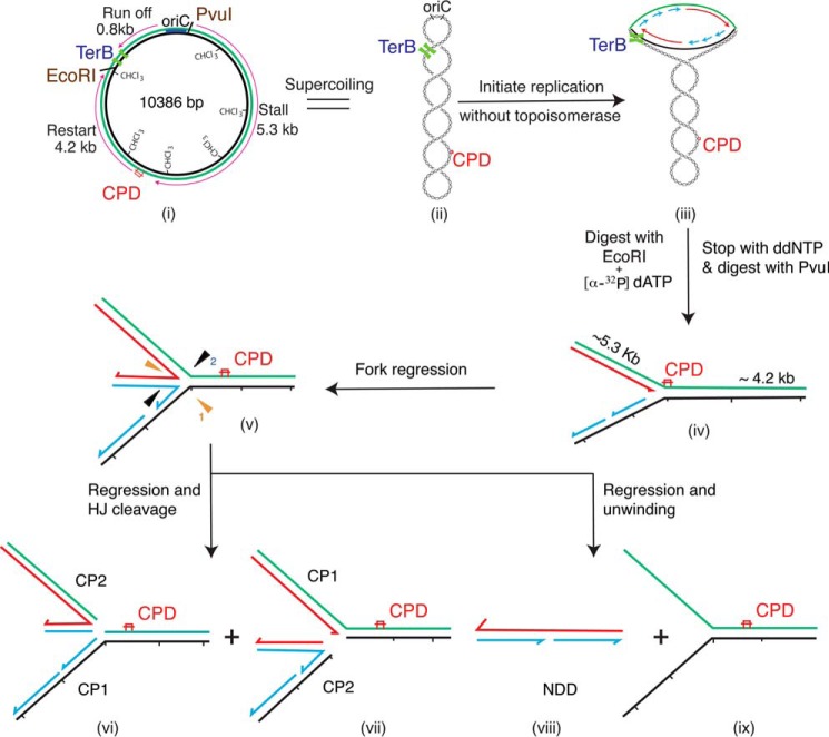
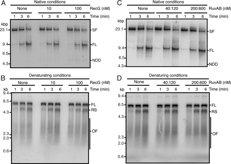
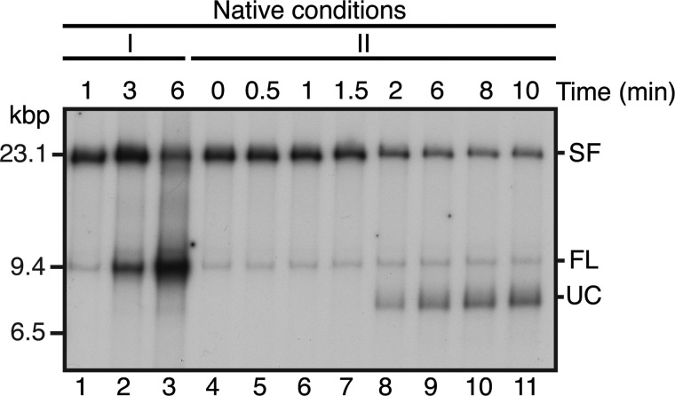
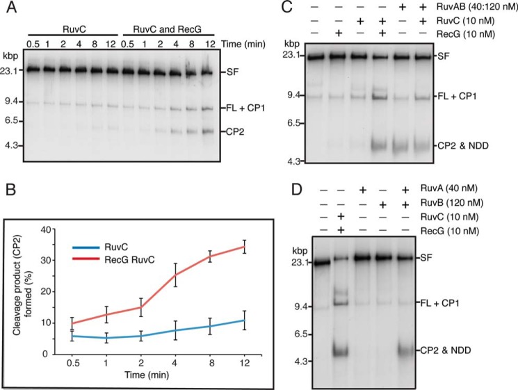
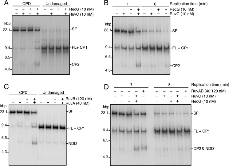
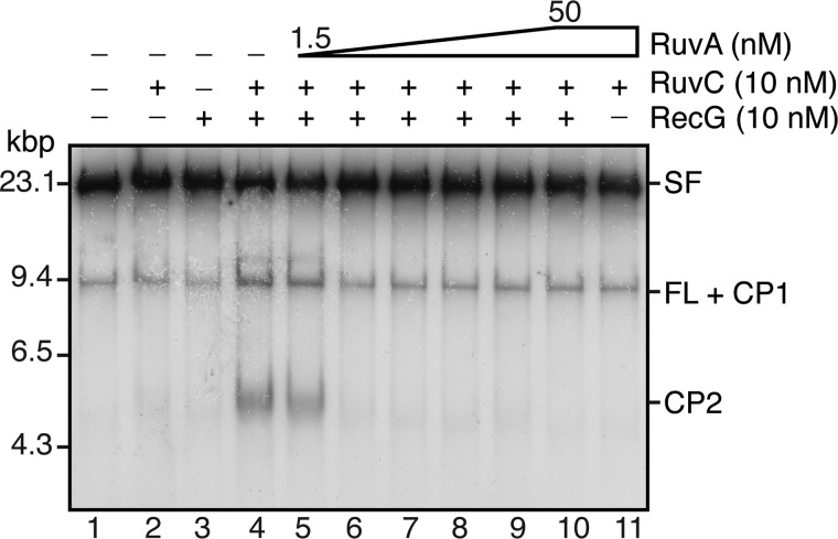
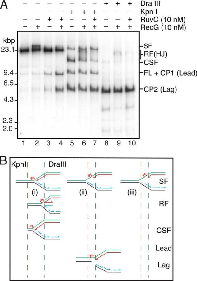
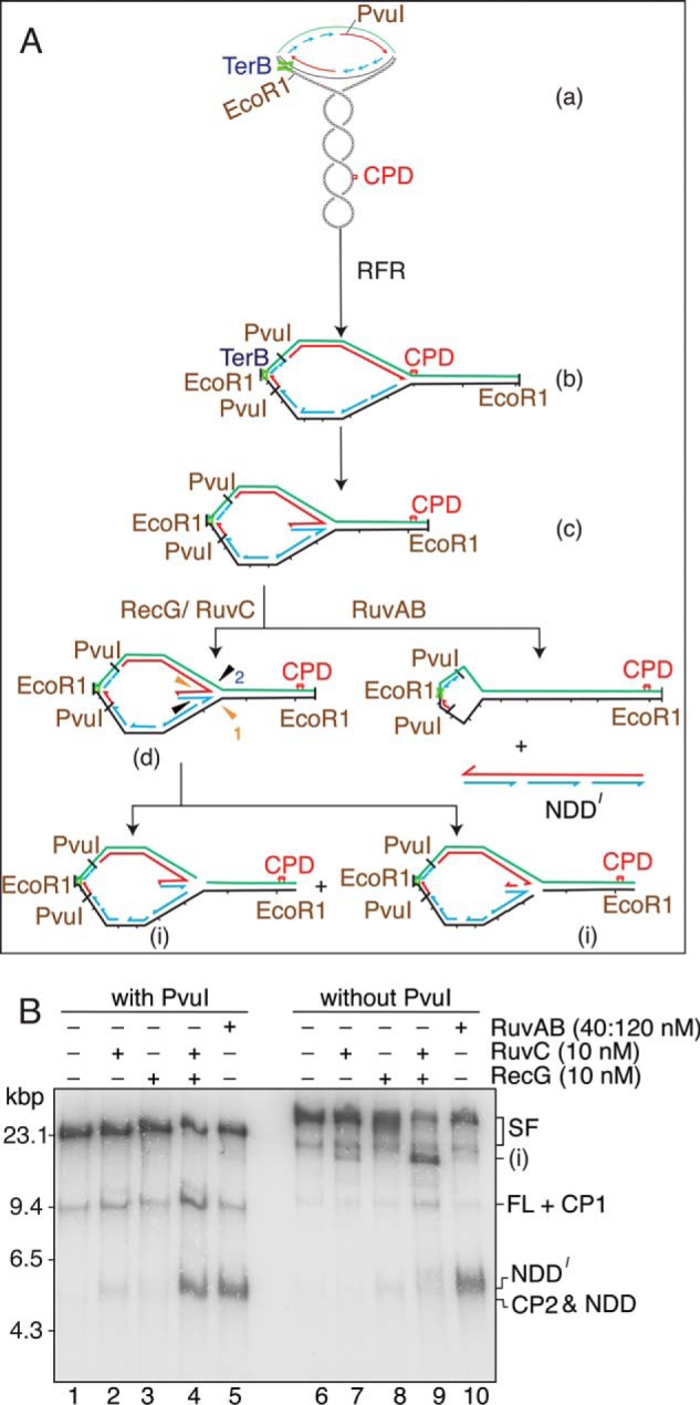
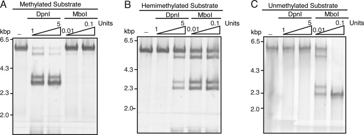
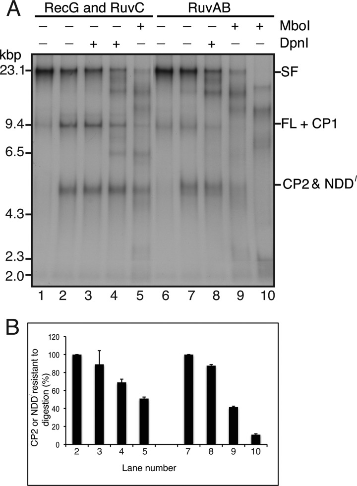
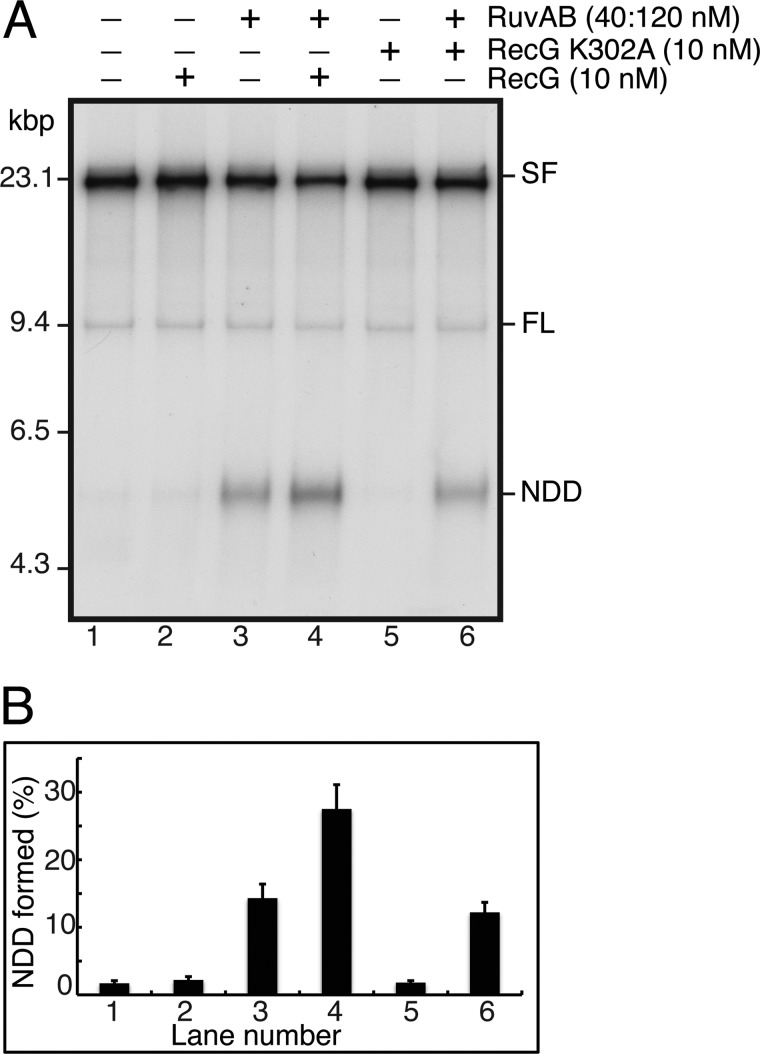
The orderly progression of replication forks formed at the origin of replication in Escherichia coli is challenged by encounters with template damage, slow moving RNA polymerases, and frozen DNA-protein complexes that stall the fork. These stalled forks are foci for genomic instability and must be reactivated. Many models of replication fork reactivation invoke nascent strand regression as an intermediate in the processing of the stalled fork. We have investigated the replication fork regression activity of RecG and RuvAB, two proteins commonly thought to be involved in the process, using a reconstituted DNA replication system where the replisome is stalled by collision with leading-strand template damage. We find that both RecG and RuvAB can regress the stalled fork in the presence of the replisome and SSB; however, RuvAB generates a completely unwound product consisting of the paired nascent leading and lagging strands, whereas RuvC cleaves the Holliday junction generated by RecG-catalyzed fork regression. We also find that RecG stimulates RuvAB-catalyzed regression, presumably because it is more efficient at generating the initial Holliday junction from the stalled fork.

Keywords: DNA Enzyme; DNA Recombination; DNA Repair; DNA Replication; Genomic Instability.


Figures

FIGURE 1.
FIGURE 1.
Reaction scheme and possible outcomes. i, template DNA. Black line, lagging-strand template; green line, leading-strand template; thin red arrow, direction of replication. The strand that is methylated is marked CH3. Replication is initiated on the supercoiled template (ii) in the absence of a topoisomerase, leading to the accumulation of an early replication intermediate where replication has been stalled because of the accumulation of positive supercoils (iii). The replication forks in the early intermediate are released by the addition of EcoRI, and labeled precursor is added at the same time. Stalled forks are accumulated by stopping DNA synthesis by the addition of ddNTPs and digesting the DNA with PvuI (iv). If the stalled forks are regressed by the addition of RecG and RuvAB, the regressed fork may be cut by the HJ resolvase RuvC (v) to give two classes of products (vi and vii), termed CP1 and CP2, that are roughly the size of full-length EcoRI-PvuI DNA and stall DNA, respectively, but differ in their strand composition (thick red arrow, nascent leading-strand DNA; thick blue arrows, nascent lagging-strand DNA). Alternatively, RuvC may not cut the regressed fork, and the nascent leading and lagging strands may be unwound from their respective template strands to give a complete NDD the same size as the stall product (viii), which will be labeled, and the duplex template DNA (ix), which will not be labeled.
FIGURE 2.
FIGURE 2.
Neither RecG nor RuvAB affects DNA replication directly. Replication reaction mixtures (20 μl) were as described under “Experimental Procedures.” The indicated concentrations of either RecG (A and B) or RuvAB (C and D) were added at the same time as EcoRI and [α-32P]dATP. Aliquots (5 μl) were removed at the indicated times post-EcoRI addition, and the DNA replication reaction was terminated by the addition of two volumes of STOP buffer. After an additional 10 min of incubation to digest the DNA products with PvuI, EDTA was added, and the reaction products were analyzed by either neutral gel electrophoresis (A and C) or denaturing alkaline gel electrophoresis (B and D). SF, stalled forks; FL, full-length EcoRI-PvuI DNA product; NDD, nascent duplex DNA; RS, restart products; OF, Okazaki fragments.
FIGURE 3.
FIGURE 3.
DnaB remains on the template DNA during the RFR incubation period. A replication reaction mixture (80 μl) was incubated as described in the legend to Fig. 2. After the 10-min incubation to digest the DNA with PvuI, the ATP concentration was restored to 1 mm, and the incubation was continued. Aliquots (15 μl) were withdrawn at the indicated times, the reactions were terminated by the addition of EDTA, and the products were analyzed by native gel electrophoresis (lanes 4–11) (II). For comparison, the products of a replication reaction that was terminated directly at the indicated times post-EcoRI cleavage are shown in lanes 1–3 (I). UC, uncoupled products (40, 41).
FIGURE 4.
FIGURE 4.
Both RecG and RuvAB generate products from stalled forks consistent with regression. A, RecG stimulates cleavage of stalled forks by RuvC. RFR reaction mixtures (120 μl) containing either 10 nm RuvC or 10 nm RuvC and 10 nm RecG were incubated at 37 °C. Aliquots (15 μl) were removed at the indicated times, and the reaction products were analyzed by native gel electrophoresis. CP1, cleavage product 1; CP2, cleavage product 2. A representative gel is shown. B, quantification of the kinetics of CP2 production by RuvC either in the presence or absence of RecG. Mean and S.D. (error bars) are shown for three experiments. C, RuvAB generates a product from stalled forks in the absence of RuvC. Standard RFR reactions containing the indicated concentrations of RecG, RuvC, and RuvAB were incubated for 10 min at 37 °C, and the products were analyzed by native gel electrophoresis. D, formation of NDD requires both RuvA and RuvB. Standard RFR reactions containing the indicated concentrations of RecG, RuvC, RuvA, RuvB, and RuvAB were incubated for 10 min at 37 °C, and the products were analyzed by native gel electrophoresis.
FIGURE 5.
FIGURE 5.
Both template damage and the presence of stalled forks are required for the generation of RFR products by RecG-RuvC and RuvAB. A, template damage is required for RecG-RuvC production of RFR products. Standard RFR reactions prepared using either undamaged template or CPD template containing the indicated concentrations of RecG and RuvC were incubated for 10 min at 37 °C, and the products were analyzed by native gel electrophoresis. B, stalled forks are required for RecG-RuvC production of RFR products. RFR reaction mixtures derived from replication reactions incubated for either 1 min or 6 min post-EcoRI addition containing the indicated concentrations of RecG and RuvC were incubated for 10 min at 37 °C, and the products were analyzed by native gel electrophoresis. C, template damage is required for RuvAB production of RFR products. Standard RFR reactions prepared using either undamaged template or CPD template containing the indicated concentrations of RuvB and RuvAB were incubated for 10 min at 37 °C, and the products were analyzed by native gel electrophoresis. D, stalled forks are required for RuvAB production of RFR products. RFR reaction mixtures derived from replication reactions incubated for either 1 min or 6 min post-EcoRI addition containing the indicated concentrations of RecG, RuvC, and RuvAB were incubated for 10 min at 37 °C, and the products were analyzed by native gel electrophoresis.
FIGURE 6.
FIGURE 6.
RuvA inhibits the generation of RFR products by RecG-RuvC. Standard RFR reaction mixtures containing the indicated concentrations of RecG, RuvC, and RuvA (increasing by a factor of 2 in lanes 5–10) were incubated for 10 min at 37 °C, and the products were analyzed by native gel electrophoresis.
FIGURE 7.
FIGURE 7.
RecG forms HJs from the stalled forks in the absence of RuvC. A, standard RFR reaction mixtures containing the 5903THF template and the indicated concentrations of RecG and RuvC were incubated for 10 min at 37 °C. DraIII (5 units) and KpnI (5 units) were then added as indicated, and the incubation continued for 10 min at 37 °C. The products were then analyzed by native gel electrophoresis. B, schematic of possible products of KpnI digestion. A stalled fork where neither the nascent leading nor lagging strand has progressed past the template lesion (i) will be cut by KpnI to give a CSF. A stalled fork where the nascent leading strand remains stalled at the lesion and lagging-strand synthesis has continued (ii) will be cut by KpnI to give separate products from the leading- and lagging-strand sister duplexes (Lead and Lag, respectively). If parental DNA unwinding proceeds in the absence of either leading- or lagging-strand synthesis, the stalled fork will not be cut by KpnI (iii). Regressed forks (RF) cut by KpnI will give products that migrate between the CSF and the SF on the gel. DraIII will cut all possible products; however, forks that have regressed past the DraIII site will retain the HJ and therefore migrate more slowly than the CSF.
FIGURE 8.
FIGURE 8.
RuvAB regresses the nascent DNA completely from the template strands. A, schematic of the reaction. The early replication intermediate (a) is digested with EcoRI but not PvuI. The stalled forks (both the counterclockwise moving fork that is stalled at the Ter site and the clockwise-moving fork that is stalled by the template damage) therefore generate a large replication bubble in the EcoRI-digested template (b). After RFR by either RecG or RuvAB, the size of this bubble is reduced (c). Note that the counterclockwise moving fork may also be regressed. RuvC can cleave the regressed fork in two different orientations (d), generating a large Y-like structure (i). RuvAB regresses the nascent DNA completely off of the template strands, generating a free nascent strand duplex (NDD′) that is a little longer than NDD because it encompasses the region from the stall point back to where the fork initiated at oriC. B, standard RFR reaction mixtures containing the indicated proteins and either digested with PvuI (with PvuI) or not treated (without PvuI) were incubated for 10 min at 37 °C and analyzed by native gel electrophoresis.
FIGURE 9.
FIGURE 9.
Digestion of differentially methylated DNA by DpnI and MboI. Duplex DNA fragments (5.3 kbp) that were either methylated on both strands (A), hemimethylated (B), or unmethylated (C), prepared as described under “Experimental Procedures,” were treated with the indicated amounts of DpnI and MboI for 10 min at 37 °C in replication reaction buffer. The products of digestion were analyzed by native agarose gel electrophoresis.
FIGURE 10.
FIGURE 10.
The RuvAB RFR product is a nascent strand duplex. A, the indicated restriction enzymes (1 unit of DpnI, 0.1 unit of MboI in lanes 5 and 10 and 0.01 unit in lane 9) were added to regression products formed by either RecG + RuvC or RuvAB in standard RFR reaction mixtures, and the incubations continued for 10 min at 37 °C. The DNA products were then analyzed by native gel electrophoresis. B, quantification of the fraction of CP2 or NDD that was resistant to digestion by the indicated restriction enzyme. The plot shows the mean and S.D. from two experiments. A representative gel is shown in A.
FIGURE 11.
FIGURE 11.
RecG stimulates RuvAB regression. A, standard RFR reaction mixtures containing the indicated concentrations of RecG, RecG K302A, and RuvAB were incubated for 10 min at 37 °C and analyzed by native gel electrophoresis. A representative gel is shown. B, quantification of the amount of NDD generated. The plot shows the mean and S.D. (error bars) from three experiments.

Similar articles

Cited by

References

    1. Hanawalt P. C. (1966) The U.V. sensitivity of bacteria: its relation to the DNA replication cycle. Photochem. Photobiol. 5, 1–12 - PubMed
    1. Skalka A. (1974) in Mechanisms of Recombination (Grell R. F., ed) pp. 421–432, Plenum Press, New York
    1. Nurse P., Zavitz K. H., Marians K. J. (1991) Inactivation of the Escherichia coli priA DNA replication protein induces the SOS response. J. Bacteriol. 173, 6686–6693 - PMC - PubMed
    1. Zavitz K. H., Marians K. J. (1991) Dissecting the functional role of PriA protein-catalysed primosome assembly in Escherichia coli DNA replication. Mol. Microbiol. 5, 2869–2873 - PubMed
    1. Cox M. M., Goodman M. F., Kreuzer K. N., Sherratt D. J., Sandler S. J., Marians K. J. (2000) The importance of repairing stalled replication forks. Nature 404, 37–41 - PubMed

Publication types

MeSH terms

LinkOut - more resources