Review
. 2014 Dec;71(24):4781-802.
doi: 10.1007/s00018-014-1703-3. Epub 2014 Aug 20.

Regulation of male sex determination: genital ridge formation and Sry activation in mice

Affiliations
Review

Regulation of male sex determination: genital ridge formation and Sry activation in mice

Satomi S Tanaka et al. Cell Mol Life Sci. 2014 Dec.

Abstract

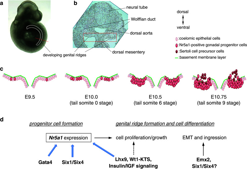
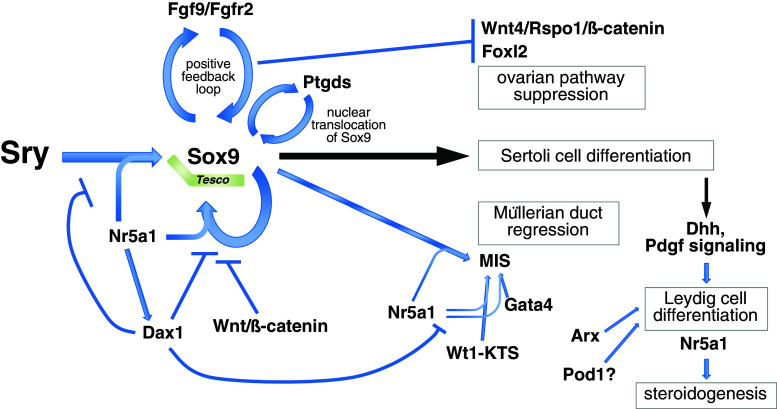
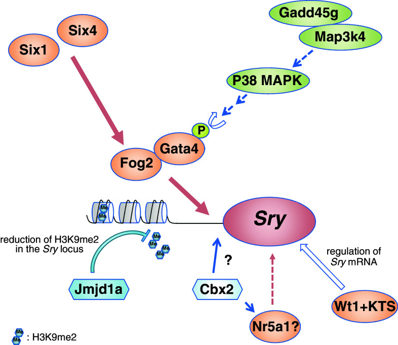
Sex determination is essential for the sexual reproduction to generate the next generation by the formation of functional male or female gametes. In mammals, primary sex determination is commenced by the presence or absence of the Y chromosome, which controls the fate of the gonadal primordium. The somatic precursor of gonads, the genital ridge is formed at the mid-gestation stage and gives rise to one of two organs, a testis or an ovary. The fate of the genital ridge, which is governed by the differentiation of somatic cells into Sertoli cells in the testes or granulosa cells in the ovaries, further determines the sex of an individual and their germ cells. Mutation studies in human patients with disorders of sex development and mouse models have revealed factors that are involved in mammalian sex determination. In most of mammals, a single genetic trigger, the Y-linked gene Sry (sex determination region on Y chromosome), regulates testicular differentiation. Despite identification of Sry in 1990, precise mechanisms underlying the sex determination of bipotential genital ridges are still largely unknown. Here, we review the recent progress that has provided new insights into the mechanisms underlying genital ridge formation as well as the regulation of Sry expression and its functions in male sex determination of mice.


Figures

Fig. 1
Fig. 1
Formation of genital ridges. In mouse embryos, genital ridges are formed on the ventral surface of the mesonephros as paired thickenings of the epithelial layer, which is accompanied by proliferation of the coelomic epithelium from the anterior portion. a Embryonic day (E) 10 mouse embryo. White dashed line indicates the location of developing genital ridges. Red dashed line indicates the position of the section in (b). b Transverse section of developing genital ridges, representing the dorsal to the top and the ventral to the bottom. c Schematic illustrations of genital ridge formation. The rectangle in b outlines the approximate position in c. Some Nr5a1 (also known as Ad4BP/Sf1)-positive gonadal progenitor cells are formed in the E9.5 coelomic epithelium. The number of Nr5a1-positive cells increases at E10.0 (around the 0 tail somite stage), and multilayered Nr5a1-positive cells are expanded at E10.5 (around the 6-tail somite stage) in the coelomic epithelium. Thereafter, Nr5a1-positive progenitor cells migrate into the dorsal inner mesenchyme region through the basement membrane layer to form the genital ridge primordium (E11.75, around the 9-tail somite stage). In XY gonads, a proportion of Nr5a1-positive daughter cells derived from the coelomic epithelium express Sry to become Sertoli cell precursors. d Scheme for the molecular network that regulates formation and development of Nr5a1-positive gonadal progenitor cells. Genital ridge formation begins from the anterior part of the coelomic epithelium, which is accompanied by Gata4 and subsequent Nr5a1 expression. Six1 and Six4 directly transactivate Nr5a1 in gonadal progenitor cells of the coelomic epithelium. Lhx9, Wt1−KTS, and insulin/insulin-like growth factor (IGF) signaling activity are required to promote gonadal progenitor cell proliferation and form the bipotential genital ridges, which are accompanied by Nr5a1 upregulation. Emx2 and possibly Six1 and Six4 contribute to regulation of the epithelial-to-mesenchymal transition (EMT) and subsequent ingression of the progenitor cells
Fig. 2
Fig. 2
Sry and the transcriptional network that governs testis determination. In mice, expression of a single genetic trigger, the Y-linked gene Sry, induces differentiation of pre-Sertoli cells. Sry directly transactivates Sox9 through the core element of the testis-specific enhancer region of Sox9 (Tesco) together with Nr5a1. Sox9 itself also contributes to the maintenance of Sox9 expression through Tesco together with Nr5a1. Excess Dax1 interferes with Sox9 upregulation, likely through inhibition of the binding of Nr5a1/Sry or Nr5a1/Sox9 proteins to Tesco. Although Dax1 interferes with the activity of Nr5a1, Dax1 expression depends on Nr5a1 activity. Sox9 upregulates Fgf9 expression, and FGF9 in turn establishes the Sox9–FGF9 positive feedback loop through FGF receptor 2 (FGFR2), which maintains a high level of Sox9 expression. The Sox9–FGF9 positive feedback loop also acts to suppress ovary-specific WNT4/R-spondin1(Rspo1)/β-catenin signaling activity. Sox9 expression also interferes with upregulation of the ovarian gene Foxl2. In addition, Sox9 upregulates Ptgds, and the signaling activity of Ptgds promotes nuclear translocation of Sox9 to facilitate Sertoli cell differentiation. Together with Nr5a1, Sox9 regulates activation of MIS that promotes regression of Müllerian ducts. MIS is also regulated synergistically by Nr5a1 and Wt1−KTS, as well as Gata4. Sertoli cells express Dhh that is required for specification of the fetal Leydig cell fate. Pdgf secreted by Sertoli cells is also required for fetal Leydig cell differentiation. Arx and probably Pod1 are involved in regulation of Leydig cell differentiation. Male steroid hormones are synthesized by Leydig cells, which is mainly regulated by Nr5a1
Fig. 3
Fig. 3
Model for Sry upregulation. Before the onset of Sry expression, a reduction in the H3K9me2 levels of the Sry locus is mediated by stage-specific upregulation of the H3K9 demethylase Jmjd1a, which may allow Sry upregulation by transcriptional factors. Interactions of Gata4 with its co-factor Fog2 are critical for Sry activation. Fog2 is upregulated in the coelomic epithelium by Six1 and Six4 before the onset of Sry expression. Gata4 is transiently activated by the Gadd45g-Map3k4-p38 MAPK pathway because of the stage-specific Gadd45g upregulation. Subsequently, the phosphorylated Gata4 and Fog2 protein complex may bind to the Sry promoter and activate Sry expression in a stage-specific manner. The Wt1+KTS isoform may contribute to the post-transcriptional regulation of Sry mRNA. The polycomb group gene Cbx2 is required for Sry upregulation, but the genetic interaction between Cbx2 and Sry is unclear. In addition, Cbx2 promotes Nr5a1 upregulation, and Nr5a1 is proposed to be one of the upstream regulators of Sry

References

    1. Anderson R, Copeland TK, Scholer H, Heasman J, Wylie C. The onset of germ cell migration in the mouse embryo. Mech Dev. 2000;91:61–68. - PubMed
    1. Ginsburg M, Snow MH, McLaren A. Primordial germ cells in the mouse embryo during gastrulation. Development. 1990;110:521–528. - PubMed
    1. Hara K, Kanai-Azuma M, Uemura M, Shitara H, Taya C, et al. Evidence for crucial role of hindgut expansion in directing proper migration of primordial germ cells in mouse early embryogenesis. Dev Biol. 2009;330:427–439. - PubMed
    1. Lawson KA, Hage WJ. Clonal analysis of the origin of primordial germ cells in the mouse. Ciba Found Symp. 1994;182:68–84. - PubMed
    1. Molyneaux KA, Stallock J, Schaible K, Wylie C. Time-lapse analysis of living mouse germ cell migration. Dev Biol. 2001;240:488–498. - PubMed

Publication types

LinkOut - more resources