. 2014 Nov 1;104(2):371-81.
doi: 10.1093/cvr/cvu195. Epub 2014 Aug 18.

Super-resolution imaging reveals that loss of the C-terminus of connexin43 limits microtubule plus-end capture and NaV1.5 localization at the intercalated disc

Affiliations

Super-resolution imaging reveals that loss of the C-terminus of connexin43 limits microtubule plus-end capture and NaV1.5 localization at the intercalated disc

Esperanza Agullo-Pascual et al. Cardiovasc Res. .

Abstract

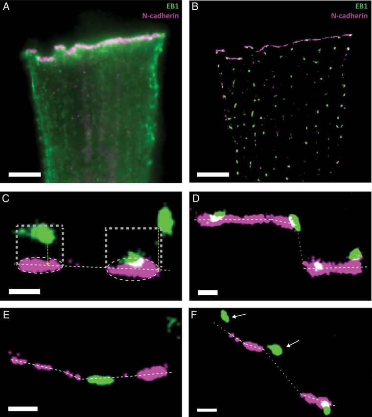
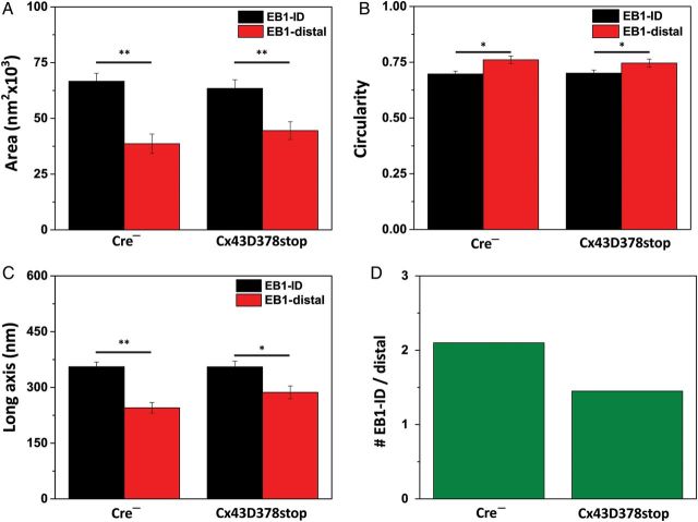
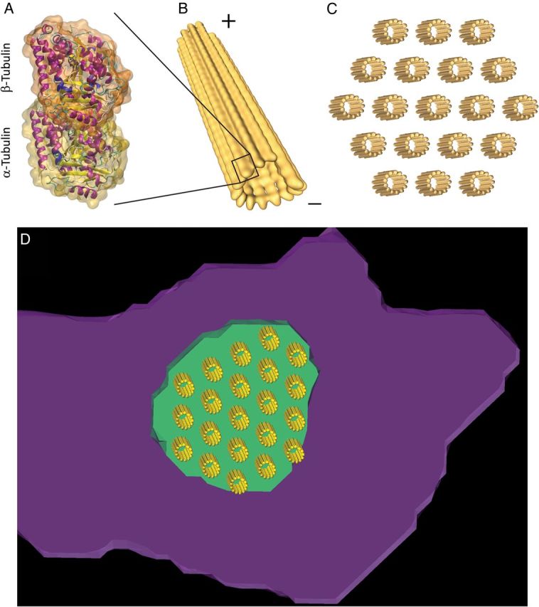
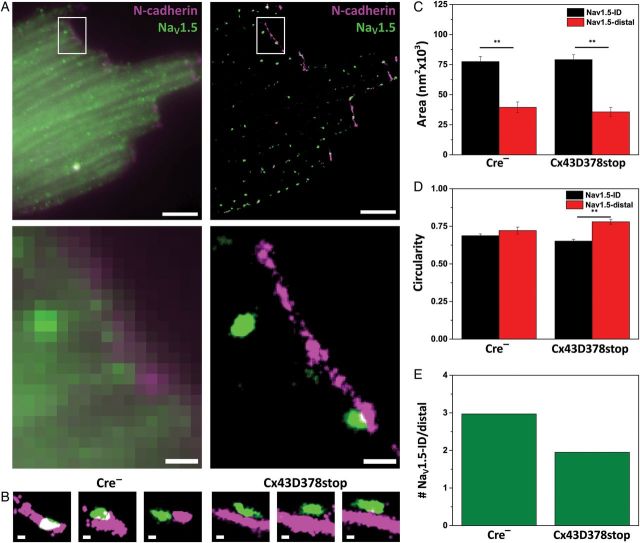
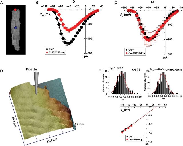
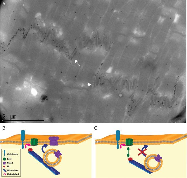
Aims: It is well known that connexin43 (Cx43) forms gap junctions. We recently showed that Cx43 is also part of a protein-interacting network that regulates excitability. Cardiac-specific truncation of Cx43 C-terminus (mutant 'Cx43D378stop') led to lethal arrhythmias. Cx43D378stop localized to the intercalated disc (ID); cell-cell coupling was normal, but there was significant sodium current (INa) loss. We proposed that the microtubule plus-end is at the crux of the Cx43-INa relation. Yet, specific localization of relevant molecular players was prevented due to the resolution limit of fluorescence microscopy. Here, we use nanoscale imaging to establish: (i) the morphology of clusters formed by the microtubule plus-end tracking protein 'end-binding 1' (EB1), (ii) their position, and that of sodium channel alpha-subunit NaV1.5, relative to N-cadherin-rich sites, and (iii) the role of Cx43 C-terminus on the above-mentioned parameters and on the location-specific function of INa.

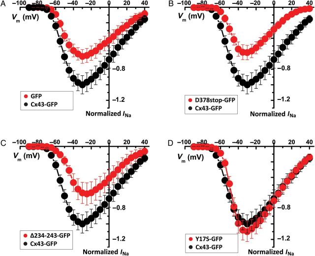
Methods and results: Super-resolution fluorescence localization microscopy in murine adult cardiomyocytes revealed EB1 and NaV1.5 as distinct clusters preferentially localized to N-cadherin-rich sites. Extent of co-localization decreased in Cx43D378stop cells. Macropatch and scanning patch clamp showed reduced INa exclusively at cell end, without changes in unitary conductance. Experiments in Cx43-modified HL1 cells confirmed the relation between Cx43, INa, and microtubules.

Conclusions: NaV1.5 and EB1 localization at the cell end is Cx43-dependent. Cx43 is part of a molecular complex that determines capture of the microtubule plus-end at the ID, facilitating cargo delivery. These observations link excitability and electrical coupling through a common molecular mechanism.

Keywords: Area composita; Connexome; Cx43; EB1; Microtubule; NaV1.5.


Figures

Figure 1
Figure 1
SRFM images of EB1 (green) and N-cadherin (purple) in adult murine ventricular myocytes. (A) TIRF image; projection of 2000 frames collected to generate the SRFM. (B) Image obtained by SRFM. (CF) White dotted lines are drawn along the major axis of the ellipsoid containing the N-cadherin plaque. Rectangular areas in (C) outline the borders of the ROI; yellow dotted lines indicate the measure of distance to the cell end for two distal clusters. Clusters overlapping with the z plane of N-cadherin (D) or located along the horizontal axis between two N-cadherin clusters (E) were referred to as EB1-ID. Clusters localized in a region where the cell end could not be defined (yellow dotted line in F) were not included for analysis. Scale bars: 500 nm.
Figure 2
Figure 2
EB1 cluster analysis. Acquisition methods detailed in Agullo-Pascual et al. For all panels, EB1-ID: black bars; EB1-distal: red bars. ‘Cx43D378stop’ refers to data obtained from myocytes collected from Cx43D378stop flox/flox-positive, αMyHC-Cre-ER(T2)-positive mice, injected with tamoxifen and euthanized 14 days after first injection. ‘Cre’ refers to Cx43D378stop flox/flox-positive, cre-negative littermates. (A) Average area occupied by individual clusters. (B) Average cluster circularity; 1.0 indicates perfect circle. (C) Average long axis of the ellipsoid fitting the cluster. (D) Proportion of EB1 clusters detected at ID for every distal cluster. For all panels, *P < 0.05; **P < 0.001. Comparisons by unpaired Student's t-test except for data in (D) (χ2 test, P < 0.05). N = 369 and 282 clusters for 32 Cre cardiomyocytes and 29 Cx43D378stop cardiomyocytes, respectively.
Figure 3
Figure 3
(A) Atomic structure of αβ tubulin heterodimer (1jff.pdb). (B) Microtubule composed of 13 protofilaments with minus- and plus-ends indicated. Protofilaments were built from αβ tubulin heterodimers associated head to tail, conferring their characteristic polarity. (C) Model of a bundle of 19 microtubules with hexagonal packing. The microtubule edge-to-edge and centre-to-centre spacing was 20 and 46 nm, respectively. (D) Partial view of one cluster of EB1 proteins (green) overlapping with a cluster of N-cadherin (purple). A model of microtubule bundle with hexagonal packing was superimposed on the SRFM signal.
Figure 4
Figure 4
Localization of NaV1.5 and N-cadherin in adult ventricular myocytes. (A) Image by TIRF (projection of 2000 frames collected for SRFM) or SRFM, left and right, respectively. White-boxed areas enlarged in bottom panels, showing increased resolution by SRFM. Purple: N-cadherin; green: NaV1.5. (B) Enlargement showing proximity between NaV1.5 and N-cadherin clusters in Cre (left) and Cx43D378stop (right) cells. (C) Average area occupied by NaV1.5 clusters at the cell end (NaV1.5-ID; black bars) or distal (NaV1.5-distal; red columns) in Cre and Cx43D378stop cells. (D) Average cluster circularity. (E) ID clusters normalized relative to the number of clusters in the distal region. n = 365 clusters in 23 cells imaged (ID: 273; distal: 92) and 441 clusters in 22 cells imaged (ID: 291; distal: 150) for Cre and Cx43D378stop. (If ID/distal ratios were calculated per cell, and then averaged: Cre:4.6 ± 0.8, n = 23; D378stop: 2.4 ± 0.3, n = 20; two cells not counted since distal clusters = 0.) Scale bars: 5 and 1 µm in (A) (upper and lower panels, respectively) and 200 nm in (B).
Figure 5
Figure 5
(A) Red and blue circles illustrate macropatch recording locations (ID or M, respectively). (B and C) IV relations of INa in macropatches from ID (B; n = 8 and 9 for Cre- and Cx43D378stop, respectively) or M regions (C; n = 5 for both groups). Black and red circles correspond to data from myocytes isolated from Cre and Cx43D378stop, respectively. (D) SICM recording of adult ventricular cell end; pipette diagram illustrates the site of recording for SPC. (E) Unitary sodium channel conductance measurements from either Cre, or Cx43D378stop cells. Number of events at Vm = −50, −60, −70, and −80 mV are, for Cre: 192, 209, 207, and 184; for Cx4D378stop: 203, 199, 193, and 150.
Figure 6
Figure 6
INa IV relations in Cx43-KD HL1 cells. Data normalized to maximum average INa at −30 mV in Cx43-GFP-expressing cells (black in all panels; n = 13). Red symbols represent data from Cx43-KD cells transfected with GFP alone (A; n = 13), Cx43D378stop (B; n = 10), a Cx43 mutant lacking the microtubule-binding domain (Cx43Δ234-243; C; n = 11), or Cx43 mutation Y17S (D; n = 11).
Figure 7
Figure 7
(A) Immunoelectron microscopy of adult ventricular ID decorated with gold particles targeting Cx43. Notice abundant clustering along gap junctions but also at areas with clear intercellular space (arrows). (B and C) Model for the interaction of microtubules (blue) with mixed clusters of junctional proteins that include Cx43 (green), N-cadherin (light blue), and PKP2 (pink). In (B), EB1 (red) anchors at the membrane to deliver cargo (a vesicle containing NaV1.5; purple). In (C), a defect in Cx43 prevents EB1 capture, impeding cargo delivery and consequently, reducing NaV1.5 population at the membrane.

Comment in

References

    1. Danik SB, Liu F, Zhang J, Suk HJ, Morley GE, Fishman GI, Gutstein DE. Modulation of cardiac gap junction expression and arrhythmic susceptibility. Circ Res. 2004;95:1035–1041. - PMC - PubMed
    1. Gutstein DE, Morley GE, Tamaddon H, Vaidya D, Schneider MD, Chen J, Chien KR, Stuhlmann H, Fishman GI. Conduction slowing and sudden arrhythmic death in mice with cardiac-restricted inactivation of connexin43. Circ Res. 2001;88:333–339. - PMC - PubMed
    1. Qu J, Volpicelli FM, Garcia LI, Sandeep N, Zhang J, Marquez-Rosado L, Lampe PD, Fishman GI. Gap junction remodeling and spironolactone-dependent reverse remodeling in the hypertrophied heart. Circ Res. 2009;104:365–371. - PMC - PubMed
    1. Akar FG, Nass RD, Hahn S, Cingolani E, Shah M, Hesketh GG, DiSilvestre D, Tunin RS, Kass DA, Tomaselli GF. Dynamic changes in conduction velocity and gap junction properties during development of pacing-induced heart failure. Am J Physiol Heart Circ Physiol. 2007;293:H1223–H1230. - PubMed
    1. Danik SB, Rosner G, Lader J, Gutstein DE, Fishman GI, Morley GE. Electrical remodeling contributes to complex tachyarrhythmias in connexin43-deficient mouse hearts. FASEB J. 2008;22:1204–1212. - PMC - PubMed

Publication types

MeSH terms