. 2014 Oct 1;104(1):103-15.
doi: 10.1093/cvr/cvu193. Epub 2014 Aug 18.

Scrib:Rac1 interactions are required for the morphogenesis of the ventricular myocardium

Affiliations

Scrib:Rac1 interactions are required for the morphogenesis of the ventricular myocardium

Veronika Boczonadi et al. Cardiovasc Res. .

Abstract

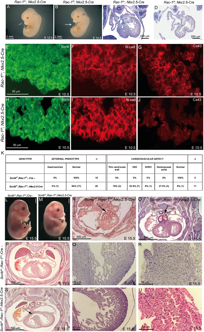
Aims: The organization and maturation of ventricular cardiomyocytes from the embryonic to the adult form is crucial for normal cardiac function. We have shown that a polarity protein, Scrib, may be involved in regulating the early stages of this process. Our goal was to establish whether Scrib plays a cell autonomous role in the ventricular myocardium, and whether this involves well-known polarity pathways.

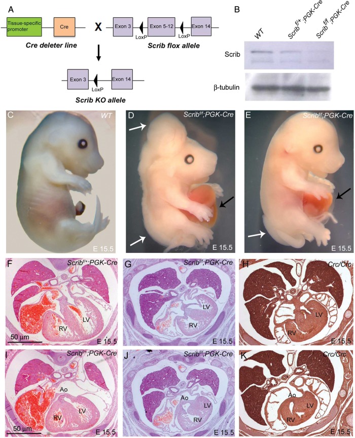
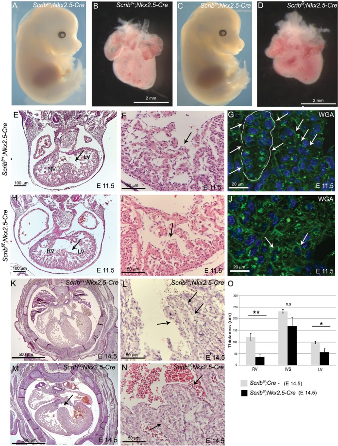
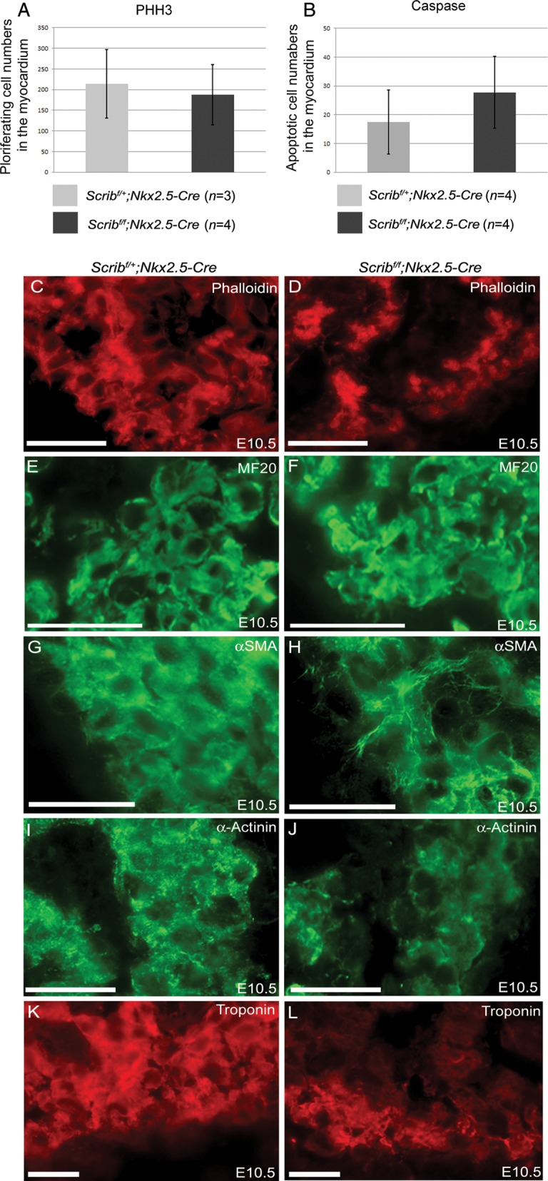
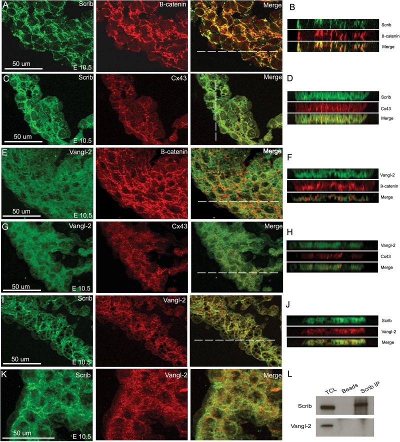
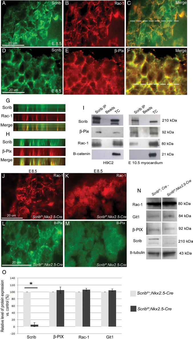
Methods and results: Deletion of Scrib in cardiac precursors utilizing Scrib(flox) mice together with the Nkx2.5-Cre driver resulted in disruption of the cytoarchitecture of the forming trabeculae and ventricular septal defects. Although the majority of mice lacking Scrib in the myocardium survived to adulthood, they developed marked cardiac fibrosis. Scrib did not physically interact with the planar cell polarity (PCP) protein, Vangl2, in early cardiomyocytes as it does in other tissues, suggesting that the anomalies did not result from disruption of PCP signalling. However, Scrib interacted with Rac1 physically in embryonic cardiomyocytes and genetically to result in ventricular abnormalities, suggesting that this interaction is crucial for the development of the early myocardium.

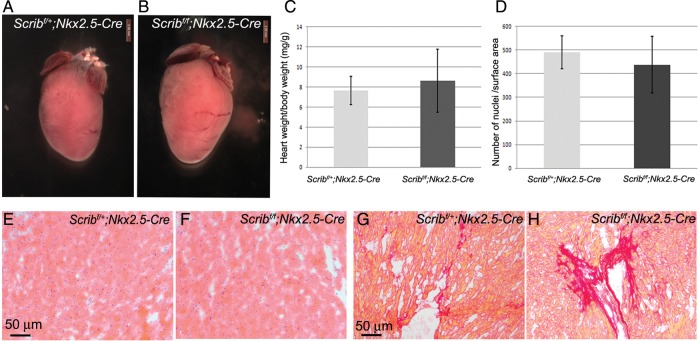
Conclusions: The Scrib-Rac1 interaction plays a crucial role in the organization of developing cardiomyocytes and formation of the ventricular myocardium. Thus, we have identified a novel signalling pathway in the early, functioning, heart muscle. These data also show that the foetus can recover from relatively severe abnormalities in prenatal ventricular development, although cardiac fibrosis can be a long-term consequence.

Keywords: Cardiac development; Cardiomyocytes; Polarity; Rac1; Scrib; Ventricular myocardium.


Figures

Figure 1
Figure 1
Cardiac anomalies in Scribf/f;PGK-Cre embryos. (A) Schematic representation of breeding strategy to obtain Scrib-deleted mice. (B) Western blotting (n = 3) showed that Scrib protein levels were markedly reduced in Scribf/f;PGK-Cre at E15.5. β-Tubulin was used as a loading control. (C–E) Scribf/f;PGK-Cre embryos display neural tube defects (white arrows) and gastroschisis (black arrows) at E14.5. (F and I) Scribf/+;PGK-Cre have normal hearts. (G and J) Scribf/f;PGK-Cre exhibit double outlet right ventricle, ventricular septal defects, and septal hyperplasia that closely resemble those seen in Crc/Crc (H and K). See Table 1 for numbers of animals analysed. Ao, aorta; LV, left ventricle; RV, right ventricle. Scale bar = 50 μm.
Figure 2
Figure 2
Defects in the ventricular myocardium in Scribf/f;Nkx2.5-Cre. (A–D) Scribf/f;Nkx2.5-Cre mouse mutants have a normal external phenotype at E14.5, whereas the heart appears smaller and immature when compared with control littermates. See Table 1 for numbers of animals analysed. (E–J) Transverse sectioning of E11.5 Scribf/f;Nkx2.5-Cre hearts (n = 5) revealed abnormalities in the trabeculae and in the formation of the interventricular septum (arrows). High-power views of the trabeculae show that the trabecular network (arrow) is less well developed in the mutant hearts than in control littermates (compare I with F). Labelling of the cell membranes with wheat germ agglutinin reveals that the cellular architecture of the trabeculae (arrows) appears more disorganized in the mutant (J) than in the control (G) (n = 3). (K–N) Transverse sections of Scribf/f;Nkx2.5-Cre hearts at E14.5 reveal peri-membranous septal defects (arrow). High-power views show that the trabeculae (arrows) appear thickened in the mutants (N) compared with control littermates (L). (O) Measurements (n = 6) revealed significant differences in the thickness of the right and left ventricular walls, but not the septum. Error bars represent standard deviation, *P < 0.05. LV, left ventricle; RV, right ventricle; WGA, wheat germ agglutinin. Scale bar: B,D = 2 mm. E,H = 100 µm, F,I,L,N = 50 µm, G,J = 20 µm, K,M = 500 µm.
Figure 3
Figure 3
Abnormal expression of cardiomyocyte markers in Scribf/f;Nkx2.5-Cre. (A and B) Neither proliferation (pHH3) nor cell death (cleaved caspase-3) was altered in myocardium from Scribf/f;Nkx2.5-Cre ventricles compared with control littermates at E10.5. (C–L) While phalloidin and MF20 staining did not show any reproducible differences between control and mutant cardiomyocytes in the E10.5 ventricle (C–F), analysis of other markers suggested that the Scribf/f;Nkx2.5-Cre ventricles were immature in comparison with control littermates; whereas striations suggesting the presence of sarcomeres (labelled by α-SMA and α-actinin) were readily apparent in control cells, these were absent in the mutant cells (H and J). In addition, there was markedly reduced expression of cardiac troponin I (K and L) in Scribf/f;Nkx2.5-Cre trabeculae. n = 3 for all experiments. Scale bar = 20 µm.
Figure 4
Figure 4
Surviving Scribf/f;Nkx2.5-Cre hearts develop cardiac fibrosis. (A–C) Six-month-old Scribf/f;Nkx2.5-Cre hearts appeared slightly larger than control littermates, although heart weight:body weight ratios showed that this was variable and not statistically significant. Error bars in C represent standard deviation. (D–F) H&E staining of myocardium shows no obvious signs of abnormalities in Scribf/f Nkx2.5-Cre hearts. Quantification of cardiomyocyte nuclei/unit surface area showed no significant difference compared with control littermates (D). (G and H) Sirius Red staining revealed increased fibrosis of the ventricular myocardium of Scribf/f;Nkx2.5-Cre mice compared with control littermates.
Figure 5
Figure 5
Scrib co-localizes with adherens junctions and gap junctions in the developing myocardium. (A–D) Scrib localizes to cardiomyocyte cell membranes at E10.5, co-localizing with β-catenin (A and B) and connexin-43 (C and D). (E–H) In contrast, Vangl2 is found in the cytoplasm of the ventricular cardiomyocytes, with reduced staining in the cell membrane where there is no evidence of co-localization with β-catenin (E and F) or connexin-43 (G and H). (I–K) Although both Scrib and Vangl2 are found in cardiomyocytes, they do not appear to co-localize at E10.5. The position of the acquired z-axis images (B, D, F, H, and J) are indicated by the horizontal white line on the composite images. (L) Co-immunoprecipitation with Scrib antibody in H9C2 cell lysate confirmed no physical interaction with Vangl2. n = 3 for all experiments. Scale bar = 50 µm.
Figure 6
Figure 6
Scrib, β-PIX, and Rac1 interact in the developing myocardium. (A–H) Immunostaining for Scrib (A and D), Rac1 (B), and β-PIX (E) in the E8.5 myocardium reveals overlapping localization at the cardiomyocyte membrane (C and F). The position of the acquired z-axis images (G and H) is indicated by the horizontal white line on the composite images (C and F). Co-localization (yellow) of Scrib/Rac1/β-PIX can be seen (G and H). (I) Co-immunoprecipitation was performed in lysates from H9C2 cells and in crude E10.5 heart homogenate (n = 3). Scrib forms protein complexes with both β-PIX and Rac1. No precipitation of β-catenin with Scrib was observed. (J–M) Rac1 and β-PIX are lost from the cell membrane in the Scrib-depleted myocardium when compared with control sections. (N and O) Western blot analysis was performed for Scrib, Rac1, Git-1, β-PIX, and β-tubulin (as a loading control), in homogenates from E10.5 control and Scribf/f;Nkx2.5-Cre hearts (n = 3 for each). The graphs display the percentage of protein expression in the mutant relative to the control. Only Scrib was significantly reduced in the Scribf/f;Nkx2.5-Cre relative to the control, *P < 0.05. Error bars represent standard deviation. Scale bar = 20 µm (A–F,J–M).
Figure 7
Figure 7
Rac1 is required for normal development of the myocardium. (A and B) Rac1f/f;Nkx2.5-Cre embryos develop cardiac oedema by E12.5 (white arrow in B). (C–D) Sectioning of Rac-1f/f;Nkx2.5-Cre at E12.5 reveals an immature heart with thin ventricular walls and a poorly developed interventricular septum. (E–J) Fluorescent immunostaining for Scrib, N-cadherin, and connexin-43 at E10.5 reveals mislocalization of Scrib and N-cadherin in the Rac1f/f;Nkx2.5-Cre, particularly in the outer region of the ventricular wall, and loss of connexin-43. (K) Table showing numbers of Scribf/+;Rac-1f/+;Nkx2.5-Cre embryos with an external or cardiac phenotype, compared with littermate controls. (L and M) The majority (16/18) of Scribf/+;Rac1f/+;Nkx2.5-Cre had no external phenotype at E15.5. (N–U) Transverse sectioning of control and Scribf/+;Rac1f/+;Nkx2.5-Cre embryos reveals double outlet right ventricle (N,O; black arrows) and ventricular septal defects in the latter (S; black arrow). The ventricular myocardium is immature in the double heterozygotes (T), compared with control littermates (Q), and the interventricular septum is poorly compacted with channels running through it (compare U with R). Scale bar: A,B,L,M = 2 mm; C,D = 200 µm, E–J,O R,T,U = 50 µm; P,S,N,O = 500 µm.

References

    1. Anderson RH, Ho SY, Redmann K, Sanchez-Quintana D, Lunkenheimer PP. The anatomical arrangement of the myocardial cells making up the ventricular mass. Eur J Cardiothorac Surg. 2005;28:517–525. - PubMed
    1. Olivotto I, Cecchi F, Poggesi C, Yacoub MH. Developmental origins of hypertrophic cardiomyopathy phenotypes: a unifying hypothesis. Nat Rev Cardiol. 2009;6:317–321. - PubMed
    1. Assémat E, Bazellières E, Pallesi-Pocachard E, Le Bivic A, Massey-Harroche D. Polarity complex proteins. Biochim Biophys Acta. 2008;1778:614–630. - PubMed
    1. Elsum I, Yates L, Humbert PO, Richardson HE. The Scribble-Dlg-Lgl polarity module in development and cancer: from flies to man. Essays Biochem. 2012;53:141. - PubMed
    1. Pearson HB, Perez-Mancera PA, Dow LE, Ryan A, Tennstedt P, Bogani D, Elsum I, Greenfield A, Tuveson DA, Simon R, Humbert PO. SCRIB expression is deregulated in human prostate cancer, and its deficiency in mice promotes prostate neoplasia. J Clin Invest. 2011;121:4257–4267. - PMC - PubMed

Publication types

MeSH terms