. 2014 Aug 19;5(4):e01497-14.
doi: 10.1128/mBio.01497-14.

Annexin A2 mediates Mycoplasma pneumoniae community-acquired respiratory distress syndrome toxin binding to eukaryotic cells

Affiliations

Annexin A2 mediates Mycoplasma pneumoniae community-acquired respiratory distress syndrome toxin binding to eukaryotic cells

Sudha R Somarajan et al. mBio. .

Abstract

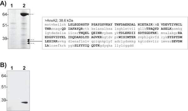
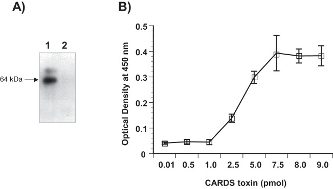
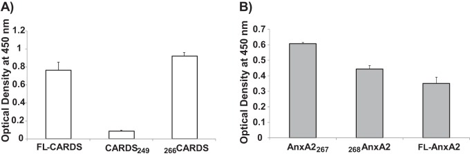
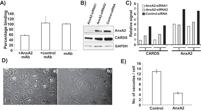
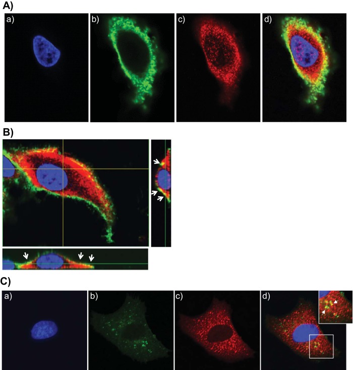
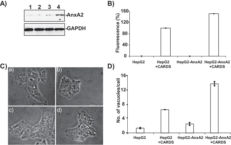
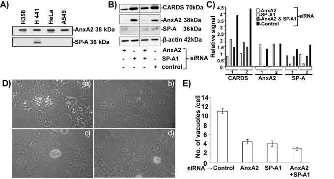
Mycoplasma pneumoniae synthesizes a novel human surfactant protein A (SP-A)-binding cytotoxin, designated community-acquired respiratory distress syndrome (CARDS) toxin, that exhibits ADP-ribosylating and vacuolating activities in mammalian cells and is directly linked to a range of acute and chronic airway diseases, including asthma. In our attempt to detect additional CARDS toxin-binding proteins, we subjected the membrane fraction of human A549 airway cells to affinity chromatography using recombinant CARDS toxin as bait. A 36-kDa A549 cell membrane protein bound to CARDS toxin and was identified by time of flight (TOF) mass spectroscopy as annexin A2 (AnxA2) and verified by immunoblotting with anti-AnxA2 monoclonal antibody. Dose-dependent binding of CARDS toxin to recombinant AnxA2 reinforced the specificity of the interaction, and further studies revealed that the carboxy terminus of CARDS toxin mediated binding to AnxA2. In addition, pretreatment of viable A549 cells with anti-AnxA2 monoclonal antibody or AnxA2 small interfering RNA (siRNA) reduced toxin binding and internalization. Immunofluorescence analysis of CARDS toxin-treated A549 cells demonstrated the colocalization of CARDS toxin with cell surface-associated AnxA2 upon initial binding and with intracellular AnxA2 following toxin internalization. HepG2 cells, which express low levels of AnxA2, were transfected with a plasmid expressing AnxA2 protein, resulting in enhanced binding of CARDS toxin and increased vacuolization. In addition, NCI-H441 cells, which express both AnxA2 and SP-A, upon AnxA2 siRNA transfection, showed decreased binding and subsequent vacuolization. These results indicate that CARDS toxin recognizes AnxA2 as a functional receptor, leading to CARDS toxin-induced changes in mammalian cells.

Importance: Host cell susceptibility to bacterial toxins is usually determined by the presence and abundance of appropriate receptors, which provides a molecular basis for toxin target cell specificities. To perform its ADP-ribosylating and vacuolating activities, community-acquired respiratory distress syndrome (CARDS) toxin must bind to host cell surfaces via receptor-mediated events in order to be internalized and trafficked effectively. Earlier, we reported the binding of CARDS toxin to surfactant protein A (SP-A), and here we show how CARDS toxin uses an alternative receptor to execute its pathogenic properties. CARDS toxin binds selectively to annexin A2 (AnxA2), which exists both on the cell surface and intracellularly. Since AnxA2 regulates membrane dynamics at early stages of endocytosis and trafficking, it serves as a distinct receptor for CARDS toxin binding and internalization and enhances CARDS toxin-induced vacuolization in mammalian cells.


Figures

FIG 1
FIG 1
CARDS toxin binds to A549 cell membrane-associated AnxA2. (A) Identification of AnxA2 bound to CARDS toxin. Membrane-enriched fractions of A549 cells were incubated with Ni-NTA alone or CARDS toxin coupled to Ni-NTA. Ni-NTA-bound membrane proteins (lane 1) or CARDS toxin-coupled Ni-NTA-bound membrane proteins (lane 2) were separated on NuPAGE (4 to 12% gradient) gels and stained with Coomassie brilliant blue G-250. Mass spectrometry analysis was performed on eluted proteins. The short dashed arrows point to protein bands that were identified as FL or processed/degraded CARDS toxin, and the long solid arrow points to AnxA2. The capital boldface letters in the AnxA2 sequence are AnxA2-specific amino acids identified by mass spectrometry. The molecular masses (in kilodaltons) of molecular mass markers are indicated to the left of the gel. (B) Immunoblot confirmation of AnxA2 bound to CARDS toxin during pulldown assay. Eluted proteins from panel A were resolved on 4 to 12% NuPAGE gels, transferred to nitrocellulose membranes, and probed with anti-AnxA2 monoclonal antibody. Eluted proteins from control uncoupled Ni-NTA beads (lane 1) show no immunoreactivity, whereas eluted proteins from CARDS toxin-coupled Ni-NTA beads (lane 2) demonstrate clear immunoreactivity at ~36-kDa range.
FIG 2
FIG 2
Binding of CARDS toxin to recombinant AnxA2. (A) Binding of CARDS toxin to AnxA2 by ligand blotting. GST-AnxA2 or BSA (2 µg each) was separated on SDS-polyacrylamide gels, transferred to nitrocellulose membranes, and incubated with CARDS toxin (7 µg/ml) for 2 h. CARDS toxin binding was detected by incubation with rabbit polyclonal anti-CARDS toxin antibody followed by incubation with goat anti-rabbit IgG and visualization with ECL. Lane 1, AnxA2; lane 2, BSA. (B) Dose-dependent binding of CARDS toxin to AnxA2. Microtiter wells were coated with 100 ng AnxA2, and increasing concentrations of CARDS toxin or BSA were added to individual wells for 1 h at room temperature. Bound protein was detected with rabbit polyclonal anti-CARDS toxin antibody and goat anti-rabbit HRP-conjugated polyclonal antibody, followed by development with TMB substrate. Wells with BSA alone served as negative controls, and the nonspecific bound values were subtracted from individual test scores. Values are means ± standard errors of the means (error bars) for triplicate wells from three separate experiments. No immunological cross-reactivity was observed between anti-CARDS toxin antibody and AnxA2.
FIG 3
FIG 3
The C terminus of CARDS toxin mediates binding to full-length (FL) and truncated AnxA2. (A) Interaction of CARDS toxin C terminus with FL AnxA2. AnxA2 (100 ng/well) was bound to individual wells on ELISA plates and incubated with equimolar concentrations of FL CARDS toxin and to N-terminal (CARDS249; amino acids [aa] 1 to 249) and C-terminal (266CARDS; aa 266 to 591) proteins. Binding was detected using anti-His tag monoclonal antibody. (B) Interaction of FL and truncated AnxA2 derivatives with FL CARDS toxin. Equimolar concentrations of FL AnxA2, truncated AnxA2267, or truncated 268AnxA2 were bound to individual wells of ELISA plates and incubated with CARDS toxin. Toxin binding was detected using rabbit polyclonal anti-CARDS toxin antibody.
FIG 4
FIG 4
AnxA2 surface accessibility promotes CARDS toxin binding to A549 cells. (A) CARDS toxin binding to A549 cells was reduced by anti-AnxA2 monoclonal antibody (mAb). A549 cells were incubated with or without anti-AnxA2 monoclonal antibody or negative-control isotype-matched monoclonal antibody for 10 min followed by incubation with CARDS toxin-mCherry protein for 30 min at 4°C. Binding of CARDS toxin-mCherry protein to A549 cells was analyzed by fluorometry as described in Materials and Methods. (B) Reduced CARDS toxin binding following suppressed AnxA2 expression. CARDS toxin (1 µg) was added to A549 cells transfected with siRNAs specific to AnxA2 or control random siRNAs and incubated for 1 h at 37°C. AnxA2 expression and CARDS toxin association with A549 cells were analyzed by immunoblotting with anti-AnxA2 monoclonal antibody and anti-CARDS polyclonal antibody. Comparative GAPDH intensities were used as loading controls. (C) Quantification of CARDS toxin associated with A549 cells upon suppression of AnxA2. Immunostained bands from panel B were quantified as detailed in Materials and Methods and normalized with GAPDH. Experiments were repeated two times (experiments 1 and 2), and the relative signal differences in binding levels of CARDS toxin and expression levels of AnxA2 are presented. (D) Decreased CARDS toxin-mediated vacuolization in A549 cells following suppressed AnxA2 expression. CARDS toxin was added to A549 cells transfected with control random siRNAs (a) or siRNAs specific to AnxA2 (b) and incubated for 24 h at 37°C. Vacuole formation was analyzed microscopically. (E) Quantification of CARDS toxin-induced vacuoles in A549-AnxA2-siRNA cells at 24 h. As described above for panel C, the cells were incubated with CARDS toxin, and the numbers of vacuoles per cell were counted and compared.
FIG 5
FIG 5
CARDS toxin colocalizes with cell surface and intracellular AnxA2. (A) Colocalization of CARDS toxin with cell surface-associated AnxA2. (a to c) A549 cells were incubated with 10 µg of CARDS toxin at 4°C for 1 h, fixed in the presence of DAPI to stain nuclei (blue) (a) and probed with rabbit polyclonal anti-CARDS toxin antibody followed by secondary goat anti-rabbit IgG conjugated with Alexa Fluor 488 (green) (b) and anti-AnxA2 monoclonal antibody (1:500) followed by secondary goat anti-mouse IgG antibody conjugated with Alexa Fluor 555 (red) (c). (d) The merged image shows colocalization of CARDS toxin and AnxA2 at the membrane surface as yellow. (B) Interaction of CARDS toxin with A549 cell surface-associated AnxA2 using confocal laser-scanning microscopy. White arrows indicate the surface colocalization of CARDS toxin with AnxA2 (yellow) based upon serial z sections (0.44 μm; z series) obtained by analyzing x-y scans. (C) Interaction of internalized CARDS toxin with cytoplasmic AnxA2. (a to c) A549 cells were incubated with 10 µg of CARDS toxin at 4°C for 1 h and shifted to 37°C for 1 h, fixed in the presence of DAPI to stain nuclei (blue) (a) and probed with specific antibodies against CARDS toxin (green) (b) and AnxA2 (red) (1:1,000) (c) as described above for panel A. White arrows indicate colocalized intracellular CARDS toxin and AnxA2. (d) Images were collected sequentially from different channels with a confocal laser-scanning microscope and merged to show colocalization (yellow). To demonstrate the colocalization of intracellular AnxA2 and CARDS toxin, a section of the merged image shown by the white square is enlarged (top right panel).
FIG 6
FIG 6
AnxA2 expression enhances CARDS toxin binding and vacuolization in transfected HepG2 cells. (A) Expression of AnxA2 in stably transfected HepG2 cells. HepG2 cells were transfected with pCDNA plasmid carrying the AnxA2 gene, and G418-resistant stable clones were isolated and screened for expression of AnxA2 (lanes 2 to 4) and compared to normal HepG2 cells (lane 1). The clone expressing the highest level of HepG2-AnxA2 (lane 4) was selected for further studies. (B) Increased binding of CARDS toxin-mCherry to HepG2-AnxA2 cells. HepG2 and HepG2-AnxA2 cells were incubated with CARDS toxin-mCherry protein for 30 min at 4°C, and binding of CARDS toxin-mCherry protein to cells was analyzed by fluorometry as described in Materials and Methods. (C) AnxA2 expression enhances CARDS toxin-mediated vacuolization. HepG2 and HepG2-AnxA2 cells were incubated with CARDS toxin (50 µg/ml) for 4 h at 37°C, and vacuole formation was analyzed microscopically. (a to d) HepG2 cells alone (a) and with CARDS toxin (b) and HepG2-AnxA2 cells alone (c) and with CARDS toxin (d) are shown. (D) Increased numbers of CARDS toxin-induced vacuoles in HepG2-AnxA2 cells at 4 h. As described above for panel C, the cells were incubated with CARDS toxin, and the numbers of vacuoles per cell were counted and compared.
FIG 7
FIG 7
CARDS toxin binding and subsequent vacuolization in siRNA-transfected H441 cells. (A) Screening of different human cell lines for the expression of AnxA2 and SP-A. Total cell lysates (5 µg for analysis of AnxA2 and 30 µg for analysis of SP-A) were separated using 4 to 12% gels, transferred to nitrocellulose membranes, and probed with anti-AnxA2 monoclonal antibody (MAb) and anti-SP-A MAb. H441 cells that expressed both AnxA2 and SP-A were used for further studies. (B) Association of CARDS toxin with the cell surface upon suppression of AnxA2 and SP-A expression. H441 cells were transfected with siRNAs specific to AnxA2, SP-A, AnxA2 and SP-A, and control random siRNAs and incubated individually with CARDS toxin (5 µg/ml) for 1 h at 4°C. Five-microgram amounts of total cell lysates were analyzed by immunoblotting with anti-AnxA2 MAb or anti-SP-A MAb and anti-CARDS toxin polyclonal antibody. Comparative β-actin intensities were used as loading controls. (C) Quantification of CARDS toxin relative to AnxA2 and SP-A in siRNA-transfected H441 cells. Using β-actin (loading control), CARDS toxin, AnxA2, and SP-A immunoblot intensities, we calculated the relative signal difference of receptor-mediated association of CARDS toxin, along with expression levels of AnxA2 and SP-A. Data are from two independent experiments (experiments 1 and 2). (D) Suppression of AnxA2 or SP-A or AnxA2 and SP-A expression decreases CARDS toxin-mediated vacuolization in H441 cells. Untreated and siRNA-transfected H441cells were incubated with CARDS toxin (50 µg/ml) for 24 h at 37°C, and vacuole formation was analyzed microscopically. (a) Control random siRNA, (b) AnxA2-siRNA, (c) SP-A1-siRNA and (d) AnxA2 and SP-A1 siRNAs. (E) Quantification of numbers of CARDS toxin-induced vacuoles in H441 and siRNA-transfected H441 cells at 24 h. As described above for panel C, the cells were incubated with CARDS toxin, and the numbers of vacuoles per cell were counted and compared as described in Materials and Methods.

References

    1. Baseman JB, Tully JG. 1997. Mycoplasmas: sophisticated, reemerging, and burdened by their notoriety. Emerg. Infect. Dis. 3:21–32. 10.3201/eid0301.970103 - DOI - PMC - PubMed
    1. Kraft M, Cassell GH, Pak J, Martin RJ. 2002. Mycoplasma pneumoniae and Chlamydia pneumoniae in asthma: effect of clarithromycin. Chest 121:1782–1788. 10.1378/chest.121.6.1782 - DOI - PubMed
    1. Biscardi S, Lorrot M, Marc E, Moulin F, Boutonnat-Faucher B, Heilbronner C, Iniguez JL, Chaussain M, Nicand E, Raymond J, Gendrel D. 2004. Mycoplasma pneumoniae and asthma in children. Clin. Infect. Dis. 38:1341–1346. 10.1086/392498 - DOI - PubMed
    1. Waites KB, Talkington DF. 2004. Mycoplasma pneumoniae and its role as a human pathogen. Clin. Microbiol. Rev. 17:697–728. 10.1128/CMR.17.4.697-728.2004 - DOI - PMC - PubMed
    1. Nisar N, Guleria R, Kumar S, Chand Chawla T, Ranjan Biswas N. 2007. Mycoplasma pneumoniae and its role in asthma. Postgrad. Med. J. 83:100–104. 10.1136/pgmj.2006.049023 - DOI - PMC - PubMed

Publication types

MeSH terms