. 2014 Sep 2;111(35):12746-51.
doi: 10.1073/pnas.1404372111. Epub 2014 Aug 19.

Phospholipase A2 regulates eicosanoid class switching during inflammasome activation

Affiliations

Phospholipase A2 regulates eicosanoid class switching during inflammasome activation

Paul C Norris et al. Proc Natl Acad Sci U S A. .

Abstract

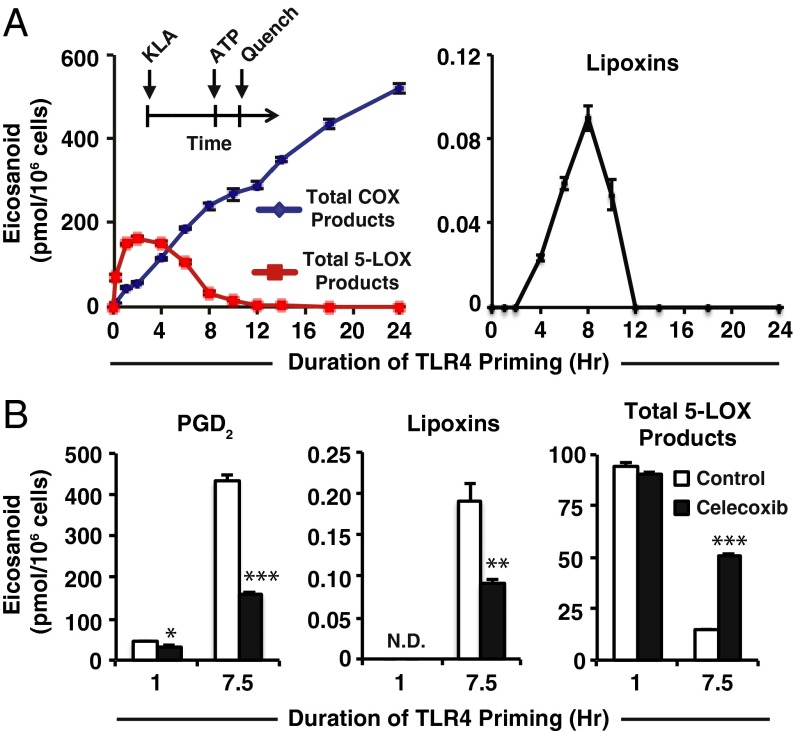
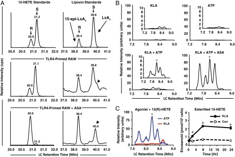
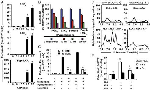
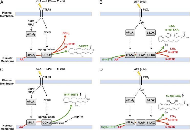
Initiation and resolution of inflammation are considered to be tightly connected processes. Lipoxins (LX) are proresolution lipid mediators that inhibit phlogistic neutrophil recruitment and promote wound-healing macrophage recruitment in humans via potent and specific signaling through the LXA4 receptor (ALX). One model of lipoxin biosynthesis involves sequential metabolism of arachidonic acid by two cell types expressing a combined transcellular metabolon. It is currently unclear how lipoxins are efficiently formed from precursors or if they are directly generated after receptor-mediated inflammatory commitment. Here, we provide evidence for a pathway by which lipoxins are generated in macrophages as a consequence of sequential activation of toll-like receptor 4 (TLR4), a receptor for endotoxin, and P2X7, a purinergic receptor for extracellular ATP. Initial activation of TLR4 results in accumulation of the cyclooxygenase-2-derived lipoxin precursor 15-hydroxyeicosatetraenoic acid (15-HETE) in esterified form within membrane phospholipids, which can be enhanced by aspirin (ASA) treatment. Subsequent activation of P2X7 results in efficient hydrolysis of 15-HETE from membrane phospholipids by group IVA cytosolic phospholipase A2, and its conversion to bioactive lipoxins by 5-lipoxygenase. Our results demonstrate how a single immune cell can store a proresolving lipid precursor and then release it for bioactive maturation and secretion, conceptually similar to the production and inflammasome-dependent maturation of the proinflammatory IL-1 family cytokines. These findings provide evidence for receptor-specific and combinatorial control of pro- and anti-inflammatory eicosanoid biosynthesis, and potential avenues to modulate inflammatory indices without inhibiting downstream eicosanoid pathways.

Keywords: enzyme coupling; lipidomics; membrane remodeling.


Conflict of interest statement

The authors declare no conflict of interest.

Figures

Fig. 1.
Fig. 1.
Duration of TLR4 priming controls purinergic 5-LOX product formation and lipoxin biosynthesis. (A, Inset) Protocol for TLR4 priming (Kdo2 lipid A, KLA) starting at time = 0 followed by ATP stimulation at indicated times and subsequent reaction quench as endpoint (further details can be found in SI Materials and Methods); eicosanoid levels from RAW264.7 (RAW) cell medium after TLR4 priming with 100 ng/mL KLA for varying durations before stimulation with 2 mM ATP for the final 10 min include total COX products (PGD2, PGE2, PGF, PGJ2, 15-deoxy PGD2, 15-deoxy PGJ2, 11-HETE, and 15-HETE); total 5-LOX products (5-HETE, LTC4, 11-trans LTC4, LTB4, 6-trans,12-epi LTB4, 6-trans LTB4, and 12-epi LTB4); lipoxins (LXA4 and 15-epi–LXA4). (B) Levels of PGD2, lipoxins (LXA4 and 15-epi–LXA4), and total 5-LOX products (as in A) from RAW medium after KLA priming for the indicated times in the absence (white bars) or presence (black bars) of 50 nM celecoxib (∼IC50) followed by stimulation with ATP for the final 30 min; PGD2 levels were decreased with celecoxib treatment vs. control with 1-h TLR4 priming (*P < 0.01) and 7.5 h TLR4 priming (***P < 0.0001); lipoxin levels were not detected (N.D.) with 1 h priming and were decreased at 7.5 h TLR4 priming with celecoxib treatment vs. control (**P < 0.005); total 5-LOX products with 1-h priming were not significantly different with celecoxib vs. control, and at 7.5-h priming were increased with celecoxib vs. control (***P < 0.0001). Data are mean values of three separate experiments ± SEM.
Fig. 2.
Fig. 2.
Chirality of lipoxins, requirement for priming, and separate effects of TLR4 and P2X7 stimulation. (A) MS monitoring was set to multiple-reaction monitoring (MRM) transition 319(−) to 219(−) m/z for 15-HETE in the first period (Left column) and 351(−) to 115(−) m/z for lipoxins in the second period (Right column); intensity is expressed in counts per second, cps). Chiral separation of (Top row) 15(R)-HETE:15(S)-HETE standards at a 1:3 concentration and 15-epi–LXA4:LXA4 standards at a 1:3 concentration; (Middle row) RAW cell medium after 7.5 h KLA and final 30 min ATP; (Bottom row) RAW cell medium after 7.5 h KLA in the presence of 1 mM ASA and final 30 min ATP. Arrowheads indicate a species ∼12 s to the Right of LXA4. A total of ∼25 million cells in a T-75 tissue culture flask containing 5 mL medium was used for both conditions [this cell quantity is considerably higher than in other experiments due to decreased signal yielded in atmospheric pressure chemical ionization (APCI) mode]; n = 1. (B) RAW medium after (Upper Left) only KLA stimulation for 8 h; (Upper Right) no stimulation for 7.5 h before ATP for final 30 min; (Lower Left) 7.5 h KLA priming before ATP stimulation for final 30 min; (Lower Right) 7.5 h KLA priming in the presence of 1 mM ASA before ATP stimulation for final 30 min. Chromatogram traces represent 70-s scheduled monitoring of MRM transition 351(−) m/z to 115(−) m/z during nonchiral reverse-phase chromatographic separation on a scale of 100 arbitrary units (Insets are on a one order of magnitude lower scale); data represent one replicate of n = 3. (C, Left) Chromatograms (as in B) of RAW medium after 30 min KLA or ATP stimulation in the presence of 1 μM 15(R)-HETE; coelution with LXA4 and 15-epi–LXA4 commercial standards are indicated with a green asterisk; (Right) 15-HETE levels from saponified phospholipids of RAW cells after KLA stimulation for the indicated times over a 24-h period represent mean values of three separate experiments ± SEM; 15-HETE levels were increased with KLA stimulation vs. control (*P < 0.001).
Fig. 3.
Fig. 3.
Millimolar ATP and cPLA2 are required for lipoxin biosynthesis in the presence of exogenous 15-HETE or TLR4 priming. (A) Extracellular eicosanoid levels from RAW cells after 30 min in the presence of 1 μM 15(R)-HETE along with the indicated concentrations of ATP (PGD2, LTC4, and lipoxin levels were higher with millimolar ATP levels vs. 0–500 μM ATP levels; *P < 0.001). (B) Extracellular eicosanoid levels from RAW cells preincubated for 30 min in the absence (control) or presence of the indicated concentrations of pyrrophenone; cells were then stimulated with ATP in the presence of 1 μM 15(R)-HETE for 30 min. (C) Eicosanoid levels in membrane phospholipids of RAW cells incubated for 30 min in the absence or presence of ATP and/or 1 μM 15(R)-HETE, and absence or presence of 500 nM pyrrophenone or 5 μM LY315920 (varespladib); 5-HETE levels increased with 15(R)-HETE + ATP-treated cells in the absence/presence of LY315920 vs. untreated cells (*P < 0.0001), but not with pyrrophenone treatment or 15(R)-HETE treatment alone; 15-HETE levels decreased in cells treated with 15(R)-HETE + ATP in the presence/absence of LY315920 vs. treatment with pyrrophenone or with 15(R)-HETE treatment alone (#P < 0.0001). (D) Bone-marrow–derived macrophages generated from GIVA cPLA2 transgenic mice (Left column) (+/+) and (Right column) (−/−) were stimulated (Upper row) with 100 ng/mL KLA in the presence of 1 mM ASA for 8 h; (Lower row) with 100 ng/mL KLA in the presence of 1 mM ASA for 7.5 h before stimulation with 2 mM ATP for the final 30 min. Data are chromatograms of extracellular media generated as described in Fig. 2B and are representative of three separate experiments. Green asterisk indicates coelution with LXA4 and 15-epi–LXA4 standards. (E) Levels of 15-HETE from membrane phospholipids in BMDM cells (from the same experiment as in D); 15-HETE levels in KLA + ASA vs. untreated cells (+/+) were increased (**P < 0.001); KLA + ASA + ATP vs. KLA + ASA-treated cells (+/+) were decreased (*P < 0.01); the same comparisons in (−/−) cells found no significant increases or decreases. Data are mean values of three separate experiments ± SEM.
Fig. 4.
Fig. 4.
Mechanism of inflammatory receptor-mediated formation of lipoxins in macrophages. (A) Macrophages expressing TLR4 (and likely other TLRs) recognize pathogen-associated molecular pattern (PAMP) species [such as lipopolysaccharide (LPS) derived from Escherechia coli, which contains the TLR4 ligand Kdo2-lipid A (KLA)] leading to activation of PLA2 hydrolysis of esterified AA and increased expression of COX-2. The majority of AA oxygenated by up-regulated COX-2 forms PGH2 (∼90–93%; via the cyclooxygenase and peroxidase active sites) and ∼4–6% is converted to 11-HETE (via the cyclooxygenase active site), and a smaller portion (∼1–3%) is converted to 15-HETE (∼30% R, ∼70% S; via the peroxidase active site). The 15-HETE is secreted from the cell, but a portion is esterified within membrane phospholipids through several possible routes putatively via fatty acyl CoA ligase. (B) Concomitantly, extracellular millimolar ATP generated by necrotized cells, PAMP recognizing cells, and recruited/activated neutrophils (reviewed in ref. 1) during a pathogenic/inflammatory assault stimulates the P2X7 receptor in macrophages. This leads to an increase in macrophage intracellular Ca2+, activating cPLA2 and 5-LOX via translocation primarily to the perinuclear membrane. LTA4 and derived metabolites, along with 5-HETE, are produced by cPLA2-mediated AA hydrolysis, assistance of FLAP, and 5-LOX metabolism. Some 5-HETE becomes esterified in cell membranes (depicted in perinuclear phospholipids but potentially in other organelles as well); the rest diffuses from the cell. Esterified 15-HETE is also hydrolyzed by cPLA2, and a portion is then converted to LXA4 and 15-epi–LXA4; hydrolysis of 15-HETE is required to enhance metabolism by 5-LOX via coupling with cPLA2 and FLAP. (C) In the presence of aspirin, acetylated COX-2 produces ∼100% 15(R)-HETE (and at significantly higher levels than with native enzyme). (D) Esterified 15(R)-HETE is subsequently released and converted to 15-epi–LXA4 at enhanced levels after P2X7 stimulation.

References

    1. Ferrari D, et al. The P2X7 receptor: A key player in IL-1 processing and release. J Immunol. 2006;176(7):3877–3883. - PubMed
    1. Ferrari D, et al. Extracellular ATP triggers IL-1 beta release by activating the purinergic P2Z receptor of human macrophages. J Immunol. 1997;159(3):1451–1458. - PubMed
    1. Solle M, et al. Altered cytokine production in mice lacking P2X(7) receptors. J Biol Chem. 2001;276(1):125–132. - PubMed
    1. Kahlenberg JM, Lundberg KC, Kertesy SB, Qu Y, Dubyak GR. Potentiation of caspase-1 activation by the P2X7 receptor is dependent on TLR signals and requires NF-kappaB-driven protein synthesis. J Immunol. 2005;175(11):7611–7622. - PubMed
    1. Suzuki K, et al. Lipopolysaccharide primes human alveolar macrophages for enhanced release of superoxide anion and leukotriene B4: Self-limitations of the priming response with protein synthesis. Am J Respir Cell Mol Biol. 1993;8(5):500–508. - PubMed

Publication types

MeSH terms