. 2014 Sep 16;111(37):13379-84.
doi: 10.1073/pnas.1324235111. Epub 2014 Aug 19.

Endothelial cell FGF signaling is required for injury response but not for vascular homeostasis

Affiliations

Endothelial cell FGF signaling is required for injury response but not for vascular homeostasis

Sunday S Oladipupo et al. Proc Natl Acad Sci U S A. .

Abstract

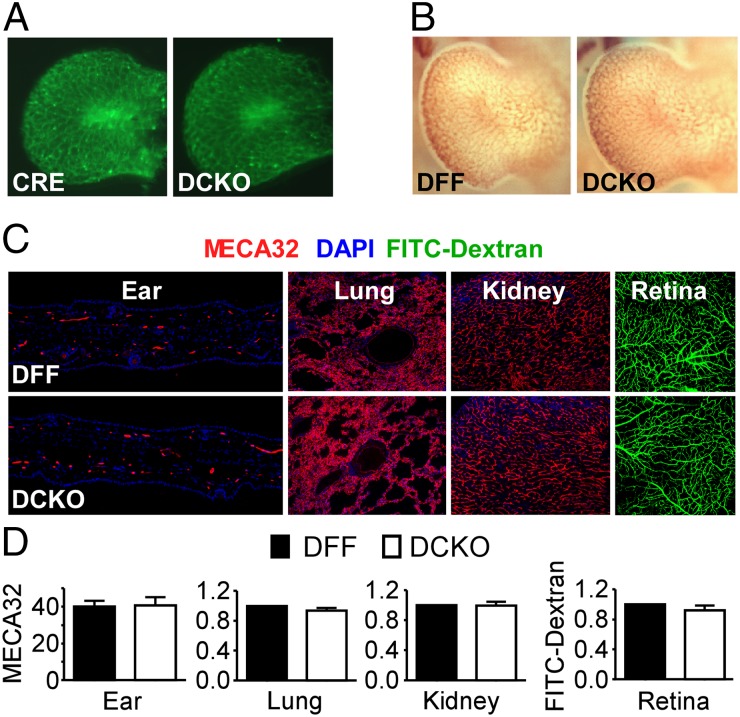
Endothelial cells (ECs) express fibroblast growth factor receptors (FGFRs) and are exquisitely sensitive to FGF signals. However, whether the EC or another vascular cell type requires FGF signaling during development, homeostasis, and response to injury is not known. Here, we show that Flk1-Cre or Tie2-Cre mediated deletion of FGFR1 and FGFR2 (Fgfr1/2(Flk1-Cre) or Fgfr1/2(Tie2-Cre) mice), which results in deletion in endothelial and hematopoietic cells, is compatible with normal embryonic development. As adults, Fgfr1/2(Flk1-Cre) mice maintain normal blood pressure and vascular reactivity and integrity under homeostatic conditions. However, neovascularization after skin or eye injury was significantly impaired in both Fgfr1/2(Flk1-Cre) and Fgfr1/2(Tie2-Cre) mice, independent of either hematopoietic cell loss of FGFR1/2 or vascular endothelial growth factor receptor 2 (Vegfr2) haploinsufficiency. Also, impaired neovascularization was associated with delayed cutaneous wound healing. These findings reveal a key requirement for cell-autonomous EC FGFR signaling in injury-induced angiogenesis, but not for vascular homeostasis, identifying the EC FGFR signaling pathway as a target for diseases associated with aberrant vascular proliferation, such as age-related macular degeneration, and for modulating wound healing without the potential toxicity associated with direct manipulation of systemic FGF or VEGF activity.

Keywords: choroidal neovascularization; neoangiogenesis; oxygen-induced retinopathy; retinopathy of prematurity.


Conflict of interest statement

The authors declare no conflict of interest.

Figures

Fig. 1.
Fig. 1.
Endothelial/hematopoietic FGFR1/2 is dispensable for vascular development and homeostasis in vivo. (A) Limb buds (embryonic day 11.5) from a Flk1-Cre, mT/mG embryo (Left) and a Flk1-Cre, mT/mG, DCKO embryo (Right) showing normal vascular patterns (green). (B) Limb bud (embryonic day 12.0) from a DFF embryo (Left) and a DCKO embryo (Right) immunostained for CD31, showing normal vascular plexus formation. (C) Representative MECA32 (+) immunofluorescence micrographs showing normal microvascular density and morphology in DFF and DCKO ear skin, lung, and kidney, and a normal retina vascular plexus formation visualized in mice perfused with FITC-dextran. (D) Meca32 (+) vessel count (external ear skin), quantitative immunofluorescence normalized to DAPI (lung and kidney), and FITC-dextran fluorescent area (retina), showing no difference in the microvascular network between DFF and DCKO mice (n = 3). All values are mean ± SD. Lung, kidney, and retina were imaged with a 10× objective and the external ear skin was imaged with a 20× objective. Data were analyzed using the unpaired Student t test.
Fig. 2.
Fig. 2.
Flk1-Cre activation is maintained in adulthood and hematopoiesis is normal in mice lacking endothelial/hematopoietic FGFR1/2. (A and B) Quantitative RT-PCR analysis of FACS CD31- and VE-cadherin–positive adult lung ECs (A) and CD45-positive adult bone marrow hematopoietic cells (B), showing efficient depletion of Fgfr1 and Fgfr2 mRNA expression in DCKO mice. Data are represented as relative expression normalized to Hprt. (C and D) FACS analysis of bone marrow hematopoietic cells revealing normal myeloid (C) or lymphoid/stem cell populations (D) in DCKO compared with DFF mice. All values are mean ± SD. Data were analyzed using the unpaired Student t test; *P < 0.05; **P < 0.01; ns, not significant. n = 2–4.
Fig. 3.
Fig. 3.
FGFR1/2 is dispensable for basal vascular permeability, BP, and vascular reactivity in vivo. (A) FITC-dextran (2 MDa) whole-mount stained DFF and DCKO adult mouse retinas showing absence of vascular leak at basal levels (representative of 8–10 retinas per group). (B) Vascular leakage assessed by EB dye permeability showing similar ear skin coloration in vehicle-treated DFF and DCKO mice but increased leakage into the skin in DCKO mice compared with DFF mice following treatment with mustard oil. (C) Quantitation of EB in ear tissue (mean ± SEM, n = 5–7, *P < 0.04). (D) Mean arterial BP. (E) Mesenteric artery (MA) vasoconstriction in response to PE (n = 4–6 animals, two vessels per animal per group). The data shown are the mean percentages of change in diameter of arteries perfused with Mops containing PE at indicated concentrations, expressed as the percentages of change in vessel diameter relative to baseline. (F) Vasodilation in response to Ach expressed as percentages of increase in arterial diameter after constriction with PE (100 μM). (G) Vasodilation in response to SNP expressed as percentages of increase in arterial diameter after constriction with PE (100 μM).
Fig. 4.
Fig. 4.
Impaired endothelial neovascularization and delayed wound healing in mice lacking endothelial FGFR1/2. (A) Representative MECA32 (+) vessels (red) showing increased neovascular growth in Flk1-Cre and DFF control mouse wound edge (2 mm) 6 d after excision wound injury compared with normal unwounded skin. Neovascularization was significantly reduced in wounded DCKO mice (Lower Right). Epidermis is marked in green (Keratin 14) and nuclei in blue (DAPI). (B) Quantitative immunofluorescence of A, MECA32-positive vessels normalized to DAPI (n = 4–10). (C) Smooth muscle actin (SMA)-positive vessel count (three to five 20× fields per mouse) (n = 3–4 mice; see Fig. S2A for corresponding representative images). (D) Western blot of wound margin tissue from Flk1-Cre, DFF, and DCKO mice showing that Vegfr2 heterozygosity (Flk1-Cre mice) has no effect on microvascular density (CD31 levels) in response to skin injury compared with DFF mice. CD31 levels are reduced in DCKO, and not in Flk1-Cre and DFF, mice following injury. VEGFR2 levels are reduced by 50% in Flk1-Cre mice compared with DFF controls and further reduced in DCKO mice. (E and F) Quantification of D using imageJ image analysis software. CD31 and VEGFR2 arbitrary density units (ImageJ) normalized to tubulin. (G) Wound healing is significantly delayed at 4 and 6 d after wounding in DCKO compared with Flk1-Cre and DFF control mice (n = 4–10). (H and I) F4/80 immunostaining (quantified in I) showing similar levels in wound-margin tissue from DFF and DCKO mice. A and H were imaged with a 10× objective. *P < 0.05, **P < 0.01, and ***P < 0.001.
Fig. 5.
Fig. 5.
Endothelial FGFR1/2 is required for neovascular response in eye injury-induced pathologic angiogenesis. (A) Representative FITC-dextran staining and (B and C) quantification shows a marked suppression of CNV in DCKO mice compared with control Flk1-Cre or Tie2-Cre and DFF mice (n = 8–10). *P < 0.05. (Scale bars: A, 100 μm.)

References

    1. Ferrara N, Kerbel RS. Angiogenesis as a therapeutic target. Nature. 2005;438(7070):967–974. - PubMed
    1. Chung AS, Ferrara N. Developmental and pathological angiogenesis. Annu Rev Cell Dev Biol. 2011;27:563–584. - PubMed
    1. Murata K, et al. Inhibition of miR-92a enhances fracture healing via promoting angiogenesis in a model of stabilized fracture in young mice. J Bone Miner Res. 2014;29(2):316–326. - PubMed
    1. Potente M, Gerhardt H, Carmeliet P. Basic and therapeutic aspects of angiogenesis. Cell. 2011;146(6):873–887. - PubMed
    1. Presta M, et al. Fibroblast growth factor/fibroblast growth factor receptor system in angiogenesis. Cytokine Growth Factor Rev. 2005;16(2):159–178. - PubMed

Publication types

MeSH terms

Substances