. 2014 Aug 5:6:197.
doi: 10.3389/fnagi.2014.00197. eCollection 2014.

Structure activity relationship of phenolic acid inhibitors of α-synuclein fibril formation and toxicity

Affiliations

Structure activity relationship of phenolic acid inhibitors of α-synuclein fibril formation and toxicity

Mustafa T Ardah et al. Front Aging Neurosci. .

Abstract

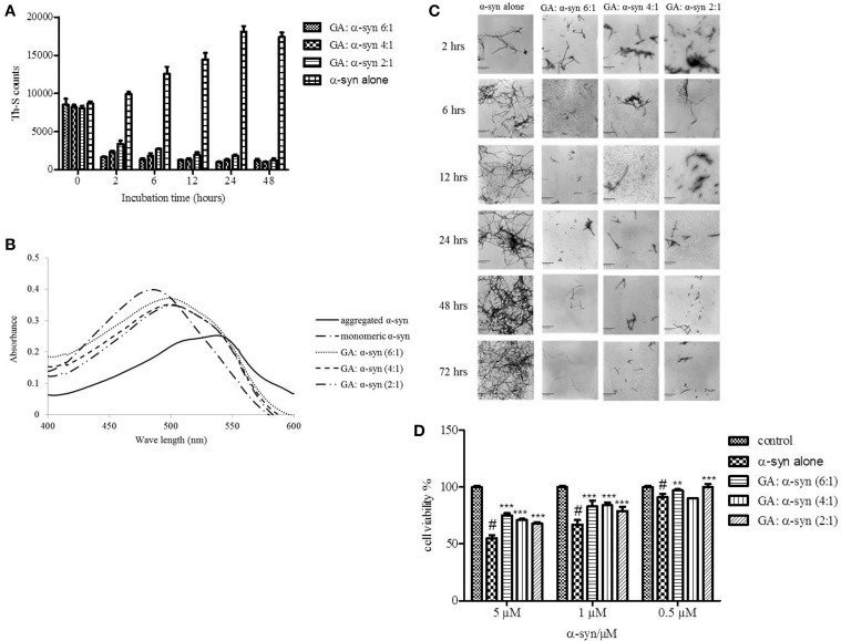
The aggregation of α-synuclein (α-syn) is considered the key pathogenic event in many neurological disorders such as Parkinson's disease (PD), dementia with Lewy bodies and multiple system atrophy, giving rise to a whole category of neurodegenerative diseases known as synucleinopathies. Although the molecular basis of α-syn toxicity has not been precisely elucidated, a great deal of effort has been put into identifying compounds that could inhibit or even reverse the aggregation process. Previous reports indicated that many phenolic compounds are potent inhibitors of α-syn aggregation. The aim of the present study was to assess the anti-aggregating effect of gallic acid (GA) (3,4,5-trihydroxybenzoic acid), a benzoic acid derivative that belongs to a group of phenolic compounds known as phenolic acids. By employing an array of biophysical and biochemical techniques and a cell-viability assay, GA was shown not only to inhibit α-syn fibrillation and toxicity but also to disaggregate preformed α-syn amyloid fibrils. Interestingly, GA was found to bind to soluble, non-toxic oligomers with no β-sheet content, and to stabilize their structure. The binding of GA to the oligomers may represent a potential mechanism of action. Additionally, by using structure activity relationship data obtained from fourteen structurally similar benzoic acid derivatives, it was determined that the inhibition of α-syn fibrillation by GA is related to the number of hydroxyl moieties and their position on the phenyl ring. GA may represent the starting point for designing new molecules that could be used for the treatment of PD and related disorders.

Keywords: Parkinson's disease; aggregation; amyloid fibrils; drug discovery; gallic acid; α-synuclein.


Figures

Figure 1
Figure 1
GA inhibits α-syn fibrillation in a concentration-dependent manner. (A) Samples of α-syn (25μM) were incubated alone or in the presence of GA (molar ration of GA: α-syn 4:1, 2:1, 1:1) for 6 days with continuous shaking at 37°C. Fibril formation was estimated by Th-S fluorescence. The assay was performed in triplicate (average of triplicate measurement ± standard deviation). (B) Congo red binding for samples of α-syn (25μM) incubated alone or in the presence of GA (molar ratio of GA: α-syn 4:1, 2:1, 1:1) for 6 days with continuous shaking at 37°C. Samples of α-syn (5μM) aged alone or with GA were mixed with Congo red at a final concentration of 5μM. The reaction samples were thoroughly mixed and placed in a 10 mm quartz cuvette. The UV absorbance spectra were recorded from 400 to 600 nm. C-F. Electron microscopy images of negatively stained samples of α-syn (25μM) aged alone or in the presence of GA (molar ratio of GA: α-syn 4:1, 2:1, 1:1) for 6 days with continuous shaking at 37°C. (C) Aged α-syn alone. (D) α-Syn aged in the presence of GA at a molar ratio of GA: α-syn 4:1. (E) α-Syn aged in the presence of GA at a molar ratio of GA: α-syn 2:1. (F) α-Syn aged in the presence of GA at a molar ratio of GA: α-syn 1:1. Scale bar, 500 nm.
Figure 2
Figure 2
GA inhibits α-syn oligomerization at a high concentration, but at lower concentrations it promotes oligomerization. (A) Samples of α-syn (25μM) aged alone or in the presence of GA at different molar ratios for 6 days with continuous shaking at 37°C were assessed for their ability to inhibit the formation of oligomers by the oligomeric ELISA assay. The assay was performed in duplicate (average of duplicate measurements ± standard deviations). (B) Immunoblot analysis of the effect of GA on α-syn oligomerization. Fresh or aged α-syn samples alone or in the presence of GA at molar ratios of GA: α-syn 1:1, 2:1 and 4:1 incubated for 6 days with continuous shaking at 37°C were separated by electrophoresis in a15% SDS-PAGE gel. Lane 1: fresh α-syn; lane 2: aged α-syn; lane 3: GA: α-syn molar ratio of 4:1, lane 4: GA:α-syn molar ratio 2:1 and lane 5: GA: α-syn molar ratio 1:1.
Figure 3
Figure 3
GA disaggregates preformed α-syn fibrils in a concentration-dependent manner. (A) Samples of aggregated α-syn were incubated for 48 h at 37°C in the absence or presence of various concentrations of GA (GA: α-syn 6:1, 4:1, 2:1). The fibril content was then measured by the Th-S binding assay. The assays were performed in triplicate (average of triplicate measurements ± standard deviations). (B) Congo red binding to samples of pre-aged α-syn (25μM) incubated alone or in the presence of GA (molar ratio of GA: α-syn 6:1, 4:1, 1:1) for 72 h with continuous shaking at 37°C. Samples of α-syn (5μM) incubated alone or with GA at different molar ratios were mixed with Congo red, at a final concentration of 5μM. The reaction samples were thoroughly mixed and placed in a 10 mm quartz cuvette. The UV absorbance spectra were recorded from 400 to 600 nm. (C) Electron microscopy images of negatively stained samples of pre-aged α-syn (25μM) incubated alone or in the presence of GA for 72 h with continuous shaking at 37°C. 1. α-Syn aged alone. 2. α-Syn aged in the presence of GA at a GA: α-syn molar ratio of 6:1. 3. α-Syn aged in the presence of GA at a GA: α-syn molar ratio of 4:1. 4. α-Syn aged in the presence of GA at a GA: α-syn molar ratio of 2:1. Scale bar, 500 nm. (D) The disaggregation of preformed α-syn fibrils by GA generated species that were less toxic to the cells. The viability of BE (2)-M17 human neuroblastoma cells was assessed by the MTT assay. The results are expressed as percentages of the control average (i.e., untreated cells). The α-syn species generated by 72 h incubation of preformed α-syn fibrils in the presence or absence of GA were added to the cells 48 h prior to MTT addition (average of 3 wells ± SD. Statistical analysis was performed using two tailed unpaired t-test, ***p < 0.001; **p < 0.01).
Figure 4
Figure 4
GA inhibits the seeded fibrillation of α-syn. (A) Samples of α-syn (100μM) seeded with short fibrillar α-syn (2μM) were incubated in the presence or absence of GA at different concentrations (10–50μM) for 6 h with continuous shaking at 37°C. The extent of fibrillation was estimated by the Th-S binding assay. The assays were performed in triplicate (average of triplicate measurements ± standard deviations). (B) Electron microscopy images of negatively stained samples of seeds alone and α-syn+ seeds incubated alone or in the presence of GA (50μM) for 6 h with continuous shaking at 37°C. Scale bar, 1000 nm.
Figure 5
Figure 5
Effect of GA on the toxicity of aggregated α-syn. (A) The viability of BE (2)-M17 human neuroblastoma cells was estimated by the MTT assay. The results are expressed as percentage of the control average (i.e., untreated cells). The cells were treated with aggregated α-syn with/without GA for 48 h prior to MTT addition (average of 3 wells ± standard deviation. Statistical analysis was performed using two-tailed unpaired t-test, ***, p < 0.001; *, p < 0.05). (B) The viability of BE (2)-M17 human neuroblastomacells was estimated by the MTT assay. The results are expressed as percentage of the control average (i.e., untreated cells). The cells were treated with α-syn oligomers in absence or presence of GA for 48 h prior to MTT addition (average of 3 wells ± standard deviation. (C) Immunoblot analysis of α-syn oligomers generated in presence or absence of GA, separated by electrophoresis in a 15% SDS-PAGE gel. (D) Immunoblot analysis of α-syn oligomers generated in presence or absence of GA, separated by electrophoresis in a 3–12% Native-PAGE gel.
Figure 6
Figure 6
GA binds to α-syn oligomers (GA: α-syn molar ratio of 4:1). (A) Gel filtration profile of the 5-day-aggregated α-syn in the presence of GA at a GA: α-syn molar ratio 4:1 (α-syn concentration = 100μM) using a superdex 200 SE column. P1 sample contains the isolated fractions corresponding to the oligomeric peak and P2 the isolated fractions corresponding to the monomeric peak. The elution was monitored at the absorbance wavelength of 215 nm. (B) Immunoblot analysis of the samples P1 and P2 separated by electrophoresis in a 15% SDS-PAGE gel. (C) UV absorbance spectra of samples P1 and P2. The UV absorbance was recorded between 200 and 600 nm employing a 10 mm quartz cuvette. (D) Electron microscopy images of negatively stained samples P1 and P2 of α-syn in the presence of GA (molar ratio of GA: α-syn 4:1) purified by SEC. Scale bar, 1000 nm.
Figure 7
Figure 7
Analysis of GA binding to monomeric α-syn by NMR spectroscopy. Proton-Nitrogen correlation (HSQC) spectra of monomeric α-syn in the presence of increasing ratios of GA: α-syn demonstrating that there are no significant changes in the positions of the NMR resonances, indicating the lack of an interaction between GA and monomeric α-syn. Protein concentration was ca. 200μM.
Figure 8
Figure 8
Effect of different benzoic acid derivatives (phenolic acids, PA) and the effect of methoxy and fluoro groups in benzoic acid derivatives on α-syn fibrillation. Samples of α-syn (25μM) were incubated alone or in the presence of different benzoic acid derivatives (PA: α-syn molar ratios of 4:1, 2:1, 1:1) for 6 days with continuous shaking at 37°C. The fibril formation was measured by the Th-S binding assay and expressed as a percentage of the fibril content of α-syn aged alone. The assay was performed in triplicate (average of triplicate measurements ± standard deviations).

References

    1. Allsop D., Swanson L., Moore S., Davies Y., York A., El-Agnaf O. M., et al. (2001). Fluorescence anisotropy: a method for early detection of Alzheimer beta-peptide (Abeta) aggregation. Biochem. Biophys. Res. Commun. 285, 58–63 10.1006/bbrc.2001.5123 - DOI - PubMed
    1. Appel-Cresswell S., Vilarino-Guell C., Encarnacion M., Sherman H., Yu I., Shah B., et al. (2013). Alpha-synuclein p.H50Q, a novel pathogenic mutation for Parkinson's disease. Mov. Disord. 28, 811–813 10.1002/mds.25421 - DOI - PubMed
    1. Argyriou A., Dermentzaki G., Papasilekas T., Moraitou M., Stamboulis E., Vekrellis K., et al. (2012). Increased dimerization of alpha-synuclein in erythrocytes in Gaucher disease and aging. Neurosci. Lett. 528, 205–209 10.1016/j.neulet.2012.08.069 - DOI - PubMed
    1. Ban J. Y., Nguyen H. T., Lee H. J., Cho S. O., Ju H. S., Kim J. Y., et al. (2008). Neuroprotective properties of gallic acid from Sanguisorbae radix on amyloid beta protein (25–35)-induced toxicity in cultured rat cortical neurons. Biol. Pharm. Bull. 31, 149–153 10.1248/bpb.31.149 - DOI - PubMed
    1. Bastianetto S., Yao Z. X., Papadopoulos V., Quirion R. (2006). Neuroprotective effects of green and black teas and their catechin gallate esters against beta-amyloid-induced toxicity. Eur. J. Neurosci. 23, 55–64 10.1111/j.1460-9568.2005.04532.x - DOI - PubMed

LinkOut - more resources