

Acute zonal occult outer retinopathy with atypical findings
- PMID: 25140180
- PMCID: PMC4129142
- DOI: 10.1155/2014/290696
Acute zonal occult outer retinopathy with atypical findings
Abstract
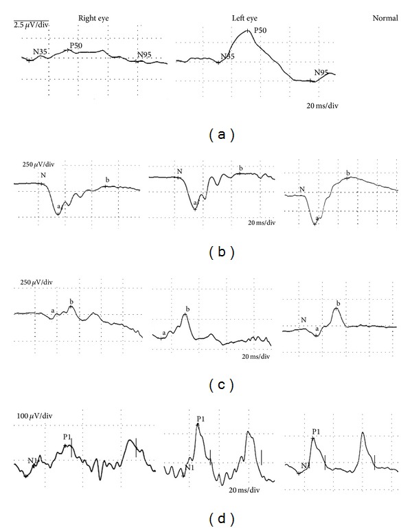
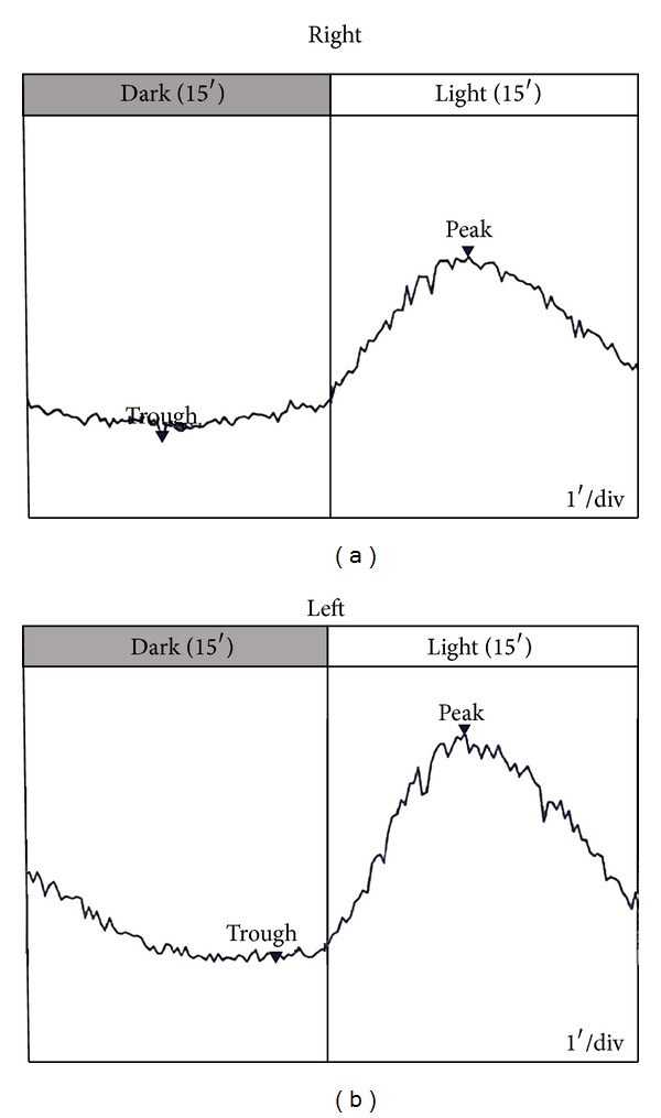
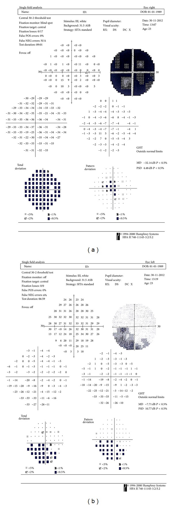
Background. To report a case of acute zonal occult outer retinopathy (AZOOR) with atypical electrophysiology findings. Case Presentation. A 23-year-old-female presented with visual acuity deterioration in her right eye accompanied by photopsia bilaterally. Corrected distance visual acuity at presentation was 20/50 in the right eye and 20/20 in the left eye. Fundus examination was unremarkable. Visual field (VF) testing revealed a large scotoma. Pattern and full-field electroretinograms (PERG and ERG) revealed macular involvement associated with generalized retinal dysfunction. Electrooculogram (EOG) light rise and the Arden ratio were within normal limits bilaterally. The patient was diagnosed with AZOOR due to clinical findings, visual field defect, and ERG findings. Conclusion. This is a case of AZOOR with characteristic VF defects and clinical symptoms presenting with atypical EOG findings.
Figures


References
-
- Monson DM, Smith JR. Acute zonal occult outer retinopathy. Survey of Ophthalmology. 2011;56(1):23–35. - PubMed
-
- Gass JD, Agarwal A, Scott IU. Acute zonal occult outer retinopathy: a long-term follow-up study. American Journal of Ophthalmology. 2002;134(3):329–339. - PubMed
-
- Bach M, Brigell MG, Hawlina M, et al. ISCEV standard for clinical pattern electroretinography (PERG): 2012 update. Documenta Ophthalmologica. 2013;126(1):1–7. - PubMed
-
- Marmor MF, Fulton AB, Holder GE, Miyake Y, Brigell M, Bach M. ISCEV Standard for full-field clinical electroretinography (2008 update) Documenta Ophthalmologica. 2009;118(1):69–77. - PubMed
LinkOut - more resources
Full Text Sources
Other Literature Sources
