Review
. 2014 Aug 7:5:124.
doi: 10.4103/2152-7806.138364. eCollection 2014.

Colloid cysts posterior and anterior to the foramen of Monro: Anatomical features and implications for endoscopic excision

Affiliations
Review

Colloid cysts posterior and anterior to the foramen of Monro: Anatomical features and implications for endoscopic excision

Waleed A Azab et al. Surg Neurol Int. .

Abstract

Background: Colloid cysts are usually located at the rostral part of the third ventricle in proximity to the foramina of Monro. Some third ventricular colloid cysts, however, attain large sizes, reach a very high distance above the roof of the third ventricle, and pose some challenges during endoscopic excision. These features led to the speculation that for such a pattern of growth to take place, the points of origin of these cysts should be at areas away from the foramina of Monro at which some anatomical "windows" exist that are devoid of compact, closely apposed forniceal structures.

Methods: A review of the literature on anatomical variations of the structures in the vicinity of the roof of the third ventricle and on reported cases with similar features was conducted.

Results: Colloid cysts may grow vertically up past the roof of the third ventricle through anatomical windows devoid of the mechanical restraint of the forniceal structures.

Conclusion: Some anatomical variations of the forniceal structures may allow unusually large sizes and superior vector of growth of a retro- or post-foraminal colloid cyst. Careful preoperative planning and knowledge of the pertinent pathoanatomy of these cysts before endoscopic excision is very important to avoid complications.

Keywords: Cavum; colloid cyst; endoscopic; foramen of Monro; fornix; third ventricle.


Figures

Figure 1
Figure 1
Sagittal MR images demonstrating variable points of forniceal “take off” from the undersurface of the corpus callosum
Figure 2
Figure 2
Illustration of the variations of the lengths of the forniceal crura and their angles [θ] of divergence from the undersurface of the corpus callosum with consequent variable dimensions of the triangular area posterior to the body of the fornix [inset]. The axial T2-weighted MR image at the level of the two forniceal bodies demonstrates this triangular area (compare to the inset)
Figure 3
Figure 3
Illustration in axial and sagittal planes of the superiorly directed vector of growth of a colloid cyst originating posterior to the foramen of Monro compared to a cyst originating in a classic location. The window between the two crura of the fornix is devoid of anatomical restraints and allows a larger size and enlargement beyond the ventricular roof
Figure 4
Figure 4
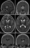
One of our cases with large colloid cyst. Sagittal (a) and coronal (b) T1-weighted MR images with contrast demonstrate the cyst to reach the inferior surface of the corpus callosum. Axial T2- weighted MR images at two consecutive cuts (c, d) demonstrating the flattened forniceal columns. Coronal T1-weighted MR image with contrast (e) demonstrates the normal internal cerebral veins at the posterior third ventricular roof becoming splayed by the lesion in the next anterior cut (f). No cavum septae pellucidi or vergae are present
Figure 5
Figure 5
Coronal T2-weighted MR image from a patient with pineal region tumor and triventricular hydrocephalus. Note the anterosuperior triangular window (asterisk) between the anterior commissure and the columns of the fornix
Figure 6
Figure 6
Illustration of the variation of the point of diverging forniceal columns and consequent formation of a larger anterosuperior window in the roof of the third ventricle which creates a route for superior direction of growth of colloid cysts originating anterior to the foramen of Monro
Figure 7
Figure 7
Serial photographs demonstrating the development of the septum pellucidum in human embryos. From Rakic and Yakovlev (1968)[23] with permission
Figure 8
Figure 8
Preoperative (a-d) and postoperative (e, f) T2-weighted MR images of one case from our records. Note the very large size of the colloid cyst reaching the corpus callosum and the splayed fornices in (b) and (c) axial images. Evidence of cavum septae pellucidi and vergae is seen in the preoperative axial cut (d) anterior and posterior to the cyst wall. The presence of cavum septae pellucidi and vergae is seen clearly in both axial (e) and coronal (f) postoperative images
Figure 9
Figure 9
Intraoperative images during endoscopic excision of the colloid cyst in Figure 6. (a) Initial endoscopic view within the lateral ventricle reveals a bulging septum pellucidum. (b) Opening the septum pellucidum leaflet exposes the cyst wall and content aspiration is started. (c) Cyst contents coming through the opening of the cyst wall. Note the calcium flakes which are commonly seen in large colloid cysts. (d) Further expansion of the cyst wall opening. (e) The foramen of Monro is seen after the cyst was decompressed and excised. (f) View of the floor of the third ventricle at the conclusion of the operation (courtesy of Professor Dr. Henry W. S. Schroeder)
Figure 10
Figure 10
Classic colloid cyst at the foramen of Monro. Preoperative T2-FLAIR (a) and T2-weighted (b) sagittal MR images. Postoperative sagittal T1-weighted image with contrast after total excision (c). Intraoperative sequential images of endoscopic excision through a transforaminal route (d-h)
Figure 11
Figure 11
Axial T2-weighted (a) and non-contrast sagittal T1- weighted MR images of the case of velum interpositum colloid cyst reported by Hingawala et al. (2009).[12] Reprinted with permission
Figure 12
Figure 12
An illustration of the areas of potential restriction and voids for extension of a colloid cyst is presented. Pink arrows are representative of areas of growth extension of the cyst above the third ventricular roof

References

    1. Apuzzo ML, Chikovani OK, Gott PS, Teng EL, Zee CS, Giannotta SL, et al. Transcallosal interfornicial approaches for lesions affecting the third ventricle: Surgical considerations and consequences. Neurosurgery. 1982;10:547–54. - PubMed
    1. Apuzzo ML, Amar AP. Transcallosal interforniceal approach. In: Apuzzo ML, editor. Surgery of the third ventricle. Baltimore: Williams and Wilkins; 1998. pp. 421–52.
    1. Born CM, Meisenzahl EM, Frodl T, Pfluger T, Reiser M, Möller HJ, et al. The septum pellucidum and its variants. An MRI study. Eur Arch Psychiatry Clin Neurosci. 2004;254:295–302. - PubMed
    1. Byrom FB, Russell DS. Ependymal cyst of the third ventricle associated with diabetes mellitus. Lancet. 1932;220:278–82.
    1. Callen PW, Callen AL, Glenn OA, Toi A. Columns of the Fornix, Not to be mistaken for the cavum septi pellucidi on prenatal sonography. J Ultrasound Med. 2008;27:25–31. - PubMed