. 2014 Dec;99(12):E2610-9.
doi: 10.1210/jc.2013-3488.

mRNA-binding protein TIA-1 reduces cytokine expression in human endometrial stromal cells and is down-regulated in ectopic endometrium

Affiliations

mRNA-binding protein TIA-1 reduces cytokine expression in human endometrial stromal cells and is down-regulated in ectopic endometrium

Hakan Mete Karalok et al. J Clin Endocrinol Metab. 2014 Dec.

Abstract

Background: Cytokines and growth factors play important roles in endometrial function and the pathogenesis of endometriosis. mRNAs encoding cytokines and growth factors undergo rapid turnover; primarily mediated by adenosine- and uridine-rich elements (AREs) located in their 3'-untranslated regions. T-cell intracellular antigen (TIA-1), an mRNA-binding protein, binds to AREs in target transcripts, leading to decreased gene expression.

Objective: The purpose of this article was to determine whether TIA-1 plays a role in the regulation of endometrial cytokine and growth factor expression during the normal menstrual cycle and whether TIA-1 expression is altered in women with endometriosis.

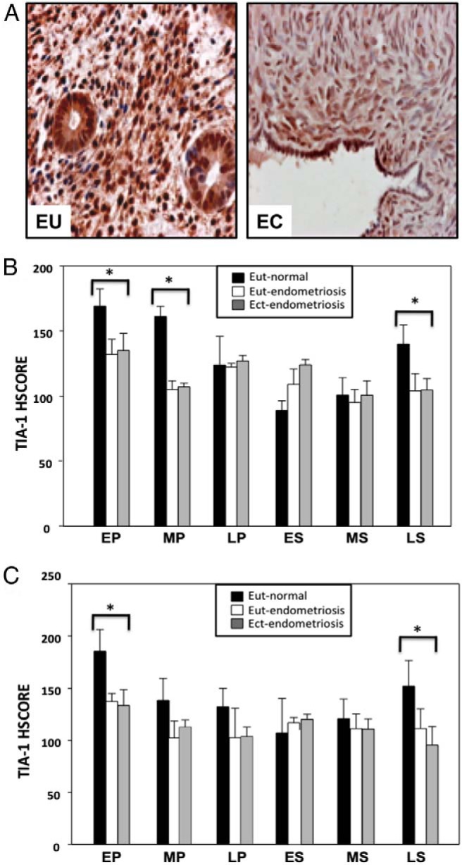
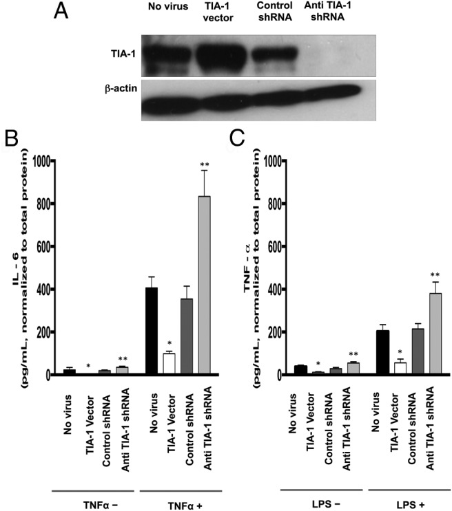
Methods: Eutopic endometrial tissue obtained from women without endometriosis (n = 30) and eutopic and ectopic endometrial tissues from women with endometriosis (n = 17) were immunostained for TIA-1. Staining intensities were evaluated by histological scores (HSCOREs). The regulation of endometrial TIA-1 expression by immune factors and steroid hormones was studied by treating primary cultured human endometrial stromal cells (HESCs) with vehicle, lipopolysaccharide, TNF-α, IL-6, estradiol, or progesterone, followed by protein blot analyses. HESCs were engineered to over- or underexpress TIA-1 to test whether TIA-1 regulates IL-6 or TNF-α expression in these cells.

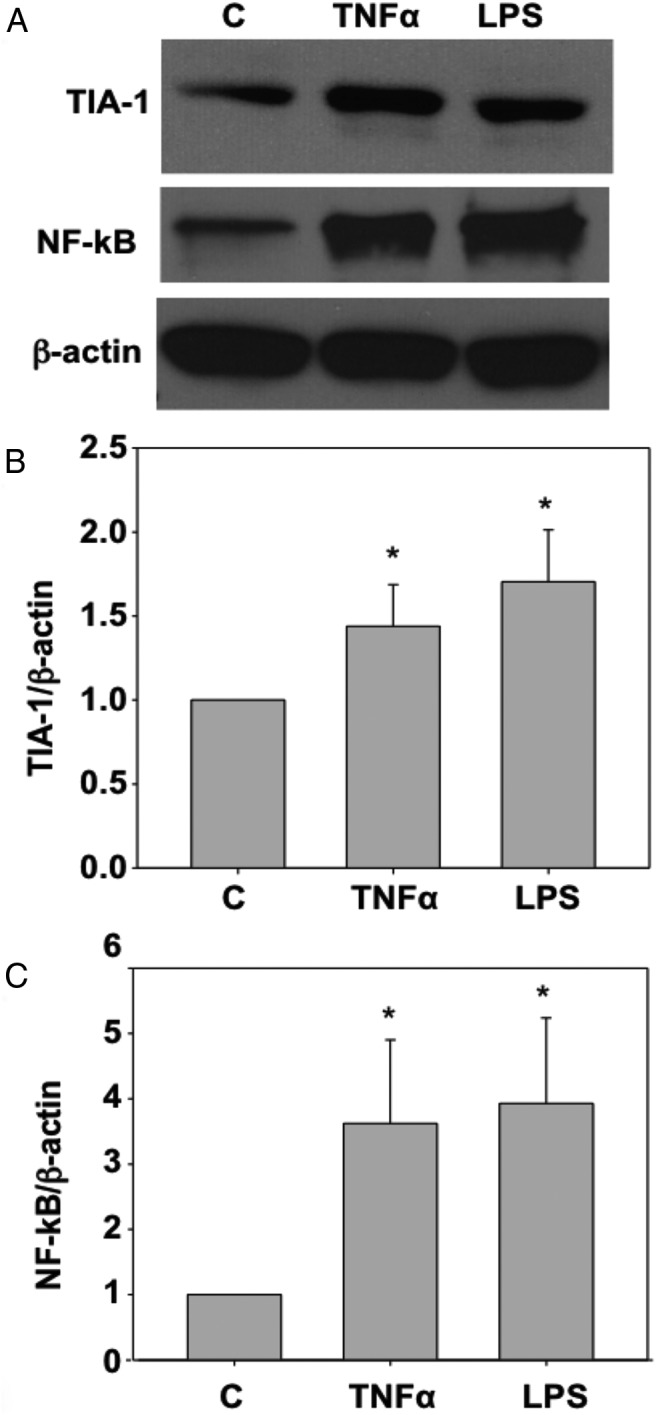
Results: We found that TIA-1 is expressed in endometrial stromal and glandular cells throughout the menstrual cycle and that this expression is significantly higher in the perimenstrual phase. In women with endometriosis, TIA-1 expression in eutopic and ectopic endometrium was reduced compared with TIA-1 expression in eutopic endometrium of unaffected control women. Lipopolysaccharide and TNF-α increased TIA-1 expression in HESCs in vitro, whereas IL-6 or steroid hormones had no effect. In HESCs, down-regulation of TIA-1 resulted in elevated IL-6 and TNF-α expression, whereas TIA-1 overexpression resulted in decreased IL-6 and TNF-α expression.

Conclusions: Endometrial TIA-1 is regulated throughout the menstrual cycle, TIA-1 modulates the expression of immune factors in endometrial cells, and downregulation of TIA-1 may contribute to the pathogenesis of endometriosis.


Figures

Figure 1.
Figure 1.
A, TIA-1 protein expression in representative samples of eutopic endometrial tissue from the EP, MP, LP, ES, MS, and LS phases of the menstrual cycle. TIA-1 immunostaining is nuclear and cytoplasmic, with a higher intensity in the nucleus. B, TIA-1 protein expression in normal eutopic endometrial stromal cells according to the menstrual cycle phase. HSCORE analysis was performed to quantitate endometrial stromal expression of TIA-1 from biopsy samples. TIA-1 protein expression is significantly higher in the EP, MP, LP, and LS phases, with the highest expression during the EP phase. P < .01: *, EP vs LP, ES, MS, and LS; **, MP vs LP, ES, and MS; ***, LP vs ES; ****, LS vs ES and MS. C, TIA-1 protein expression in eutopic endometrial glandular cells according to the menstrual cycle phase. HSCORE analysis was performed to quantitate the endometrial glandular expression of TIA-1 from biopsy samples. TIA-1 protein expression is significantly higher in the LS and EP phases, with the highest expression during the EP phase. P < .01: *, EP vs LP, ES, and MS; **, LS vs ES.
Figure 2.
Figure 2.
TIA-1 protein expression in normal primary HESCs treated with E2 and progesterone. Cultured HESCs were treated with vehicle, 3 different doses of estrogen (E1, 100 pM; E2, 1 nM; E3, 10 nM) or 3 different doses of progesterone (P1, 10 nM; P2, 100 nM; P3, 1 μΜ) for 24 hours. Total protein was extracted, and TIA-1 protein expression was analyzed by Western blot. A band corresponding to TIA-1 was observed at ∼45 kDa. Reprobing the blot with an antibody against β-actin provided the internal loading control. Bars represent means + SD. P = nonsignificant for control vs E1, E2, and E3 or P1, P2, and P3.
Figure 3.
Figure 3.
A, TIA-1 and NF-κB protein expression levels in primary HESCs treated with TNF-α or LPS. Cultured HESCs were treated with vehicle, TNF-α (10 ng/mL), or LPS (1000 ng/mL) for 24 hours each. Total protein was extracted and TIA-1 protein was analyzed by Western blot. A band corresponding to TIA-1 was observed at ∼45 kDa. Reprobing the blot with an antibody against NF-κB and then β-actin provided the positive control for immune stimulation and the internal loading control, respectively. Bars represent means + SD. B and C, TIA-1 (B) and NF-κB (C) expression in HESCs increased significantly in response to TNF-α or LPS treatment. *, P < .05 for TNF-α or LPS vs control.
Figure 4.
Figure 4.
A, Representative micrographs of TIA-1 protein immunostaining in normal eutopic endometrium (EU) from a woman without endometriosis and in ectopic endometrium from a woman with endometriosis (EC) in the EP phase. TIA-1 expression is cytoplasmic and nuclear in both the glandular and stromal components of normal endometrium and ectopic endometrium. B, TIA-1 protein expression in stromal cells of eutopic endometrium from women without endometriosis (■; Eut-normal) and eutopic (□; Eut-endometriosis) and ectopic (▩; Ect-endometriosis) endometrium from women with endometriosis. HSCORE analysis was performed to quantitate endometrial stromal cell expression of TIA-1 from biopsy samples. *, P < .005, normal endometrium vs eutopic or ectopic endometrium from women with endometriosis in the same menstrual cycle phase. C, TIA-1 protein expression in glandular cells of eutopic endometrium from women without endometriosis (■; Eut-normal) and eutopic (□; Eut-endometriosis) and ectopic (▩; Ect-endometriosis) endometrium from women with endometriosis. HSCORE analysis was performed to quantitate endometrial glandular expression of TIA-1 from biopsy samples. *, P < .005; normal endometrium vs eutopic or ectopic endometrium from women with endometriosis in the same menstrual cycle phase.
Figure 5.
Figure 5.
A, TIA-1 expression in HESCs after lentiviral-mediated overexpression and knockdown. Cells were transduced with vehicle (No virus), TIA-1–expressing lentiviral particles (TIA-1 vector), control shRNA-expressing lentiviral particles (Control shRNA) or anti-TIA-1 shRNA-expressing lentiviral particles (Anti TIA-1 shRNA). Total protein was extracted, and TIA-1 protein expression was analyzed by Western blot. A band corresponding to TIA-1 was observed at ∼45 kDa. Reprobing the blot with an antibody against β-actin provided the internal loading control. B, IL-6 expression by HESCs after lentiviral-mediated overexpression and knockdown of TIA-1 with or without TNF-α pretreatment. Cells were transduced with vehicle (No virus), TIA-1–expressing lentiviral particles (TIA-1 Vector), control shRNA–expressing lentiviral particles (Control shRNA), or anti-TIA-1 shRNA-expressing lentiviral particles (Anti TIA-1 shRNA). Cells were grown to preconfluence and then treated with vehicle or TNF-α (10 ng/mL) in serum-free medium for 24 hours. Culture medium was collected, and IL-6 levels were detected in the supernatants of cells by ELISAs. *, P < .05, TIA-1 vector vs no virus or control shRNA; **, P < .05, anti TIA-1 shRNA vs no virus or control shRNA. C, TNF-α expression by HESCs after lentiviral-mediated overexpression and knockdown of TIA-1 with or without LPS pretreatment. Cells were transduced with vehicle (No virus), TIA-1–expressing lentiviral particles (TIA-1 Vector), control shRNA-expressing lentiviral particles (Control shRNA), or anti-TIA-1 shRNA-expressing lentiviral particles (Anti TIA-1 shRNA). Cells were grown to preconfluence and then were treated with vehicle or LPS (1000 ng/mL) in serum-free medium for 24 hours. Culture medium was collected, and TNF-α levels were detected in the supernatants of cells by ELISAs. *, P < .05, TIA-1 vector vs no virus or control shRNA; **, P < .05; anti TIA-1 shRNA vs no virus or control shRNA

References

    1. Seli E, Berkkanoglu M, Arici A. Pathogenesis of endometriosis. Obstet Gynecol Clin North Am. 2003;30:41–61. - PubMed
    1. Wheeler JM. Epidemiology of endometriosis-associated infertility. J Reprod Med. 1989;34:41–46. - PubMed
    1. Gao X, Outley J, Botteman M, Spalding J, Simon JA, Pashos CL. Economic burden of endometriosis. Fertil Steril. 2006;86:1561–1572. - PubMed
    1. Simoens S, Hummelshoj L, D'Hooghe T. Endometriosis: cost estimates and methodological perspective. Hum Reprod Update. 2007;13:394–404. - PubMed
    1. Seli E, Arici A. Endometriosis: Interaction of immune and endocrine systems. Semin Reprod Med. 2003;21:135–144. - PubMed

Publication types

MeSH terms