

An efficient approach for differentiating Alzheimer's disease from normal elderly based on multicenter MRI using gray-level invariant features
- PMID: 25140532
- PMCID: PMC4139346
- DOI: 10.1371/journal.pone.0105563
An efficient approach for differentiating Alzheimer's disease from normal elderly based on multicenter MRI using gray-level invariant features
Abstract
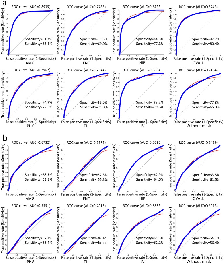
Machine learning techniques, along with imaging markers extracted from structural magnetic resonance images, have been shown to increase the accuracy to differentiate patients with Alzheimer's disease (AD) from normal elderly controls. Several forms of anatomical features, such as cortical volume, shape, and thickness, have demonstrated discriminative capability. These approaches rely on accurate non-linear image transformation, which could invite several nuisance factors, such as dependency on transformation parameters and the degree of anatomical abnormality, and an unpredictable influence of residual registration errors. In this study, we tested a simple method to extract disease-related anatomical features, which is suitable for initial stratification of the heterogeneous patient populations often encountered in clinical data. The method employed gray-level invariant features, which were extracted from linearly transformed images, to characterize AD-specific anatomical features. The intensity information from a disease-specific spatial masking, which was linearly registered to each patient, was used to capture the anatomical features. We implemented a two-step feature selection for anatomic recognition. First, a statistic-based feature selection was implemented to extract AD-related anatomical features while excluding non-significant features. Then, seven knowledge-based ROIs were used to capture the local discriminative powers of selected voxels within areas that were sensitive to AD or mild cognitive impairment (MCI). The discriminative capability of the proposed feature was measured by its performance in differentiating AD or MCI from normal elderly controls (NC) using a support vector machine. The statistic-based feature selection, together with the knowledge-based masks, provided a promising solution for capturing anatomical features of the brain efficiently. For the analysis of clinical populations, which are inherently heterogeneous, this approach could stratify the large amount of data rapidly and could be combined with more detailed subsequent analyses based on non-linear transformation.
Conflict of interest statement
Figures
References
-
- Hardy J, Allsop D (1991) Amyloid deposition as the central event in the aetiology of Alzheimer's disease. Trends in Pharmacological Sciences 12: 383–388. - PubMed
-
- Tiraboschi P, Hansen LA, Thal LJ, Corey-Bloom J (2004) The importance of neuritic plaques and tangles to the development and evolution of AD. Neurology 62: 1984–1989. - PubMed
-
- Waldemar G, Dubois B, Emre M, Georges J, McKeith IG, et al. (2007) Recommendations for the diagnosis and management of Alzheimer's disease and other disorders associated with dementia: EFNS guideline. Eur J Neurol 14: e1–26. - PubMed
-
- Fox NC, Schott JM (2004) Imaging cerebral atrophy: normal ageing to Alzheimer's disease. Lancet 363: 392–394. - PubMed
-
- Higdon R, Foster NL, Koeppe RA, DeCarli CS, Jagust WJ, et al. (2004) A comparison of classification methods for differentiating fronto-temporal dementia from Alzheimer's disease using FDG-PET imaging. Statistics in Medicine 23: 315–326. - PubMed
Publication types
MeSH terms
Grants and funding
LinkOut - more resources
Full Text Sources
Other Literature Sources
Medical
