

Mipu1 overexpression protects macrophages from oxLDL-induced foam cell formation and cell apoptosis
- PMID: 25141035
- PMCID: PMC4248253
- DOI: 10.1089/dna.2014.2501
Mipu1 overexpression protects macrophages from oxLDL-induced foam cell formation and cell apoptosis
Abstract
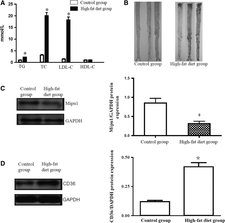
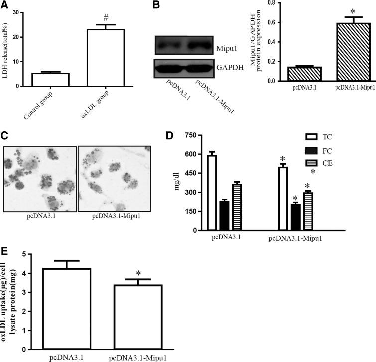
Mipu1 (myocardial ischemic preconditioning upregulated protein 1) is a novel N-terminal Kruppel-associated box (KRAB)/C2H2 zinc finger superfamily protein, that displays a powerful effect in protecting H9c2 cells from oxidative stress-induced cell apoptosis. The present study aims to investigate the effect of Mipu1 overexpression on oxidized low-density lipoprotein (oxLDL)-induced foam cell formation, cell apoptosis, and its possible mechanisms. New Zealand healthy rabbits were used to establish atherosclerosis model, and serum levels of triglycerides, total cholesterol, high-density lipoprotein cholesterol, and low-density lipoprotein cholesterol were detected by an automatic biochemical analyzer. Sudan IV staining was used to detect atherosclerotic lesions. The RAW264.7 macrophage cell line was selected as the experimental material. Oil red O staining, high-performance liquid chromatography, and Dil-labeled lipoprotein were used to detect cholesterol accumulation qualitatively and quantitatively, respectively. Flow cytometry was used to determine cell apoptosis. Real-time quantitative polymerase chain reaction (PCR) was used to detect the mRNA expression of the main proteins that are associated with the transport of cholesterol, such as ABCA1, ABCG1, SR-BI, and CD36. Western blot analysis was used to detect the protein expression of Mipu1. There were atherosclerotic lesions in the high-fat diet group with Sudan IV staining. High-fat diet decreased Mipu1 expression and increased CD36 expression significantly at the 10th week compared with standard-diet rabbits. Mipu1 overexpression decreased oxLDL-induced cholesterol accumulation, oxLDL uptake, cell apoptosis, and cleaved caspase-3. Mipu1 overexpression inhibited the oxLDL-induced CD36 mRNA and protein expression, but it did not significantly inhibit the mRNA expression of ABCA1, ABCG1, and SR-BI. Mipu1 overexpression inhibits oxLDL-induced foam cell formation and cell apoptosis. Mipu1 overexpression reduces the lipid intake of macrophages and might be associated with the downregulation of CD36 expression in the presence of oxLDL.
Figures


References
-
- Amezaga N., Sanjurjo L., Julve J., Aran G., Perez-Cabezas B., Bastos-Amador P., Armengol C., Vilella R., Escola-Gil J.C., Blanco-Vaca F., et al. (2014). Human scavenger protein AIM increases foam cell formation and CD36-mediated oxLDL uptake. J Leukoc Biol 95,509–520 - PubMed
-
- Arcaro G., Cretti A., Balzano S., Lechi A., Muggeo M., Bonora E., and Bonadonna R.C. (2002). Insulin causes endothelial dysfunction in humans: sites and mechanisms. Circulation 105,576–582 - PubMed
-
- Ashraf M.Z., and Gupta N. (2011). Scavenger receptors: implications in atherothrombotic disorders. Int J Biochem Cell Biol 43,697–700 - PubMed
-
- Boyle J.J., Johns M., Kampfer T., Nguyen A.T., Game L., Schaer D.J., Mason J.C., and Haskard D.O. (2012). Activating transcription factor 1 directs Mhem atheroprotective macrophages through coordinated iron handling and foam cell protection. Circ Res 110,20–33 - PubMed
Publication types
MeSH terms
Substances
LinkOut - more resources
Full Text Sources
Other Literature Sources
Research Materials
