. 2015 Feb;61(2):648-59.
doi: 10.1002/hep.27387. Epub 2015 Jan 5.

Chronic passive venous congestion drives hepatic fibrogenesis via sinusoidal thrombosis and mechanical forces

Affiliations

Chronic passive venous congestion drives hepatic fibrogenesis via sinusoidal thrombosis and mechanical forces

Douglas A Simonetto et al. Hepatology. 2015 Feb.

Abstract

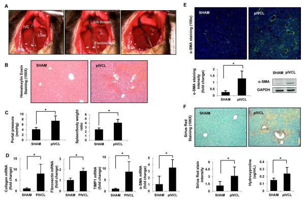
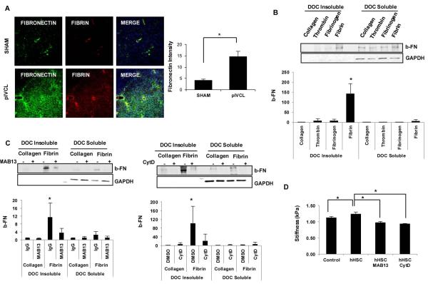
Chronic passive hepatic congestion (congestive hepatopathy) leads to hepatic fibrosis; however, the mechanisms involved in this process are not well understood. We developed a murine experimental model of congestive hepatopathy through partial ligation of the inferior vena cava (pIVCL). C57BL/6 and transgenic mice overexpressing tissue factor pathway inhibitor (SM22α-TFPI) were subjected to pIVCL or sham. Liver and blood samples were collected and analyzed in immunohistochemical, morphometric, real-time polymerase chain reaction, and western blot assays. Hepatic fibrosis and portal pressure were significantly increased after pIVCL concurrent with hepatic stellate cell (HSC) activation. Liver stiffness, as assessed by magnetic resonance elastography, correlated with portal pressure and preceded fibrosis in our model. Hepatic sinusoidal thrombosis as evidenced by fibrin deposition was demonstrated both in mice after pIVCL as well as in humans with congestive hepatopathy. Warfarin treatment and TFPI overexpression both had a protective effect on fibrosis development and HSC activation after pIVCL. In vitro studies show that congestion stimulates HSC fibronectin (FN) fibril assembly through direct effects of thrombi as well as by virtue of mechanical strain. Pretreatment with either Mab13 or Cytochalasin-D, to inhibit β-integrin or actin polymerization, respectively, significantly reduced fibrin and stretch-induced FN fibril assembly.

Conclusion: Chronic hepatic congestion leads to sinusoidal thrombosis and strain, which in turn promote hepatic fibrosis. These studies mechanistically link congestive hepatopathy to hepatic fibrosis.


Figures

Figure 1
Figure 1. Characterization of a Murine Model of Congestive Fibrosis by Partial Ligation of the Inferior Vena Cava
Surgical model of congestive hepatopathy in C57BL/6 mice through partial ligation of the suprahepatic inferior vena cava (pIVCL) is shown (A). The IVC was circumferentially isolated and a sterile wire was placed on the anterior surface of the IVC (A, left). A 6.0 silk thread was then tightly tied around both the IVC and the wire (A, right), which was subsequently removed. Six weeks postoperatively livers were harvested and subjected to analysis. Hematoxylin-Eosin staining (100X) on paraffin sections demonstrate areas of centrilobular necrosis, vascular extravasation and sinusoidal dilatation (B). Portal pressure was directly measured via cannulation of portal vein and spleen/body ratio was calculated as a surrogate of portal hypertension (C). Hepatic expression of fibrogenic markers is increased after pIVCL compared to SHAM (D). Hepatic stellate cell activation after pIVCL is also demonstrated by increased α-SMA immunofluorescence (200X) and protein levels (E). Significant increase in hepatic fibrosis 6 weeks post-pIVCL is shown by Sirius red staining (100X) and hydroxyproline content (F). Data represent mean ± STDEV; n=8-9, *p<0.05.
Figure 2
Figure 2. Anticoagulation Disrupts Congestive Fibrosis
Immunofluorescence for fibrin (green) was significantly increased in the liver after pIVCL compared to controls, and spatially associated with α-SMA expression (red) (200X) (A). Mice received warfarin (1 μg/mL) in drinking water post-pIVCL for 6 weeks until sacrifice. Control animals received plain water for 6 weeks. International normalized ratio (INR) of two times the controls was successfully achieved with this dosage (B, left upper panel). Portal pressure was measured and livers were perfused with 10 mL of 1X PBS through the portal vein to remove circulating fibrinogen. Spleen and liver were then harvested and body ratios calculated (B). Immunofluorescence for fibrin (green) and α-SMA (red) were significantly decreased with warfarin treatment (C). Warfarin-treated animals showed significant reduction in hepatic fibrosis compared to untreated pIVCL group (D). Data represent mean ± STDEV; n=10, *p<0.05.
Figure 3
Figure 3. Tissue Factor Pathway Inhibitor Transgenic Mice Show Reduced Fibrosis after Partial IVC Ligation
Tissue factor pathway inhibitor (TFPI) overexpression was obtained via smooth muscle-specific promoter SM22α (SM22α-TFPI) in C57BL/6 mice, to achieve elevated systemic levels of TFPI. SM22α-TFPI mice were submitted to SHAM and pIVCL surgeries and wild type mice were used as controls. Western blot analysis is shown of fibrin, α-SMA and β-actin from liver lysates of SHAM and pIVCL in WT, SM22α-TFPI and warfarin-treated WT mice. Lysates are shown from two distinct mice from each group. Bovine fibrinogen (1 mg/ml) and thrombin (0.2 U/ml) were mixed to form fibrin in vitro, which was used as positive control (A). Fibrin (green) and α-SMA (red) immunofluorescence were reduced in SM22α-TFPI mice after pIVCL compared to wild type animals. Hepatic fibrosis was significantly reduced as demonstrated by sirius red staining and hydroxyproline assay (C). Portal pressure and spleen/body ratio were not affected by TFPI overexpression (D). Data represent mean ± STDEV; n=7-10, *p<0.05.
Figure 4
Figure 4. Fibrin Stimulates Fibronectin Fibril Assembly by HSC
Fibronectin (green) and fibrin (red) immunofluorescence were performed from frozen liver sections after 6 weeks of SHAM and pIVCL. Fibronectin significantly increased after pIVCL compared to SHAM and co-localized with fibrin at sites of congestion-related injury (A). Serum starved hHSC were plated on collagen-, fibrinogen- and fibrin-coated or uncoated dishes. Thrombin (0.2 U) was added to serum-free media of cells on uncoated dishes. Biotinylated fibronectin (10 μg) was added for 6 hours and cells were collected for deoxycholic acid (DOC) solubility assay as described on methods. Insoluble and soluble fractions were resolved on a 4–12% SDS-PAGE gel. GAPDH is shown as loading control. Fibrin significantly increased insoluble fraction of fibronectin compared to collagen, fibrinogen and thrombin (B). hHSC plated on fibrin- or collagen- coated dishes were pre-treated with rat anti-human CD29 (Mab13; 1:200) or cytochalasin D (1:1000) for 30 minutes prior to addition of b-FN. Mab13 and CytD significantly reduced assembly of exogenous fibronectin by hHSC in response to fibrin (C). Fibrin gels containing hHSC were constructed and Mab13 or CytD added to serum-free media. Fibrin gels with no cells were used as controls. MR elastography was obtained after 5 days for measurement of shear stiffness (kPa) (D). In vitro experiments were performed 3 times with similar results. Data represent mean ± STDEV; *p<0.05.
Figure 5
Figure 5. Liver Stiffness Correlates with Portal Hypertension in Congestive Hepatopathy
Mice underwent SHAM or pIVCL surgeries for 2, 4 or 6 weeks. On day of sacrifice, MR elastogram was performed using a custom birdcage coil. RE wave images were acquired with a multifrequency 3-D/3-axis EPI MRE sequence (120 and 200Hz) with a resolution of 0.3×0.3×2 mm3 (A). Yellow dashed line indicates total liver areas (upper panel). Portal pressure was directly measured, via cannulation of portal vein, and livers were harvested for hydroxyproline assay. Liver stiffness (kPa) correlated with portal pressure after pIVCL (R2=0.7162 at 120Hz and R2=0.7593 at 200Hz), to a much greater extent than with fibrosis as measured by hydroxyproline content (R2=0.0982 at 120Hz and R2=0.0184 at 200Hz) (B). Liver stiffness (C) and portal pressure (D) significantly increased after pIVCL at 2, 4 and 6 weeks compared to SHAM groups. Fibrosis was only significantly increased at 6 weeks after pIVCL compared to SHAM (E). Data represent mean ± STDEV; n=4-5, *p<0.05.
Figure 6
Figure 6. Mechanical Forces Stimulate Fibronectin Fibril Assembly by HSC
Primary human and murine HSC were seeded on 6-well plates of flexible silicone bottom coated with collagen type IV at 0.3 ×10⁶ cells/well. After incubation for 24 hours, cells were serum-starved overnight and then subjected to cyclic uniform stretch. Amplitudes of 10% strain were used at a rate of 30 cycles/min at 37 °C and 5% CO2 in a humidified incubator. Serum-starved HSCs were submitted to cyclic stretch for 24 hours. HSC supernatant were collected for western blot analysis of fibronectin. Comassie blue was used for loading control (A). Biotinylated fibronectin (10 μg) was added and cells were submitted to cyclic stretch for 6 hours. Streptavidin immunofluorescence (green) demonstrates deposition of b-FN fibrils on the surface of HSCs. Nucleus staining (DAPI) is in blue (B). b-FN at 10 and 50 μg/mL was added to serum-starved HSC. Cells were cyclic stretched for 6 hours and collected for DOC-solubility assay. Cyclic stretch significantly increased insoluble fibronectin compared to non-stretched cells (C). Pre-treatment with rat anti-human CD29 (Mab13; 1:200) or cytochalasin D (1:1000) for 30 minutes prior to cyclic stretch significantly reduced assembly of exogenous fibronectin by hHSC (D, E). Experiments were performed 3 times with similar results. Data represent mean ± STDEV; *p<0.05.
Figure 7
Figure 7. Intrahepatic Thrombosis is increased in Humans with Congestive Hepatopathy
Liver samples were obtained from healthy individuals (controls) and from patients with congestive hepatopathy secondary to congestive heart failure or corrective surgery (Fontan procedure) for complex congenital heart disease. Fibrin (green) and α-SMA (red) immunofluorescence are significantly increased in congestive hepatopathy compared to controls. Fibrosis is also significantly increased as demonstrated by Sirius red staining (n=20, controls; n=12, Fontan procedure; n=14, congestive heart failure). Patient demographics are shown in Supplementary Table 1 and 2. Data represent mean ± STDEV; *p<0.05.
Figure 8
Figure 8. Proposed Model of Fibrosis Development in Congestive Hepatopathy
In chronic hepatic congestion, sinusoidal dilatation leads to stretch of adjacent hepatic stellate cells, and stasis promotes intravascular thrombosis with fibrin clot formation. Cyclic mechanical stretch induces release of fibronectin by HSC and both fibrin and stretch stimulate fibronectin fibril assembly via β1-integrin and actin-dependent mechanism.

Comment in

References

    1. Ahmed A. American College of Cardiology/American Heart Association Chronic Heart Failure Evaluation and Management guidelines: relevance to the geriatric practice. Journal of the American Geriatrics Society. 2003;51:123–126. - PubMed
    1. de Leval MR, Deanfield JE. Four decades of Fontan palliation. Nature reviews. Cardiology. 2010;7:520–527. - PubMed
    1. Asrani SK, Asrani NS, Freese DK, Phillips SD, Warnes CA, Heimbach J, Kamath PS. Congenital heart disease and the liver. Hepatology. 2012;56:1160–1169. - PubMed
    1. Wanless IR, Liu JJ, Butany J. Role of thrombosis in the pathogenesis of congestive hepatic fibrosis (cardiac cirrhosis) Hepatology. 1995;21:1232–1237. - PubMed
    1. Wanless IR, Wong F, Blendis LM, Greig P, Heathcote EJ, Levy G. Hepatic and portal vein thrombosis in cirrhosis: possible role in development of parenchymal extinction and portal hypertension. Hepatology. 1995;21:1238–1247. - PubMed

Publication types

MeSH terms