. 2014 Oct 3;289(40):27625-39.
doi: 10.1074/jbc.M114.582429. Epub 2014 Aug 20.

Stress-dependent proteolytic processing of the actin assembly protein Lsb1 modulates a yeast prion

Affiliations

Stress-dependent proteolytic processing of the actin assembly protein Lsb1 modulates a yeast prion

Moiez Ali et al. J Biol Chem. .

Abstract

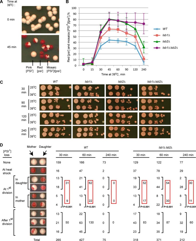
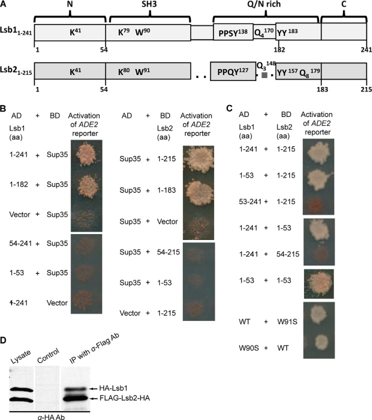
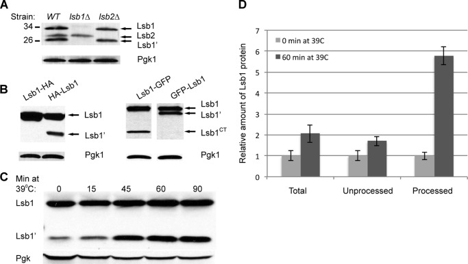
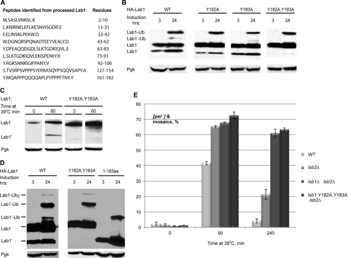
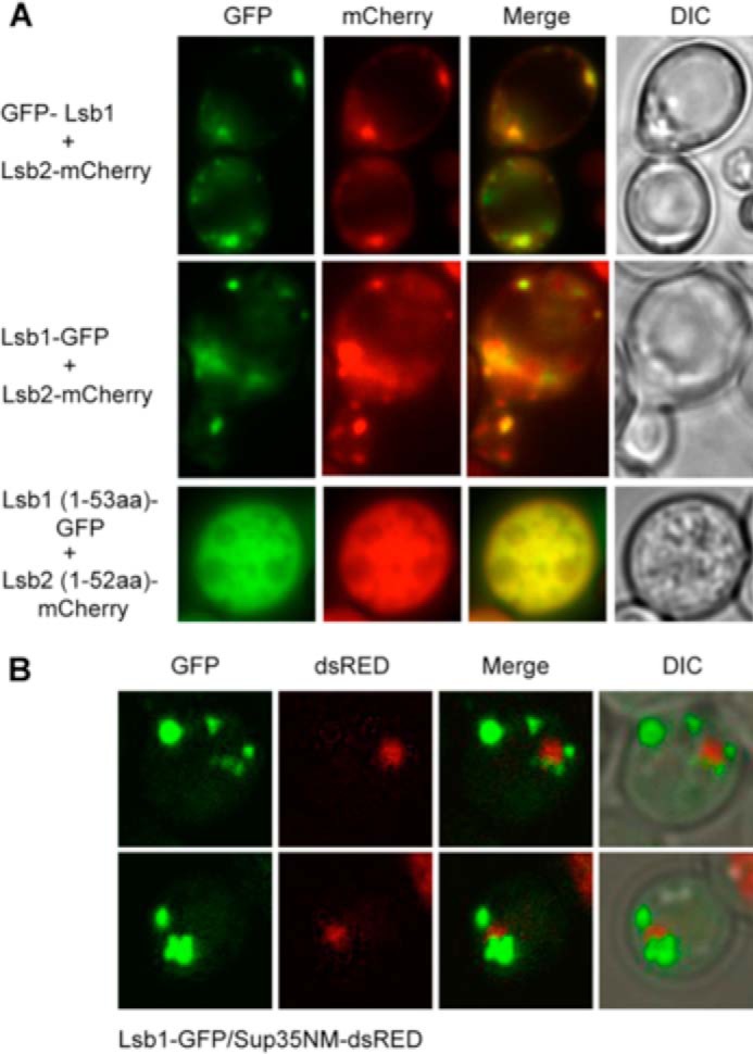
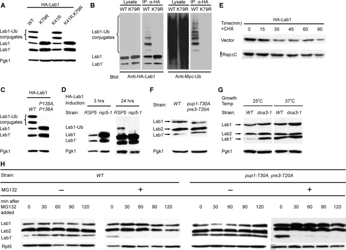
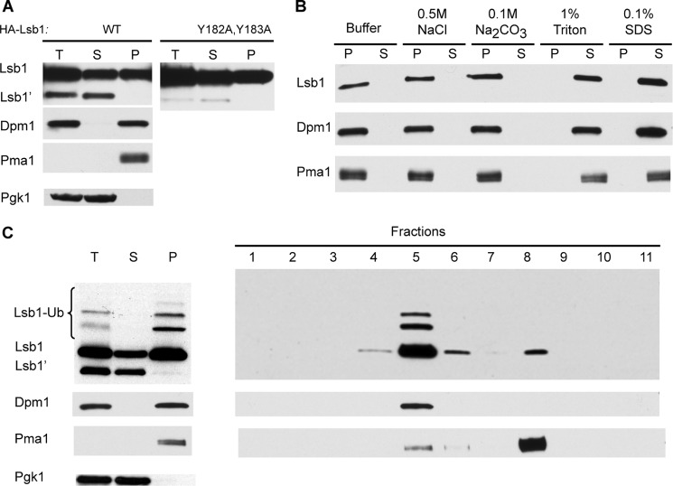
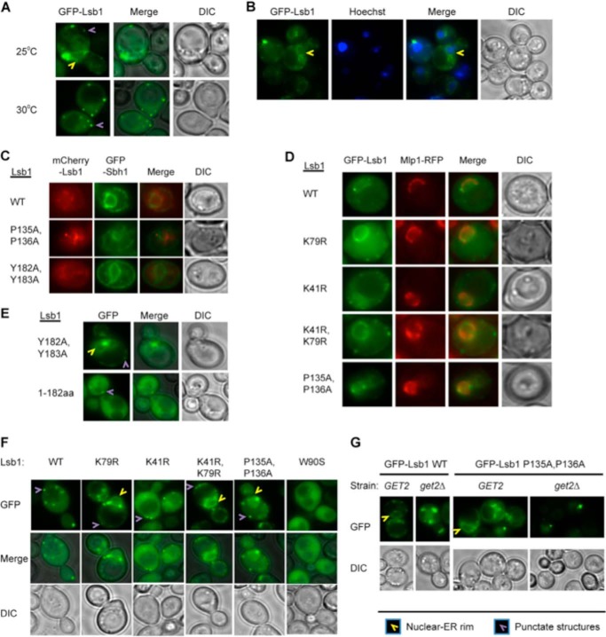
Yeast prions are self-propagating amyloid-like aggregates of Q/N-rich protein that confer heritable traits and provide a model of mammalian amyloidoses. [PSI(+)] is a prion isoform of the translation termination factor Sup35. Propagation of [PSI(+)] during cell division under normal conditions and during the recovery from damaging environmental stress depends on cellular chaperones and is influenced by ubiquitin proteolysis and the actin cytoskeleton. The paralogous yeast proteins Lsb1 and Lsb2 bind the actin assembly protein Las17 (a yeast homolog of human Wiskott-Aldrich syndrome protein) and participate in the endocytic pathway. Lsb2 was shown to modulate maintenance of [PSI(+)] during and after heat shock. Here, we demonstrate that Lsb1 also regulates maintenance of the Sup35 prion during and after heat shock. These data point to the involvement of Lsb proteins in the partitioning of protein aggregates in stressed cells. Lsb1 abundance and cycling between actin patches, endoplasmic reticulum, and cytosol is regulated by the Guided Entry of Tail-anchored proteins pathway and Rsp5-dependent ubiquitination. Heat shock-induced proteolytic processing of Lsb1 is crucial for prion maintenance during stress. Our findings identify Lsb1 as another component of a tightly regulated pathway controlling protein aggregation in changing environments.

Keywords: Actin; Endoplasmic Reticulum (ER); Heat Shock; Prion; Proteasome; Rsp5; Sup35; Ubiquitylation (Ubiquitination).


Figures

FIGURE 1.
FIGURE 1.
Effects of Lsb proteins on [PSI+]. A, [PSI+] destabilization by HS. B, lsb1Δ affects [PSI+] destabilization during HS. In panels A and B, yeast cultures were grown to early exponential stage at 25 °C, shifted to 39 °C for the specified periods of time, plated on YPD and incubated at 25 °C. [PSI+] (pink), [psi] (red), and mosaic [PSI+]/[psi] colonies were detected by visual inspection. The percentages of colonies in which prion was destabilized by heat shock (red [psi] and [PSI+]/[psi] mosaic) are indicated. Averages of six experiments are shown with error bars correspond to standard deviations. C, the lsb1Δ and/or lsb2Δ do not affect viability of the exponential [PSI+] culture at 39 °C. The control strain LSB1, LSB2 (OT55), and isogenic lsb1Δ and/or lsb2Δ strains were pre-grown at 25 °C, and aliquots were placed at 25 and 39 °C. Serial decimal dilutions were prepared after the indicated periods of time, and 2.5 μl of each dilution were spotted onto YPD medium. Plates were photographed after 6 days at 30 °C. D, analysis of the first cell divisions after HS at 39 °C. After resumption of cell division, mother and daughter cells were separated by micromanipulation. Only pairs where both mother and daughter were viable are presented (for viability data, see Table 2). For each type of mother/daughter [PSI+] distribution, numbers are shown. The probability of random deviation of the observed results from those expected in case of equal segregation of [PSI+] between the mother and daughter cells, calculated by using the χ-square approach, is indicated.
FIGURE 2.
FIGURE 2.
Lsb proteins interact with each other and with the prion domain of Sup35. A, structural organization of the Lsb1 and Lsb2 proteins. Conserved lysine (K), tryptophan (W), glutamine (Q), and tyrosine (Y) as well as putative Rsp5 binding sites PPSY (Lsb1) and PPQY (Lsb2) are shown. Superscript and subscript numbers correspond to amino acid positions and the number of repeated residues in a stretch, respectively. Black bars with numbers indicate the numbers of amino acids used to make truncations. B and C, yeast two-hybrid assay. Lsb proteins (full size or truncated versions) and the Sup35 prion domain consisting of the first 113 codons of SUP35 fused to activation (AD) and DNA binding (BD) domains of Gal4 were used. Two-hybrid interaction is detected by activation of the GAL-ADE2 reporter construct, resulting in growth on −Ade. B, the C-terminal region of Lsb1/Lsb2 is not required for interaction with Sup35. C, the N-terminal region of Lsb1/Lsb2 is required and sufficient for their interactions with each other. Mutation of the conservative tryptophan residue does not affect this interaction. D, biochemical detection of Lsb1/Lsb2s interaction. Yeast lysates prepared from cells expressing HA-Lsb1 and FLAG-Lsb2-HA from plasmid PCUP1 promoter were incubated with anti-FLAG-agarose. Bound proteins were analyzed by SDS-PAGE and anti-HA immunoblotting. Lysate of cells expressing HA-Lsb1 and Myc-Lsb2-HA proteins was used as a control. 10% of total protein used for immunoprecipitation was loaded as the “lysate.” The processed form of Lsb1 (see Fig. 3) is not distinguishable because of the overlap with more abundant Lsb2.
FIGURE 3.
FIGURE 3.
Processing of Lsb1. A, Lsb1, but not Lsb2, is processed. Lsb1 and Lsb2 expressed from endogenous promoter at the chromosomal foci were identified in cell lysates by anti-Lsb Ab. B, Lsb1 undergoes processing at the C terminus as detected by HA-tag/anti-HA Ab (left) and GFP-tag/anti-GFP Ab (right). N terminally or C terminally tagged proteins were expressed from plasmid PCUP1 promoter in cells growing at 30 °C. C, HS induces processing of Lsb1 expressed from chromosomal endogenous promoter and detected by anti-Lsb Ab. Pgk1 protein detected by anti-Pgk1 Ab was used as a loading control (A–C). D, densitometry was used to determine the relative levels of total, processed, and unprocessed form of Lsb1 protein after 60 min at 39 °C. Average measurements of levels of Lsb1 forms at 0 min at 39 °C for each blot were set to a value 1.0 and compared with measurement of the same form of protein after 60 min at 39 °C. The error bar represents mean ± S.D. for 6 independent experiments in each case.
FIGURE 4.
FIGURE 4.
Lsb1 is processed at residues Tyr-182,Tyr-183. A, detection of the C-terminal peptide of processed Lsb1 protein by LC-MS/MS. Y.MQAPPPQQQQAPLPYPPPFTNY.Y was identified as the potential C-terminal peptide. B, Y182A,Y183A substitution blocks Lsb1 processing. HA-tagged wild type, single, and double mutants of Lsb1 were induced from PCUP1 for the indicated period of time and analyzed by immunoblotting with anti-HA Ab. C, thermal stress induces processing of wild type but not the Y182A,Y183A mutant. Lsb1 proteins are expressed from the endogenous promoter. Protein levels were analyzed at the indicated time points using anti-Lsb Ab. D, molecular weights of the unprocessed and processed forms of Lsb1 are equal to molecular weights of the full size Y182A,Y183A mutant Lsb1 protein and truncated Lsb1 protein (1–183 amino acids), respectively, as judged from the SDS-PAGE gel. Wild type, mutant, and truncated proteins, tagged with HA at N termini, were expressed from the copper-inducible promoter for the indicated periods of time. Protein extracts were run on SDS-PAGE gel and detected with the anti-HA antibody. Pgk1 protein detected by anti-Pgk1 Ab was used as a loading control (B–D). E, double substitution Y182A,Y183A in LSB1 (in combination with lsb2Δ) affects [PSI+] destabilization and recovery during HS the same way as lsb1Δ. Yeast were grown and treated by HS as in Fig. 1B.
FIGURE 5.
FIGURE 5.
Colocalization of Lsb proteins with each other and Sup35. A, Lsb1 and Lsb2 wild type or truncated proteins tagged with GFP (Lsb1) and mCherry (Lsb2) at the N or C terminus as indicated on the figure were co-expressed from the PCUP1 promoter for 3 h at 30 °C in [psirnq] strain. Fluorescent microscopy images of live cells are shown. B, aggregates of Sup35NM-dsRED are surrounded by puncta of Lsb1-GFP. Both proteins were co-expressed from the PCUP1 promoter for 24 h in the [psiRNQ+] strain. Projection of 15 z-focal planes collected with 0.4-μm step size on Zeiss LSM510 confocal microscope (Carl Zeiss, Inc., Thornwood, NY) with a ×100 1.3 NA Zeiss oil immersion objective. DIC, differential interference contrast.
FIGURE 6.
FIGURE 6.
Effects of ubiquitin-proteasome system on Lsb1 processing. A, substitution of a single lysine residue, K79R, in Lsb1 prevents accumulation of high molecular weight (Mr) conjugates. HA-Lsb1, its derivatives, and Myc-Ub were expressed from the copper-inducible promoter PCUP1. Lsb1 was detected with anti-HA Ab. B, high Mr conjugates represent ubiquinated Lsb1. HA-Lsb1 and Myc-Ub were expressed from PCUP1. Lsb1 was immunoprecipitated with anti-HA Ab. 10% of total protein used for immunoprecipitation was loaded as the “lysate.” High Mr conjugates of HA-Lsb1 react to anti-Myc Ab, confirming that they contain Myc-Ub. C, double P135A,P136A substitution in the potential Rsp5 binding site prevents Lsb1 ubiquitination. HA-Lsb1 wild type, mutant, and Myc-Ub were expressed from PCUP1. Lsb1 was detected with anti-HA Ab. D, E3 enzyme Rsp5 is involved in Lsb1 ubiquitination. HA-Lsb1 was expressed from PCUP1 in wild type and rsp5-1 temperature-sensitive mutant cells growing at 37 °C. HA-Lsb1 accumulates in mutant cells after 3 h of induction (left panel). Ubiquitinated Lsb1 can be detected in the wild type, but not in mutant cells after 24 h of induction (right panel). Lsb1 protein was identified by anti-HA Ab. E, partial stabilization of Lsb1 in cells deficient in Rsp5 activity. HA-tagged Lsb1 was expressed from the PCUP1 promoter in cells transformed either with vector (control) or plasmid expressing mutant Rsp5ΔC from PGAL promoter. Cells were grown in the presence of copper and galactose and protein levels of Lsb1 were analyzed at the indicated time points after the addition of cycloheximide (CHX). F, processing of Lsb1 is not affected in the mutant strain deficient in the trypsin-like (pre3-T20A) and postacidic/post-glutamic-like (pup1-T30A) activities of the proteasome. G, Lsb1 is processed in the mutant strain deficient in chymotrypsin-like activity of the proteasome (pre2-1/doa3-1). Cells were grown overnight, diluted in the morning, and incubated with shaking at 30 °C (F) or at the temperature indicated (G). Equal amounts of cells were collected, lysed by boiling, and protein was detected by Western blot with anti-Lsb Ab. H, the Lsb1 processing is inhibited by MG132. Processed Lsb1 is not detected after inhibition of chymotryptic peptidase activity of the proteasome by the addition of the proteasome inhibitor MG132 to the wild-type (WT) strain or to the mutant, pup1-T30A,pre3-T20A defective in the other peptidase activities of the proteasome. Cells incubated with MG132 for the indicated periods of time were collected, lysed by boiling, and analyzed by Western blot with anti-Lsb Ab. Pgk1 protein detected by anti-Pgk1 Ab (A, C, D, F, and G) and Rpt5 protein detected by anti-Rpt5 Ab (H) were used as a loading control.
FIGURE 7.
FIGURE 7.
Association of Lsb1 with membranes. A, total extracts (T) of cells expressing HA-tagged Lsb1 (WT or Y182A,Y183A mutant) from PCUP1 were separated into soluble (S) and pellet (P) fractions by centrifugation, separated by SDS-gel electrophoresis, and probed with anti-HA Ab. Unprocessed (Lsb1) and processed (Lsb1′) forms are indicated. B, the pellet fraction obtained from HA-Lsb1 expressing cells (A) was washed with detergents, salts, and buffer as indicated, separated into pellet and soluble fractions as in A, and probed with anti-HA Ab. C, sucrose gradient of pelleted Lsb1 protein. Left panel, total extracts of cells expressing HA-tagged Lsb1 were separated into soluble and pellet fractions and analyzed as in A. Right panel, the pellet fraction was subjected to sucrose gradient and fractions were analyzed by SDS-PAGE and anti-HA immunoblotting to detect Lsb1. A–C, protein markers of plasma membrane (Pma1), ER (Dpm1), and cytoplasm (Pgk1) were detected with protein-specific antibodies.
FIGURE 8.
FIGURE 8.
Association of Lsb1 with nuclear-ER rim. A–G, representative images of live cells are shown. At least 100 cells were analyzed for each experiment. Wild type GFP-Lsb1 and its mutant derivatives were expressed from the copper-inducible promoter, PCUP1. A, GFP-Lsb1 forms a rim and small dots at the cell periphery in 40–50% of cells growing at 25 °C. At 30 °C, GFP-Lsb1 is not present in the rim but is distributed over the cytoplasm and forms small dots in 96% of cells. B, GFP-Lsb1 rim, formed at 25 °C, is colocalized with the nuclear-ER rim. Live cells expressing GFP-Lsb1 from PCUP1 were stained with Hoechst to indicate the positions of the nuclei. C, Lsb1 colocalizes with ER marker protein Sbh1. Proteins were expressed from plasmid promoters PCUP1- mCherry-Lsb1 and PGAL-GFP-Sbh1 for 18 h. Colocalization was detected in all cells. D, GFP-Lsb1 expressed from the PCUP1 promoter colocalizes with nuclear envelope protein Mlp1-RFP expressed from endogenous promoter. Colocalization was detected in all cells. E, GFP-Lsb1 mutant deficient in processing (Y182A,Y183A) is present in the nuclear nuclear-ER rim in 40–50% of cells, whereas the truncated version (1–183 amino acids) corresponding to the processed form of Lsb1 is observed only in the cytoplasm in all cells growing at 25 °C. F, ubiquitination affects localization of Lsb1 to the nuclear-ER rim. GFP-Lsb1 WT and mutants not deficient in ubiquitination (K41R and W90S) do not associate with the nuclear-ER rim in almost all cells growing at 30 °C. However, Lsb1 mutants deficient in ubiquitination (K79R, and K41R,K79R, and P135A,P136A) associate with the nuclear-ER rim in 60–80% of cells growing at 30 °C. The W90S mutant, defective in interaction with Las17, does not form small dots as has been reported previously. G, deletion of get2Δ abolishes localization of wild type and ubiquitination-deficient (P135A,P136A) mutant GFP-Lsb1 to nuclear-ER rim at 25 °C.

References

    1. Chernoff Y. O. (2007) Stress and prions: lessons from the yeast model. FEBS Lett. 581, 3695–3701 - PMC - PubMed
    1. Li L., Kowal A. S. (2012) Environmental regulation of prions in yeast. PLoS Pathog. 8, e1002973. - PMC - PubMed
    1. Verghese J., Abrams J., Wang Y., Morano K. A. (2012) Biology of the heat shock response and protein chaperones: budding yeast (Saccharomyces cerevisiae) as a model system. Microbiol. Mol. Biol. Rev. 76, 115–158 - PMC - PubMed
    1. Chernova T. A., Wilkinson K. D., Chernoff Y. O. (2014) Physiological and environmental control of yeast prions. FEMS Microbiol. Rev. 38, 326–344 - PMC - PubMed
    1. Kopito R. R. (2000) Aggresomes, inclusion bodies and protein aggregation. Trends Cell Biol. 10, 524–530 - PubMed

Publication types

MeSH terms