. 2014 Oct 15;25(20):3133-46.
doi: 10.1091/mbc.E14-03-0815. Epub 2014 Aug 20.

Loss of γ-cytoplasmic actin triggers myofibroblast transition of human epithelial cells

Affiliations

Loss of γ-cytoplasmic actin triggers myofibroblast transition of human epithelial cells

Susana Lechuga et al. Mol Biol Cell. .

Abstract

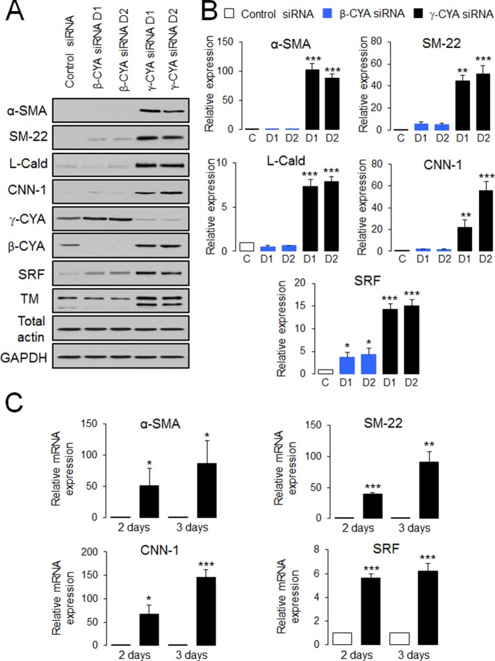
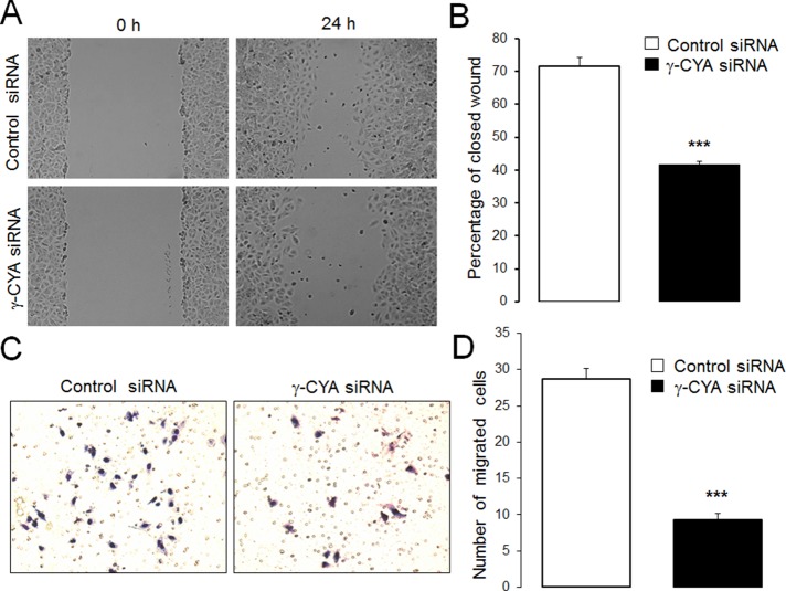
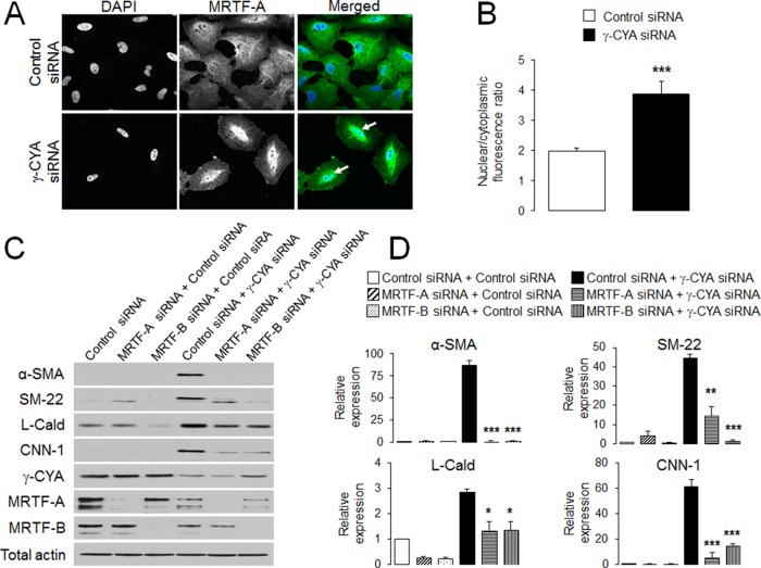
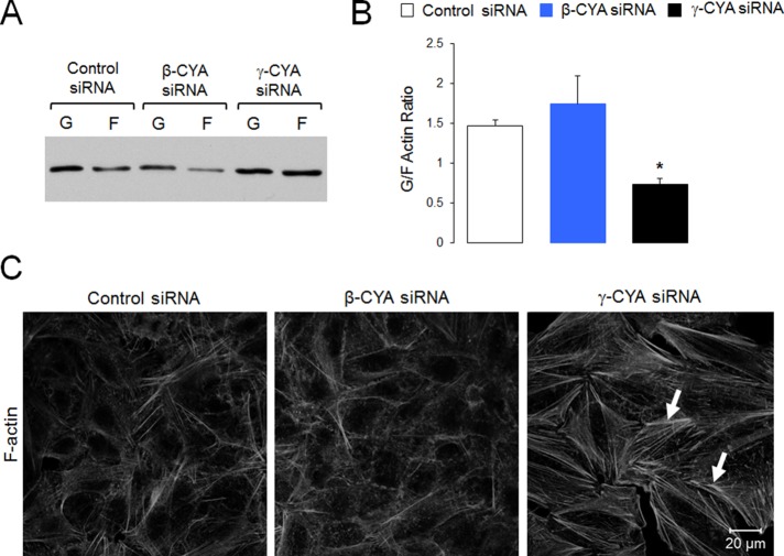
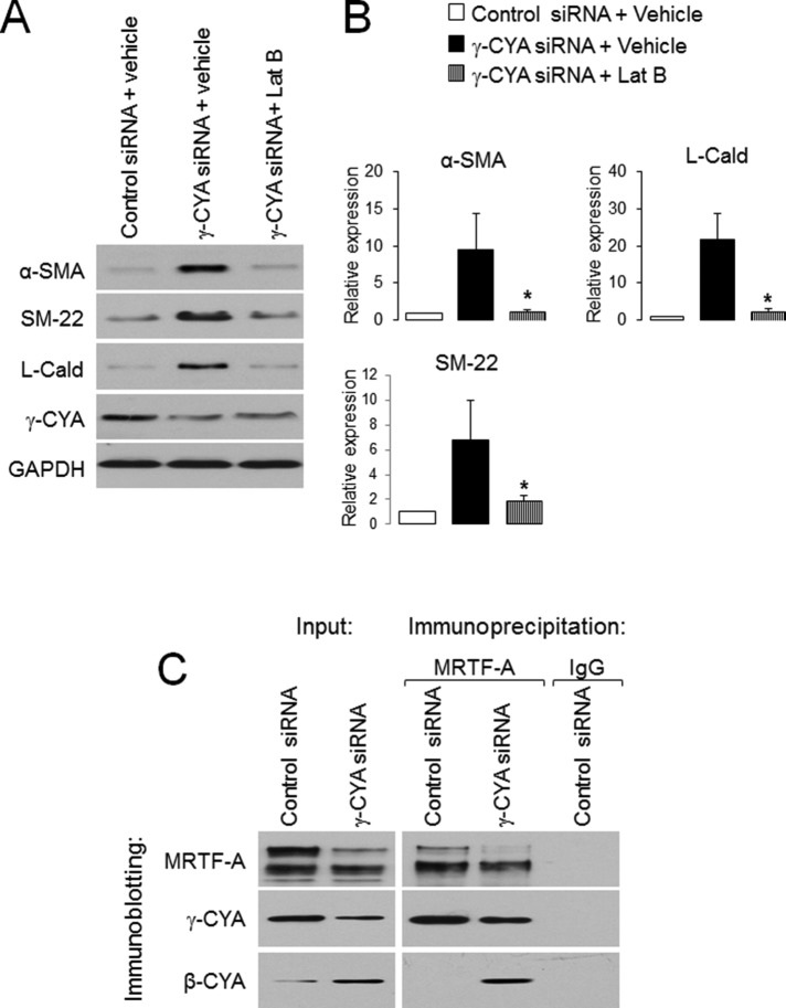
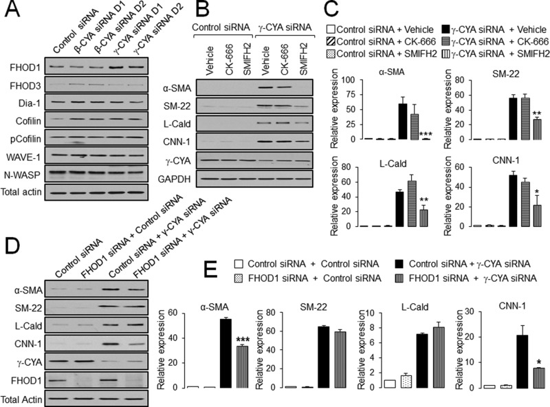
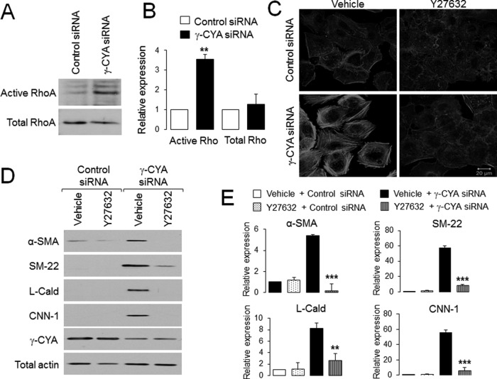
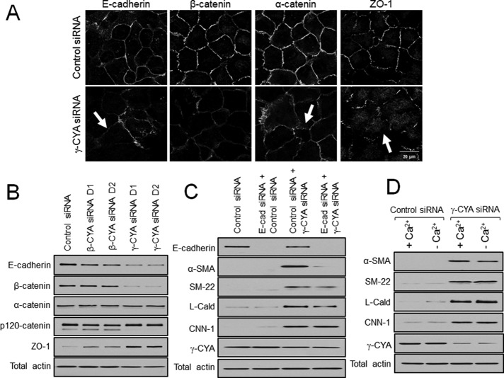
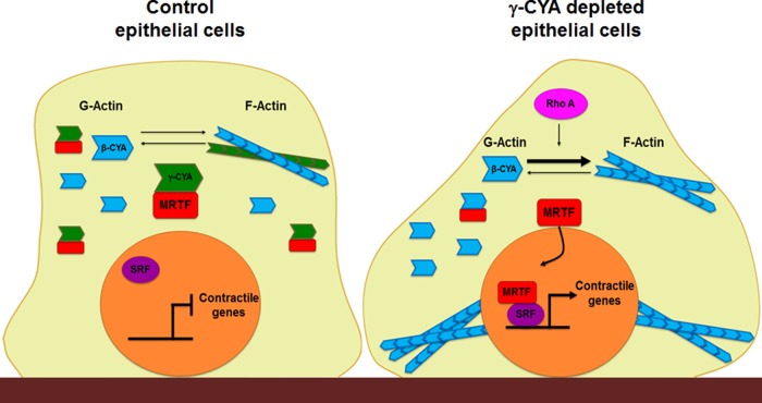
Transdifferentiation of epithelial cells into mesenchymal cells and myofibroblasts plays an important role in tumor progression and tissue fibrosis. Such epithelial plasticity is accompanied by dramatic reorganizations of the actin cytoskeleton, although mechanisms underlying cytoskeletal effects on epithelial transdifferentiation remain poorly understood. In the present study, we observed that selective siRNA-mediated knockdown of γ-cytoplasmic actin (γ-CYA), but not β-cytoplasmic actin, induced epithelial-to-myofibroblast transition (EMyT) of different epithelial cells. The EMyT manifested by increased expression of α-smooth muscle actin and other contractile proteins, along with inhibition of genes responsible for cell proliferation. Induction of EMyT in γ-CYA-depleted cells depended on activation of serum response factor and its cofactors, myocardial-related transcriptional factors A and B. Loss of γ-CYA stimulated formin-mediated actin polymerization and activation of Rho GTPase, which appear to be essential for EMyT induction. Our findings demonstrate a previously unanticipated, unique role of γ-CYA in regulating epithelial phenotype and suppression of EMyT that may be essential for cell differentiation and tissue fibrosis.


Figures

FIGURE 1:
FIGURE 1:
Downregulation of γ-CYA selectively stimulates expression of EMyT markers in lung epithelial cells. A549 epithelial cells were transfected with control or β-CYA– or γ-CYA–specific siRNA duplexes (D1 and D2), and expression of EMyT markers was analyzed by immunoblotting (A, B) and quantitative real-time RT-PCR (C) at different times after transfection. Immunoblots are quantified by densitometric analysis, and protein expression is calculated relative to the control siRNA–treated group. mRNA expression of all EMyT markers is normalized by the expression of a housekeeping gene. Data are presented as mean ± SE (n = 3); *p < 0.05, **p < 0.005, ***p < 0.0005.
FIGURE 2:
FIGURE 2:
Depletion of γ-CYA inhibits epithelial cell migration. (A, B) Control and γ-CYA–depleted A549 cell monolayers were mechanically wounded on day 3 posttransfection. To examine the rate of cell motility, the cell-free area was measured at 0 and 24 h after wounding; percentage of closed wound area was calculated as described in Materials and Methods. (C, D) Control and γ-CYA–depleted A549 cells were subjected to the Matrigel invasion assay. Data are presented as mean ± SE (n = 3); ***p ≤ 0.0005.
FIGURE 3:
FIGURE 3:
SRF mediates induction of EMyT markers in γ-CYA–depleted epithelial cells. (A, B) A549 cells transfected with either control or γ-CYA siRNAs were immunolabeled for SRF and counterstained with a nuclear dye, DAPI, on day 4 posttransfection. Intensity of nuclear SRF signal was quantified with image analysis. (C) A549 cells stably expressing SRE luciferase reporter were transfected with either control or γ-CYA siRNAs and cotransfected with a Renilla luciferase plasmid. Dual luciferase assay was performed 2 d posttransfection. The luminescence signal of SRF-dependent promoters was normalized by the Renilla luciferase signal. (D, E) A549 cells were subjected to sequential transfections with one of the following siRNA pairs: control–control, SRF–control, control–γ-CYA, and SRF–γ-CYA. Expression of targeted proteins and EMyT markers was determined by immunoblotting on day 3 after the second transfection. Data are presented as mean ± SE (n = 3); **p < 0.005, ***p < 0.0005.
FIGURE 4:
FIGURE 4:
MRTF is essential for the induction of EMyT markers in γ-CYA–depleted epithelial cells. (A, B) A549 cells transfected with either control or γ-CYA siRNAs were immunolabeled for MRTF-A and counterstained with a nuclear dye, DAPI. Intensity of nuclear and cytoplasmic MRTF-A, determined by image analysis, was used to calculate the nuclear/cytoplasmic ratio. Arrows indicate nuclear localization of MRTF-A in γ-CYA–depleted cells. (C, D) A549 cells were subjected to sequential transfections with one of the following siRNA pairs: control–control, MRTF-A–control, MRTF-B–control, control–γ-CYA, MRTF-A–γ-CYA, and MRTF-B–γ-CYA. Expression of targeted proteins and EMyT markers was determined by immunoblotting on day 3 after the second transfection. Data are presented as mean ± SE (n = 3); *p < 0.05, **p < 0.005, ***p < 0.0005.
FIGURE 5:
FIGURE 5:
Depletion of γ-CYA but not β-CYA induces actin polymerization. A549 cells transfected with control or cytoplasmic actin isoform–specific siRNAs were subjected to detergent fractionation to determine the G-/F-actin ratio (A, B) or fluorescence labeling with Alexa 555–phalloidin to visualize actin filaments (C). Arrows highlight basal stress fibers in γ-CYA–depleted cells. Data are presented as mean ± SE (n = 3); *p < 0.05.
FIGURE 6:
FIGURE 6:
EMyT induction in γ-CYA–depleted epithelial cells is controlled by actin polymerization, and γ-CYA preferentially interacts with MRTF. (A, B) γ-CYA–depleted A549 cells were treated for 24 h with either vehicle or the F-actin–depolymerizing drug Lat B (1 μM), and expression of EMyT markers was determined by immunoblotting. Data are presented as mean ± SE (n = 3); *p < 0.05. (C) Association of MRTF-A with γ-CYA and β-CYA in control and γ-CYA–depleted A549 cells was examined using immunoprecipitation with anti–MRTF-A antibody.
FIGURE 7:
FIGURE 7:
Upregulation of EMyT markers in γ-CYA-depleted cells depends on formin-mediated actin polymerization. (A) A549 cells were transfected with control or β-CYA– or γ-CYA–specific siRNA duplexes, and expression of different actin-polymerizing proteins was analyzed by immunoblotting. (B, C) Control and γ-CYA–depleted A549 cells were treated for 24 h with either vehicle or pharmacological inhibitors of Arp2/3 complex (CK-666) and formin-dependent actin polymerization (SMIFH2). Effects of the inhibitors on induction of EMyT marker were determined by immunoblotting. (D, E) A549 cells were subjected to sequential transfections with one of the following siRNA pairs: control–control, FHOD1–control, control–γ-CYA, and FHOD1–γ-CYA. Expression of targeted proteins and EMyT markers was determined by immunoblotting on day 3 after the second transfection. Data are presented as mean ± SE (n = 3); *p < 0.05, **p < 0.005, ***p < 0.0005.
FIGURE 8:
FIGURE 8:
Rho activation is essential for EMyT induction in γ-CYA–depleted cells. (A, B) Level of total and GTP-bound active RhoA in control and γ-CYA–depleted A549 cells was determined by a rhotekin pull-down assay and immunoblotting. (C–E) Control and γ-CYA–depleted A549 cells were treated for 24 h with either vehicle or ROCK inhibitor, Y27632, and assembly of stress fibers (C), as well as expression of different EMyT markers (D, E), was examined by fluorescence analysis and immunoblotting. Data are presented as mean ± SE (n = 3); **p < 0.005, ***p < 0.0005.
FIGURE 9:
FIGURE 9:
Disassembly of intercellular junctions does not contribute to induction of EMyT in γ-CYA–depleted epithelial cells. A549 cells were transfected with either control or cytoplasmic actin isoform–specific siRNAs. The integrity of cell–cell contacts (A) and expression of junctional proteins (B) were examined on day 4 posttransfection. Arrows highlight disassembly of intercellular junctions in γ-CYA–depleted cells. (C) A549 cells were subjected to sequential transfections with one of the following siRNA pairs: control–control, E-cadherin–control, control–γ-CYA, and E-cadherin–γ-CYA. Expression of targeted proteins and EMyT markers was determined by immunoblotting on day 3 after the second transfection. (D) Control and γ-CYA–depleted A549 cells were either incubated in normal cell culture medium (+Ca2+) or subjected to 24 h of extracellular calcium depletion (–Ca2+) and analyzed for expression of EMyT markers.
FIGURE 10:
FIGURE 10:
Schematic diagram of key molecular events that are likely to mediate myogenic transdifferentiation of γ-CYA–depleted epithelial cells. In control cells (left), MRTF preferentially interacts with monomeric γ-CYA in the cytoplasm, which prevents nuclear translocation and activation of this transcriptional regulator. As a result, nuclear SRF does not stimulate expression of contractile genes, and the myogenic transcriptional program remains inactive. Loss of γ-CYA triggers RhoA activation and Rho-mediated actin polymerization, which allow nuclear translocation of MRTF and subsequent formation of active MRTF/SRF complexes. This stimulates transcription of contractile/cytoskeletal genes, leading to EMyT.

Similar articles

Cited by

References

    1. Baarlink C, Wang H, Grosse R. Nuclear actin network assembly by formins regulates the SRF coactivator MAL. Science. 2013;340:864–867. - PubMed
    1. Baranwal S, Naydenov NG, Harris G, Dugina V, Morgan KG, Chaponnier C, Ivanov AI. Nonredundant roles of cytoplasmic beta- and gamma-actin isoforms in regulation of epithelial apical junctions. Mol Biol Cell. 2012;23:3542–3553. - PMC - PubMed
    1. Beach JR, Hussey GS, Miller TE, Chaudhury A, Patel P, Monslow J, Zheng Q, Keri RA, Reizes O, Bresnick AR, et al. Myosin II isoform switching mediates invasiveness after TGF-beta-induced epithelial-mesenchymal transition. Proc Natl Acad Sci USA. 2011;108:17991–17996. - PMC - PubMed
    1. Belyantseva IA, Perrin BJ, Sonnemann KJ, Zhu M, Stepanyan R, McGee J, Frolenkov GI, Walsh EJ, Friderici KH, Friedman TB, et al. Gamma-actin is required for cytoskeletal maintenance but not development. Proc Natl Acad Sci USA. 2009;106:9703–9708. - PMC - PubMed
    1. Bergeron SE, Zhu M, Thiem SM, Friderici KH, Rubenstein PA. Ion-dependent polymerization differences between mammalian beta- and gamma-nonmuscle actin isoforms. J Biol Chem. 2010;285:16087–16095. - PMC - PubMed

Publication types