. 2014 Oct;93(10):988-92.
doi: 10.1177/0022034514548222. Epub 2014 Aug 20.

ENAM mutations with incomplete penetrance

Affiliations

ENAM mutations with incomplete penetrance

F Seymen et al. J Dent Res. 2014 Oct.

Abstract

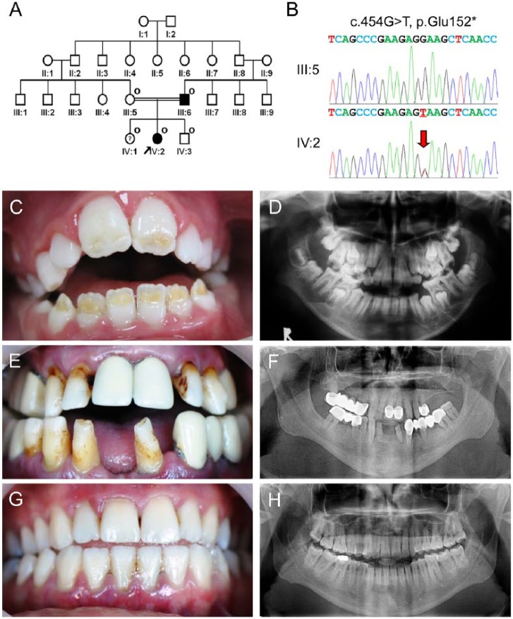
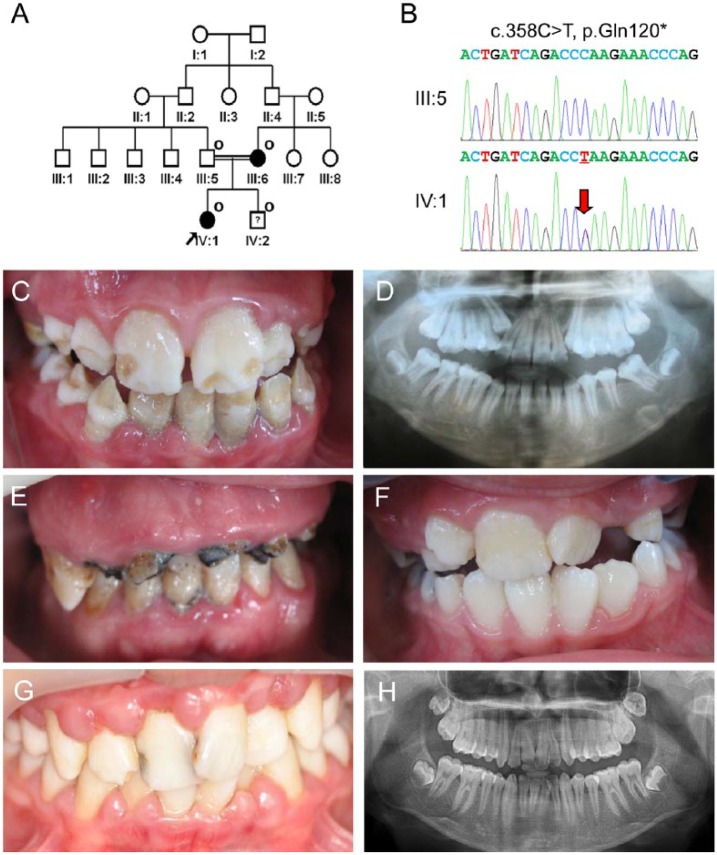
Amelogenesis imperfecta (AI) is a genetic disease affecting tooth enamel formation. AI can be an isolated entity or a phenotype of syndromes. To date, more than 10 genes have been associated with various forms of AI. We have identified 2 unrelated Turkish families with hypoplastic AI and performed mutational analysis. Whole-exome sequencing identified 2 novel heterozygous nonsense mutations in the ENAM gene (c.454G>T p.Glu152* in family 1, c.358C>T p.Gln120* in family 2) in the probands. Affected individuals were heterozygous for the mutation in each family. Segregation analysis within each family revealed individuals with incomplete penetrance or extremely mild enamel phenotype, in spite of having the same mutation with the other affected individuals. We believe that these findings will broaden our understanding of the clinical phenotype of AI caused by ENAM mutations.

Keywords: amelogenesis imperfecta; enamel; enamelin; expressivity; hypoplastic; tooth.


Conflict of interest statement

The authors declare no potential conflicts of interest with respect to the authorship and/or publication of this article.

Figures

Figure 1.
Figure 1.
Clinical and mutational analysis of family 1. (A) Pedigree of family 1. The “O” symbol indicates members recruited for this study. The proband is indicated with a black arrow. The question symbol (?) indicates that this individual is clinically normal but has the mutation. (B) Comparison of the ENAM exon 7 sequencing chromatograms for the unaffected family member (III:5) with the wild-type (top) sequence, and the mutated allele in the proband (IV:2) reveals a G-to-T transversion: c.454G>T, p.Glu152*. A red arrow indicates the mutated nucleotide. (C) Frontal clinical photo of the proband. Yellow dentin color can be seen through the hypoplastic enamel located on the incisal half of the permanent anterior teeth. (D) Panoramic radiograph of the proband reveals unusual crown of the developing second permanent molars due to hypoplastic enamel. (E) Frontal clinical photo of the affected father shows the characteristic horizontal hypoplastic grooves. (F) Panoramic radiograph of the affected father shows multiple prosthetics and thin enamel in remaining teeth. (G) Frontal clinical photo of a sister of the proband. Despite having the mutation, her dentition has no enamel defects. (H) Panoramic radiograph of a sister of the proband reveals normal-looking tooth structures.
Figure 2.
Figure 2.
Clinical and mutational analysis of family 2. (A) Pedigree of family 2. The “O” symbol indicates members recruited for this study. The proband is indicated with a black arrow. The question symbol (?) indicates that this individual is clinically normal but has the mutation. (B) Comparison of the ENAM exon 7 sequencing chromatograms for the unaffected family member (III:5) with the wild-type (top) sequence, and the mutated allele in the proband (IV:1) reveals a C-to-T transition: c.358C>T, p.Gln120*. (C) Frontal clinical photo of the proband shows a localized form of enamel regions with some normal-looking areas. (D) Panoramic radiograph of the proband shows an irregular form of crowns. (E) Frontal clinical photo of the affected mother. She has several remaining teeth with gross destruction. (F) Frontal clinical photo of a brother of the proband at age 8 yr. He has normal-looking teeth, even though he also has the mutation. However, he has minor enamel defects, resembling an enamel fracture or dental caries, on the incisal corners of the maxillary right lateral incisor. (G) Frontal clinical photo of a brother of the proband at age 14 yr. Dental caries of maxillary central incisors and gingivitis can be seen. (H) Panoramic radiograph of a brother of the proband at age 14 yr.

References

    1. Chan HC, Mai L, Oikonomopoulou A, Chan HL, Richardson AS, Wang SK, et al. (2010). Altered enamelin phosphorylation site causes amelogenesis imperfecta. J Dent Res 89:695-699. - PMC - PubMed
    1. Chan HC, Estrella NM, Milkovich RN, Kim JW, Simmer JP, Hu JC. (2011). Target gene analyses of 39 amelogenesis imperfecta kindreds. Eur J Oral Sci 119(Suppl 1):311-323. - PMC - PubMed
    1. Chaussain C, Bouazza N, Gasse B, Laffont AG, Opsahl Vital S, Davit-Beal T, et al. (2014). Dental caries and enamelin haplotype. J Dent Res 93:360-365. - PubMed
    1. El-Sayed W, Parry DA, Shore RC, Ahmed M, Jafri H, Rashid Y, et al. (2009). Mutations in the beta propeller WDR72 cause autosomal-recessive hypomaturation amelogenesis imperfecta. Am J Hum Genet 85:699-705. - PMC - PubMed
    1. Gutierrez SJ, Chaves M, Torres DM, Briceno I. (2007). Identification of a novel mutation in the enamalin gene in a family with autosomal-dominant amelogenesis imperfecta. Arch Oral Biol 52:503-506. - PubMed

Publication types

MeSH terms

LinkOut - more resources