. 2014 Sep 2;111(35):12871-6.
doi: 10.1073/pnas.1323292111. Epub 2014 Aug 20.

Primary cilia signaling mediates intraocular pressure sensation

Affiliations

Primary cilia signaling mediates intraocular pressure sensation

Na Luo et al. Proc Natl Acad Sci U S A. .

Abstract

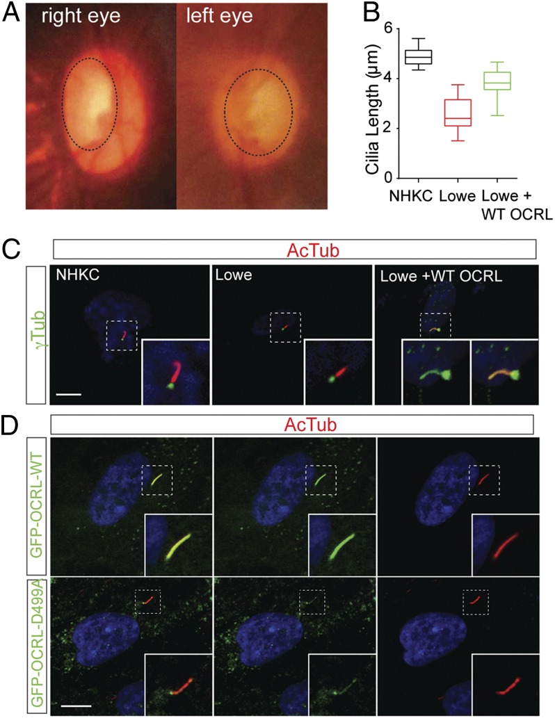
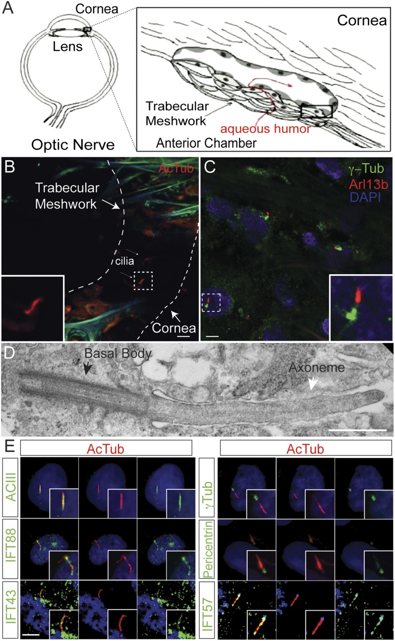
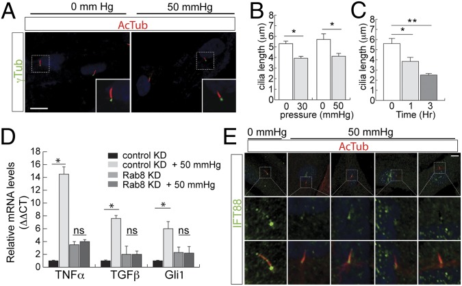
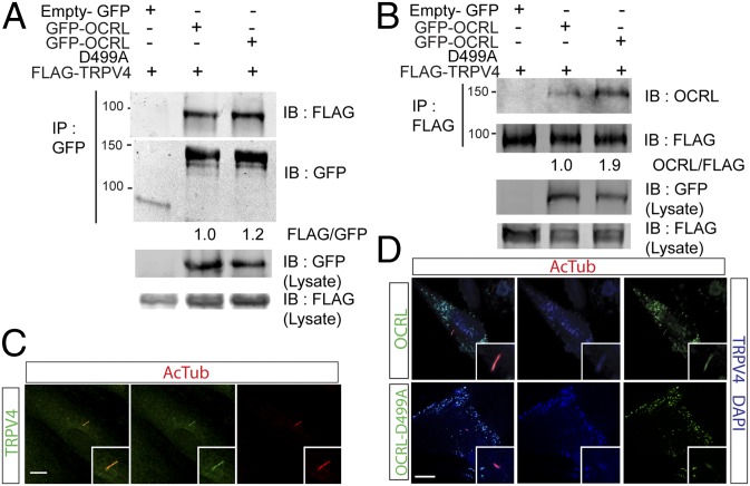
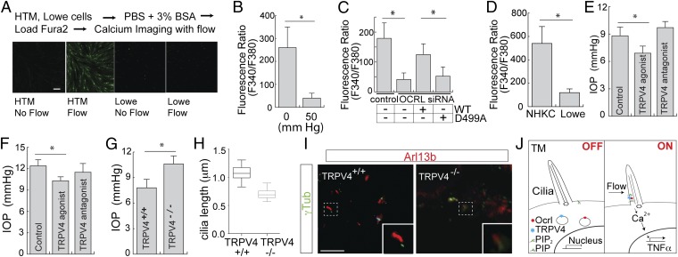
Lowe syndrome is a rare X-linked congenital disease that presents with congenital cataracts and glaucoma, as well as renal and cerebral dysfunction. OCRL, an inositol polyphosphate 5-phosphatase, is mutated in Lowe syndrome. We previously showed that OCRL is involved in vesicular trafficking to the primary cilium. Primary cilia are sensory organelles on the surface of eukaryotic cells that mediate mechanotransduction in the kidney, brain, and bone. However, their potential role in the trabecular meshwork (TM) in the eye, which regulates intraocular pressure, is unknown. Here, we show that TM cells, which are defective in glaucoma, have primary cilia that are critical for response to pressure changes. Primary cilia in TM cells shorten in response to fluid flow and elevated hydrostatic pressure, and promote increased transcription of TNF-α, TGF-β, and GLI1 genes. Furthermore, OCRL is found to be required for primary cilia to respond to pressure stimulation. The interaction of OCRL with transient receptor potential vanilloid 4 (TRPV4), a ciliary mechanosensory channel, suggests that OCRL may act through regulation of this channel. A novel disease-causing OCRL allele prevents TRPV4-mediated calcium signaling. In addition, TRPV4 agonist GSK 1016790A treatment reduced intraocular pressure in mice; TRPV4 knockout animals exhibited elevated intraocular pressure and shortened cilia. Thus, mechanotransduction by primary cilia in TM cells is implicated in how the eye senses pressure changes and highlights OCRL and TRPV4 as attractive therapeutic targets for the treatment of glaucoma. Implications of OCRL and TRPV4 in primary cilia function may also shed light on mechanosensation in other organ systems.


Conflict of interest statement

Conflict of interest statement: Y.S. is an inventor on a patent related to the work described, which is managed by Indiana University School of Medicine.

Figures

Fig. 1.
Fig. 1.
OCRL mutation in a Lowe syndrome patient with congenital glaucoma. (A) Optic nerve photos of the glaucoma patient. Photograph demonstrating glaucomatous optic nerve cupping in affected patient (cupping represented with dashed line). (B and C) Ciliary defect in a Lowe syndrome patient. After cilia induction by serum-starvation for 48 h, normal control (NHKC), Lowe syndrome patient (Lowe), and WT-GFP-OCRL transduced patient keratinocytes (Lowe + WT) were measured for ciliary length by staining with antiacetylated α-tubulin (red) and γ-tubulin (green) antibodies. Representative figure is shown in C. Average of three independent experiments, n = 50 cilia. Error bars represent SD. ANOVA, P < 0.001. (Scale bar, 10 µm.) (D) Primary cilia in HTM cells transfected with wild-type and mutant OCRL. HTM cells transfected with GFP-OCRL-WT or GFP-OCRL-D499A, followed by serum-starvation for 48 h, were then stained with acetylated α-tubulin. (Scale bar, 5 μm.)
Fig. 2.
Fig. 2.
Primary cilia are conserved in mammalian TM. (A) Schematic of aqueous humor outflow in the eye. Aqueous humor is generated in the ciliary body, and drains out of the trabecular meshwork. (B and C) Primary cilia in normal and glaucomatous human TM. TM removed from human donor eyes (B) and surgical TM specimens during glaucoma surgery (C) were immunostained with anti-Arl13b (red), acetylated α-tubulin (AcTub, red), γ-tubulin (γ-Tub, green), and DAPI (blue). (Scale bar, 5 μm.) (D) Electron micrograph of a primary cilium in HTM in a glaucomatous eye. TM specimen removed during glaucoma surgery was imaged by transmission electron microscopy. (Scale bar, 500 nm.) (E) Characterization of primary cilia in cultured HTM cells. Serum-starved to induce ciliogenesis, HTM cells were immunostained with antiacetylated α-tubulin (AcTub, red), IFT88 (green), IFT43 (green), IFT57 (green), Adenylate cyclase III (green), pericentrin (green), and γ-tubulin (γ-Tub, green) antibodies. DAPI in blue. (Scale bar, 5 μm.)
Fig. 3.
Fig. 3.
Cilia shortening in response to pressure. (A and B) Elevated pressure resulted in ciliary shortening in HTM cells. HTM cells were serum-starved for 48 h, cultured in atmospheric (0 mmHg) or 50-mmHg pressure for 60 min, followed by immunostaining for acetylated α-tubulin. Cilia shown in Inset; cilia length was measured and shown in B. Error bars represent SD. n > 50 cilia, three independent experiments, paired t test. *P < 0.05. (Scale bar in A, 5 μm.) (C) HTM cells were serum-starved for 48 h, cultured in 50-mmHg pressure, for 0, 1, or 3 h, followed by immunostaining for acetylated α-tubulin. Cilia length was measured and shown. Error bars represent SD. n > 50 cilia, three independent experiments unpaired t test, *P < 0.01, **P < 0.001. (D) Pressure-mediated transcriptional activation requires primary cilia. Cilia formation in HTM cells (control and stable Rab8 knockdown) were induced by serum-starvation for 48 h followed by treatment with or without 50-mmHg pressure for 3 h. Levels of mRNA of TNF-α, GLI1, and TGF-β were measured. Three independent experiments with error bars represent SD. *P < 0.001; ns, not significant. (E) Altered distribution of IFT88 with elevated pressure. HTM cells were serum-starved to induce ciliogenesis, followed by treatment with or without elevated pressure (50 mmHg). Representative images of immunostaining with antibodies for IFT88 (green), acetylated α-tubulin (red), and DAPI (blue), showing the loss of IFT88 in the distal tip of cilia under elevated pressure conditions. (Scale bar, 10 μm.)
Fig. 4.
Fig. 4.
OCRL and TRPV4 interact and localize to the cilia. (A) OCRL coimmunoprecipitates TRPV4. HEK293 cells transfected with FLAG-TRPV4 and empty GFP vector, GFP-OCRL-WT, or GFP-OCRL-D499A were subjected to immunoprecipitation with GFP-Trap_A beads. Immunoblot for GFP and FLAG-TRPV4 was performed. Ratio of FLAG/GFP are shown as indicated. (B) TRPV4 coimmunoprecipitates OCRL. HEK293 cells transfected with FLAG-TRPV4 and empty GFP vector, GFP-OCRL-WT, or GFP-OCRL-D499A were subjected to immunoprecipitation with anti-FLAG beads. Immunoblot for GFP and FLAG-TRPV4 was performed. Ratio of OCRL/FLAG are shown as indicated. (C) TRPV4 localizes with HTM cilia. HTM cells serum-starved for 48 h were immunostained with anti-TRPV4 (green) and antiacetylated α-tubulin (red) antibodies. DAPI in blue. (Scale bar, 5 μm.) (D) OCRL-WT but not D499A mutant colocalize with TRPV4 in cilia. After serum-starvation for 48 h, HTM cells transfected with FLAG-TRPV4 and GFP-OCRL-WT or GFP-OCRL-D499A were immunostained with antiacetylated α-tubulin (red) and anti-FLAG antibodies. OCRL and OCRL-D499A in green. FLAG-TRPV4 and DAPI in blue. (Scale bar, 5 μm.)
Fig. 5.
Fig. 5.
OCRL and TRPV4 regulate calcium flux in HTM cells and IOP in mice. (A) Calcium flow experiment with HTM and Lowe cells. HTM or Lowe cells were loaded with Fura-2 AM dye for 1 h, and 16 μL/s flow was applied to the cells. Representative images are shown. (Scale bar, 10 μm.) (B) Defective TRPV4-mediated calcium signaling in HTM cells under elevated pressure. HTM cells were serum-starved (48 h) to induce ciliogenesis, followed by treatment with 0 or 50 mmHg for 3 h, Fura-2 AM dye was loaded for 1 h, followed with TRPV4 agonist GSK1016790A (0.1 µM). Ratio of F340/380 (value × 1000) that indicates calcium mobilization is shown, with average from three independent experiments. Error bars represent SD, paired t test, *P < 0.001. (C) OCRL is required for TRPV4-mediated calcium signaling in HTM cells. HTM cells were treated with OCRL siRNA or scrambled control siRNA, with or without wild-type OCRL or OCRL-D499A rescue. TRPV4 agonist GSK1016790A (0.1 µM) treatment was performed and calcium mobilization by F340/380 (value × 1000) was measured and shown. n > 50 cells, Representative of three independent experiments, error bars represent SD. paired t test, *P < 0.001. (D) Lowe syndrome patient cells exhibit defective TRPV4-mediated calcium flux. NHKC or Lowe keratinocytes were serum-starved, loaded with Fura-2 AM and then treated with TRPV4 agonist; F340/380 ratio (value × 1000) was determined. Average of three independent experiments, error bars represent SD, paired t test, *P < 0.001. (E) TRPV4 agonist but not antagonist lowers IOP in a rat model. Eight-d-old wpk−/− rats were treated with sham, TRPV4 agonist (GSK 1016790A, 20 ng⋅mL⋅d), or antagonist (HC 067047, 50 ng⋅mg⋅d) for 8 d. At day 17, IOP measurements (mmHg) were performed using a Tonolab tonometer. Error bars represent SD. n = 4, unpaired t test, *P < 0.05. (F) TRPV4 agonist but not an antagonist lowers IOP in mouse. Nine-week-old C57BL/6 WT mice were treated with sham, TRPV4 agonist GSK 1016790A or antagonist HC 067047 for 4 d. IOP was measured using a Tonolab tonometer 24 h after treatment. Error bars represent SD. n = 20, three independent experiments, unpaired t test, *P < 0.01. (G) TRPV4−/− mice exhibited higher IOP than TRPV4+/+ mice. IOP measurements of 7-mo-old TRPV4+/+ (n = 4) and TRPV4−/− (n = 6) were determined. Error bars represent SD, paired t test, *P < 0.05. (H and I) Shortened cilia in TM cells of TRPV4−/−. Primary cilia in paraffin-embedded sections from both TRPV4+/+ and TRPV4−/− mouse eyes were stained with acetylated α-tubulin (n = 5 per group, total of 50 cilia per group were measured). Quantification of cilia length (H) and representative images (I) are shown. Error bars represent SD, paired t test, *P < 0.05. (Scale bar, 5 μm.) (J) Model for OCRL and TRPV4 function in the cilia in TM cells.

References

    1. Loi M. Lowe syndrome. Orphanet J Rare Dis. 2006;1:16. - PMC - PubMed
    1. Zhang X, Jefferson AB, Auethavekiat V, Majerus PW. The protein deficient in Lowe syndrome is a phosphatidylinositol-4,5-bisphosphate 5-phosphatase. Proc Natl Acad Sci USA. 1995;92(11):4853–4856. - PMC - PubMed
    1. Ungewickell AJ, Majerus PW. Increased levels of plasma lysosomal enzymes in patients with Lowe syndrome. Proc Natl Acad Sci USA. 1999;96(23):13342–13344. - PMC - PubMed
    1. Rbaibi Y, et al. OCRL1 modulates cilia length in renal epithelial cells. Traffic. 2012;13(9):1295–1305. - PMC - PubMed
    1. Luo N, et al. Compensatory role of inositol 5-phosphatase INPP5B to OCRL in primary cilia formation in oculocerebrorenal syndrome of Lowe. PLoS ONE. 2013;8(6):e66727. - PMC - PubMed

Publication types

MeSH terms