. 2014 Aug 20;34(34):11212-21.
doi: 10.1523/JNEUROSCI.1245-14.2014.

Chromophore supply rate-limits mammalian photoreceptor dark adaptation

Affiliations

Chromophore supply rate-limits mammalian photoreceptor dark adaptation

Jin-shan Wang et al. J Neurosci. .

Abstract

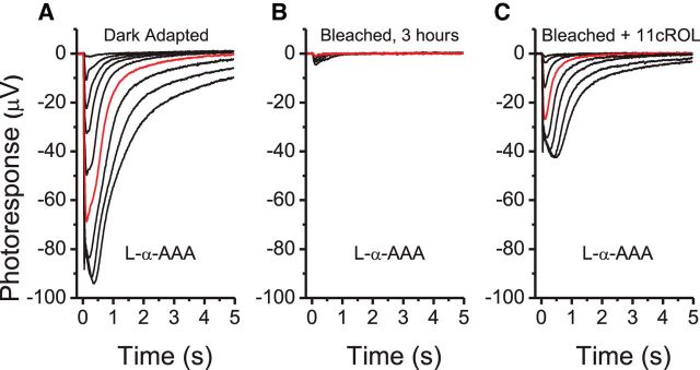
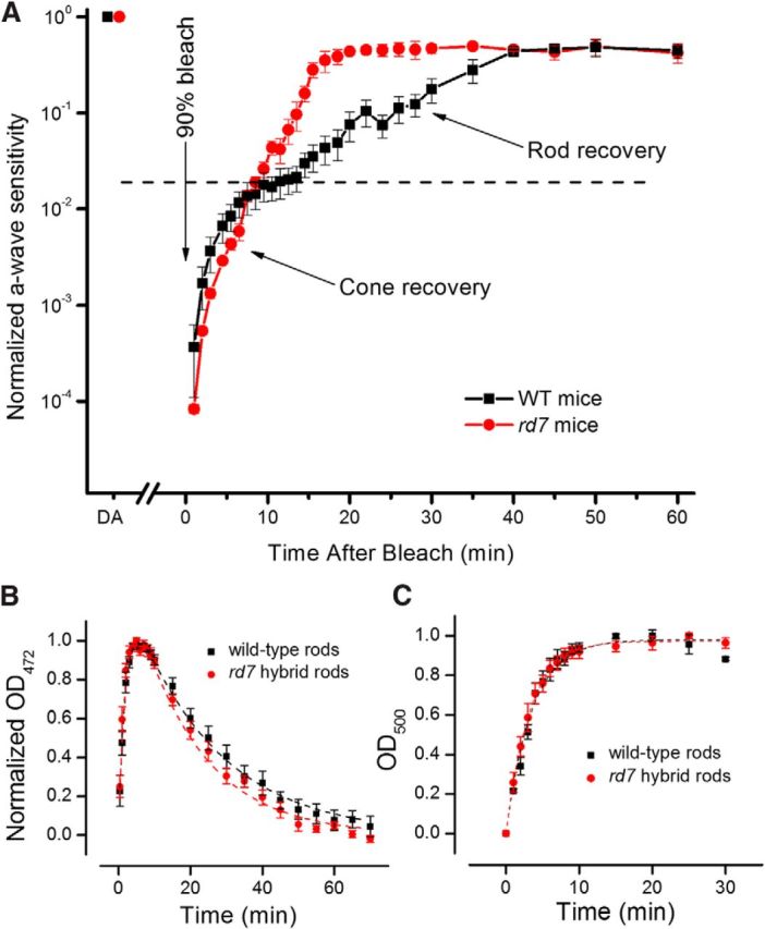
Efficient regeneration of visual pigment following its destruction by light is critical for the function of mammalian photoreceptors. Here, we show that misexpression of a subset of cone genes in the rd7 mouse hybrid rods enables them to access the normally cone-specific retina visual cycle. The rapid supply of chromophore by the retina visual cycle dramatically accelerated the mouse rod dark adaptation. At the same time, the competition between rods and cones for retina-derived chromophore slowed cone dark adaptation, indicating that the cone specificity of the retina visual cycle is key for rapid cone dark adaptation. Our findings demonstrate that mammalian photoreceptor dark adaptation is dominated by the supply of chromophore. Misexpression of cone genes in rods may represent a novel approach to treating visual disorders associated with mutations of visual cycle proteins or with reduced retinal pigment epithelium function due to aging.

Keywords: dark adaptation; photoreceptors; pigment regeneration; retina; retinol dehydrogenase; visual cycle.


Figures

Figure 1.
Figure 1.
Flash response families from single-cell suction electrode recordings of wild-type (A) and rd7 (B) rods. Red traces in both parts show responses to 14.8 photon μm−2 test flash. C, Intensity-response data from individual wild-type (n = 16) and rd7 (n = 8) rods fit with Equation 1. Half-saturating flash intensities, I0, are given in Table 1. D, Left, Comparison of the kinetics of normalized dim flash responses from wild-type (black) and rd7 (red) rods (n = 8; flash intensity = 14.8 photon μm−2). Right, Comparison of the kinetics of saturated flash responses from wild-type (black) and rd7 (red) rods (n = 8; flash intensity = 2170 photon μm−2). E, background adaptation in wild-type (n = 13) and rd7 (n = 6) rods. Data are fit with Equation 2. DA, dark adapted.
Figure 2.
Figure 2.
Suction electrode recordings from individual wild-type rods (A), cones (B), and rd7 rods (C). Top, left, Families of flash responses in dark-adapted state. Right, Families of flash responses from photoreceptors bleached at 90% in isolated retina removed from RPE and then incubated in darkness for 3 h. Red traces indicate flash intensities of 655 photon μm−2 for wild-type rods (A) and rd7 rods (C) and 2170 photon μm−2 for cones (B). Bottom, The corresponding intensity-response data for dark-adapted (black squares) and bleach-adapted (red circles) photoreceptors fit with Equation 1. For wild-type rods (A), Sf decreased from 2.07 ± 0.10 × 10⁻¹ pA photon−1 μm2 (n = 16) to 1.53 ± 0.26 × 10⁻³ pA photon−1 μm2 (n = 8), reflecting a 135-fold desensitization induced by the bleach. For cones (B), Sf decreased from 7.34 ± 0.25 × 10⁻⁴ pA photon−1 um2 (n = 8) to 1.75 ± 0.21 × 10⁻⁴ pA photon−1 um2 (n = 8), reflecting a 4.2-fold desensitization induced by the bleach. For rd7 rods (C), Sf decreased from 1.84 ± 0.18 × 10⁻¹ pA photon−1 um2 (n = 6) to 1.16 ± 0.14 × 10⁻² pA photon−1 um2 (n = 6), reflecting a 16-fold desensitization induced by the bleach. Data are shown as mean ± SEM.
Figure 3.
Figure 3.
Families of transretinal responses from wild-type rods (A), cones (B), and rd7 rods (C) in dark-adapted retinae (left) and retinae exposed to a 90% bleach followed by a 3 h recovery in darkness (right). Red traces represent responses to test flashes of 2170 photon μm−2 intensity. Flash strengths increased with a step of ∼ 0.5 log units (505 nm light). Photoreceptors faced the stimulating light.
Figure 4.
Figure 4.
Time course of recovery from a bleach in isolated retina for cones (black squares) and rd7 rods (red circles) determined from transretinal recordings. Sensitivity was normalized to its dark-adapted level before the bleach. The recovery of sensitivity was fit by a single exponential function with a time constant of 1.7 ± 0.3 min (black line; n = 10) for cones and 2.4 ± 0.2 min (red line; n = 8) for rd7 rods. For comparison, the steady-state level of desensitization in wild-type rods following an identical bleach is also shown (dashed line). Data are shown as mean ± SEM.
Figure 5.
Figure 5.
Effect of treatment with the Müller cell-specific gliotoxin l-α-AAA on rd7 rod response amplitude and sensitivity following a bleach. Transretinal recordings from rd7 rods in dark-adapted retina (A), following a 90% bleach and 3 h dark recovery (B), and after a subsequent treatment with 100 μm 11-cis-retinol (11cROL; C). Red traces in all parts correspond to a test flash of 2170 photon μm−2 intensity.
Figure 6.
Figure 6.
MSP measurements of rhodopsin levels in wild-type and rd7 rods. Absorption spectra of wild-type (A) and rd7 (B) rod outer segments measured in dark-adapted conditions (black), 1 min after ∼90% bleach (blue), and 3 h after the 90% bleach (red). Normalized absorption spectra (mean ± SEM) of rd7 rod outer segments measured in dark-adapted conditions (black squares), after ∼90% bleach and 3 h incubation (red circles) in l-α-AAA (n = 3; C), in l-α-AAA with 20 μm 11-cis-retinol (n = 3; D), and in l-α-AAA with 20 μm 11-cis-retinal (n = 3; E).
Figure 7.
Figure 7.
Time course of dark adaptation of wild-type (black squares; n = 6) and rd7 (red circles; n = 6) retinae measured with in vivo ERG recordings (A). The a-wave sensitivity was normalized to its dark-adapted level before the bleach. B, Production and decay of Met III as a function of time after an extensive bleach (>90%) in wild-type (black squares, n = 9) and rd7 (red circles, n = 8) mice rod photoreceptors. Met III was assessed as the change in spectral absorbance at 472 nm. Fitted to the data are double exponential functions with the time constants τ1 = 2.0, τ2 = 23.7 min (WT), and τ1 = 2.1, τ2 = 18.0 min (rd7). All retinae were incubated in l-α-AAA for 3 h prior the start of the experiment. C, Pigment regeneration as a function of time after an extensive bleach (>90%) and exogenous treatment with 11-cis-retinal (50 μm in 1% BSA) in WT (black squares; n = 9) and rd7 (red circles; n = 7) mice rod photoreceptors. The pigment regeneration was measured by MSP as the change in spectral absorbance at 500 nm. The two datasets were not significantly different (p = 0.18) and fitting a single exponential function to the averaged data produced time constants of τ = 3.5 min (wild-type) and τ = 3.2 min (rd7). All retinae were incubated in l-α-AAA for 3 h prior the start of the experiment. The exogenous treatments were made onstage after the decay of photoproducts was complete. Data are shown as mean ± SEM.

Similar articles

Cited by

References

    1. Akhmedov NB, Piriev NI, Chang B, Rapoport AL, Hawes NL, Nishina PM, Nusinowitz S, Heckenlively JR, Roderick TH, Kozak CA, Danciger M, Davisson MT, Farber DB. A deletion in a photoreceptor-specific nuclear receptor mRNA causes retinal degeneration in the rd7 mouse. Proc Natl Acad Sci U S A. 2000;97:5551–5556. doi: 10.1073/pnas.97.10.5551. - DOI - PMC - PubMed
    1. Calvert PD, Krasnoperova NV, Lyubarsky AL, Isayama T, Nicoló M, Kosaras B, Wong G, Gannon KS, Margolskee RF, Sidman RL, Pugh EN, Jr, Makino CL, Lem J. Phototransduction in transgenic mice after targeted deletion of the rod transducin alpha-subunit. Proc Natl Acad Sci U S A. 2000;97:13913–13918. doi: 10.1073/pnas.250478897. - DOI - PMC - PubMed
    1. Chen J, Rattner A, Nathans J. The rod photoreceptor-specific nuclear receptor Nr2e3 represses transcription of multiple cone-specific genes. J Neurosci. 2005;25:118–129. doi: 10.1523/JNEUROSCI.3571-04.2005. - DOI - PMC - PubMed
    1. Coleman PA, Miller RF. Do N-methyl-D-aspartate receptors mediate synaptic responses in the mudpuppy retina? J Neurosci. 1988;8:4728–4733. - PMC - PubMed
    1. Corbo JC, Cepko CL. A hybrid photoreceptor expressing both rod and cone genes in a mouse model of enhanced S-cone syndrome. PLoS Genet. 2005;1:e11. doi: 10.1371/journal.pgen.0010011. - DOI - PMC - PubMed

Publication types

MeSH terms