

Myxoid Cellular Neurothekeoma: A New Entity of S100-Negative, CD68-Positive Myxoid Neurothekeoma
- PMID: 25143683
- PMCID: PMC4135109
- DOI: 10.5021/ad.2014.26.4.510
Myxoid Cellular Neurothekeoma: A New Entity of S100-Negative, CD68-Positive Myxoid Neurothekeoma
Abstract
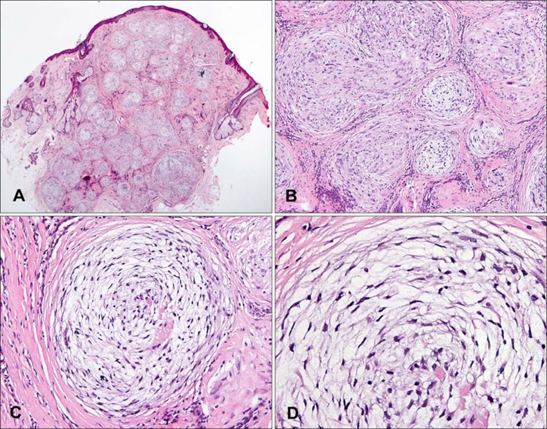
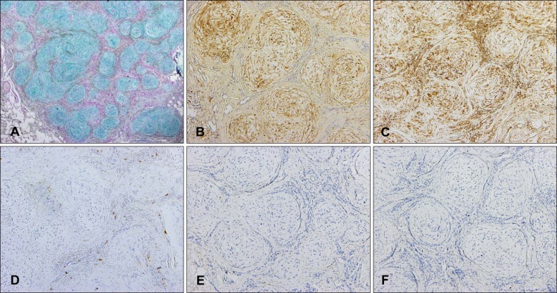
Neurothekeoma is a rare cutaneous neoplasm, occurring as a cutaneous papule or nodule on the face, shoulders, and upper extremities. Neurothekeoma has been subclassified as either the myxoid, cellular, or mixed type, depending on the amount of myxoid matrix and on immunohistochemical analysis. We observed a clinical case with conflicting histopathological and immunohistochemical findings. In this case, microscopic examination showed the typical presentation of myxoid neurothekeoma; however, immunohistochemical staining was negative for S100 protein and positive for CD68, which is the characteristic pattern of cellular neurothekeoma. We report a very rare form of myxoid cellular neurothekeoma of the face in a young woman.
Keywords: CD68; Neurothekeoma; S100 protein.
Figures

References
-
- Papadopoulos EJ, Cohen PR, Hebert AA. Neurothekeoma: report of a case in an infant and review of the literature. J Am Acad Dermatol. 2004;50:129–134. - PubMed
-
- Fetsch JF, Laskin WB, Hallman JR, Lupton GP, Miettinen M. Neurothekeoma: an analysis of 178 tumors with detailed immunohistochemical data and long-term patient follow-up information. Am J Surg Pathol. 2007;31:1103–1114. - PubMed
-
- Yang YW, Shih IH, Huang YH, Kuo TT, Hong HS. Mixed-type neurothekeoma presenting with an unusual clinical appearance of multiple satellite lesions on the back. Dermatol Surg. 2005;31:720–722. - PubMed
-
- Oh SH, Lee HJ, Chang SE, Lee MW, Choi JH, Moon KC, et al. A case of cellular neurothekeoma. Korean J Dermatol. 2006;44:1126–1129.
-
- Ryu DJ, Kim HJ, Jung JY, Kwon YS, Lee JH. A case of myxoid neurothekeoma on the hand. Korean J Dermatol. 2009;47:982–985.
Publication types
LinkOut - more resources
Full Text Sources
Other Literature Sources
