Review
. 2014 Aug 15;4(5):406-25.
eCollection 2014.

The role of molecular imaging in diagnosis of deep vein thrombosis

Affiliations
Review

The role of molecular imaging in diagnosis of deep vein thrombosis

Sina Houshmand et al. Am J Nucl Med Mol Imaging. .

Abstract

Venous thromboembolism (VTE) mostly presenting as deep venous thrombosis (DVT) and pulmonary embolism (PE) affects up to 600,000 individuals in United States each year. Clinical symptoms of VTE are nonspecific and sometimes misleading. Additionally, side effects of available treatment plans for DVT are significant. Therefore, medical imaging plays a crucial role in proper diagnosis and avoidance from over/under diagnosis, which exposes the patient to risk. In addition to conventional structural imaging modalities, such as ultrasonography and computed tomography, molecular imaging with different tracers have been studied for diagnosis of DVT. In this review we will discuss currently available and newly evolving targets and tracers for detection of DVT using molecular imaging methods.

Keywords: FDG-PET/CT; SPECT; deep vein thrombosis; molecular imaging; venous thromboembolism.


Figures

Figure 1
Figure 1
A schematic view depicting elements of the venous thrombus and binding sites for different radiotracers. 1. FDG taken up by metabolically active inflammatory cells and platelets. 2. Radiolabeled platelets indicating sites of aggregated platelets. 3. GP IIb/IIIa cyclic RGD peptides (Apcitide, DMP 444, Bitistatin) targeting GP IIb/IIIa receptors on activated platelets. 4. TP850 pentapeptide targeting fibrin α chain. 5. 59D8, T2G1, GC4, 64C5 targeting fibrin β chain. 6. Cyclic fibrin binding peptide EP-2104R. 7. DI-80B3 targeting D-domain of the fibrin. 8. Fibronectin-binding domain targeting lysine residue in fibrin. 9. Recombinant tissue plasminogen activatior (rt-PA) binding to C-terminal lysine residue of fibrin.
Figure 2
Figure 2
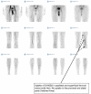
99mTc-DI-80B3 (ThromboView®) images of right leg proximal deep vein thrombosis. Reprinted from [24]. Copyright (2012), with permission from Elsevier.
Figure 3
Figure 3
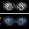
New deep vein thrombosis in right leg. FDG MIP image (A), axial PET (B), and axial PET/CT (C) show high metabolic activity (red arrows) in a dilated vein consistent with a fresh new clot.
Figure 4
Figure 4
Suspected recurrence of deep vein thrombosis in right leg. Axial CT (A) shows a dilated vein with no metabolic activity in combined axial PET/CT (B, red arrows) consistent with inactive old changes; no new clot.
Figure 5
Figure 5
The FDG-PET image above demonstrates relatively intense uptake of FDG in superior vena cava of an 8-year-old patient with neuroendocrine malignancy, which was interpreted to represent active thrombotic lesion in this location. Metastatic uptake in the cervical lymph nodes on the left side is also visible.
Figure 6
Figure 6
A: A 77-year-old female patient presented with Staphylococcus aureus bacteremia presumed to originate from a peripherally inserted central catheter. The catheter had been removed and the patient underwent FDG PET/CT to establish the extent of disease. Maximum intensity projection image showed unexpected FDG accumulation in the left brachial vein proximal to the removed catheter (green arrow) and in the right lung (red arrows). B, C: Transaxial image of FDG-avid left brachial vein (green arrow). Corresponding CT image revealed stranding in surrounding fat suggestive of inflammation. Subsequent ultrasound confirmed deep venous thrombosis (DVT) of the left brachial vein. D, E: FDG PET showed intense FDG uptake peripherally in the parenchyma of the right lung with barely visible findings on CT (red arrows). Adapted with permission from Lippincott Williams and Wilkins/Wolters Kluwer Health: Clinical Nuclear Medicine [150], copyright (2013).

References

    1. Silverstein MD, Heit JA, Mohr DN, Petterson TM, O’Fallon WM, Melton LJ 3rd. Trends in the incidence of deep vein thrombosis and pulmonary embolism: a 25-year population-based study. Arch Intern Med. 1998;158:585–593. - PubMed
    1. Spencer FA, Goldberg RJ, Lessard D, Reed G, Emery C, Gore JM, Pacifico L, Weitz JI. Factors associated with adverse outcomes in outpatients presenting with pulmonary embolism: the Worcester Venous Thromboembolism Study. Circ Cardiovasc Qual Outcomes. 2010;3:390–394. - PMC - PubMed
    1. White RH, Zhou H, Murin S, Harvey D. Effect of ethnicity and gender on the incidence of venous thromboembolism in a diverse population in California in 1996. Thromb Haemost. 2005;93:298–305. - PubMed
    1. Beckman MG, Hooper WC, Critchley SE, Ortel TL. Venous thromboembolism: a public health concern. Am J Prev Med. 2010;38:S495–501. - PubMed
    1. Siegel R, Naishadham D, Jemal A. Cancer statistics, 2013. CA Cancer J Clin. 2013;63:11–30. - PubMed

LinkOut - more resources