

The value of (68)Ga-DOTATATE PET/CT in diagnosis and management of neuroendocrine tumors compared to current FDA approved imaging modalities: a review of literature
- PMID: 25143861
- PMCID: PMC4138137
The value of (68)Ga-DOTATATE PET/CT in diagnosis and management of neuroendocrine tumors compared to current FDA approved imaging modalities: a review of literature
Abstract
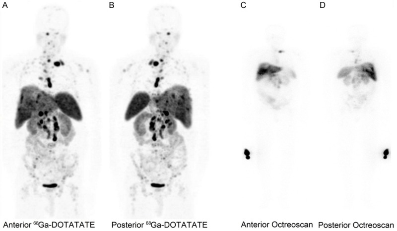
Neuroendocrine tumors (NETs) are rare group of neoplasms arising from nervous and endocrine systems. Somatostatin analogue imaging is a functional imaging modality of choice for evaluating the NETs. Recent availability of positron emitting radioisotope labeled somatostatin analogues to image neuroendocrine cancers, has raised the interests to use this new imaging modality in management of patients with NETs. (68)Ga-DOTATATE PET/CT has demonstrated superiority in lesion detection compared to Octreoscan, MIBG scintigraphy and MRI. In this article, we reviewed the published studies evaluating the role of (68)Ga-DOTATATE PET in diagnosis and management of patients with neuroendocrine tumors and comparing it to current FDA approved imaging modalities including Octreoscan, MIBG scintigraphy, (18)F FDG PET/CT, CT and MRI.
Keywords: DOTATATE; Gallium 68; hybrid imaging; neuroendocrine tumors; positron emission tomography.
Figures
References
-
- Yao JC, Hassan M, Phan A, Dagohoy C, Leary C, Mares JE, Abdalla EK, Fleming JB, Vauthey JN, Rashid A, Evans DB. One hundred years after carcinoid: epidemiology of and prognostic factors for neuroendocrine tumors in 35,825 cases in the United States. J Clin Oncol. 2008;26:3063–72. - PubMed
-
- Lamberts SW, Bakker WH, Reubi JC, Krenning EP. Somatostatin-receptor imaging in the localization of endocrine tumors. N Engl J Med. 1990;323:1246–9. - PubMed
-
- Krenning EP, Bakker WH, Breeman WA, Koper JW, Kooij PP, Ausema L, Lameris JS, Reubi JC, Lamberts SW. Localisation of endocrine-related tumours with radioiodinated analogue of somatostatin. Lancet. 1989;1:242–4. - PubMed
-
- Chopra A. 99mTc-ethylenediamine N,N’-diacetic acid/hydrazinonicotinamide-Tyr3-octreotide. In: Chopra A, editor. Molecular Imaging and Contrast Agent Database (MICAD) Bethesda: NIH; 2007.
-
- Al-Nahhas A, Win Z, Szyszko T, Singh A, Khan S, Rubello D. What can gallium-68 PET add to receptor and molecular imaging? Eur J Nucl Med Mol Imaging. 2007;34:1897–1901. - PubMed
Publication types
LinkOut - more resources
Full Text Sources