Cerebellar integrity in the amyotrophic lateral sclerosis-frontotemporal dementia continuum
- PMID: 25144223
- PMCID: PMC4140802
- DOI: 10.1371/journal.pone.0105632
Cerebellar integrity in the amyotrophic lateral sclerosis-frontotemporal dementia continuum
Abstract
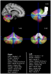
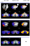
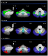
Amyotrophic lateral sclerosis (ALS) and behavioural variant frontotemporal dementia (bvFTD) are multisystem neurodegenerative disorders that manifest overlapping cognitive, neuropsychiatric and motor features. The cerebellum has long been known to be crucial for intact motor function although emerging evidence over the past decade has attributed cognitive and neuropsychiatric processes to this structure. The current study set out i) to establish the integrity of cerebellar subregions in the amyotrophic lateral sclerosis-behavioural variant frontotemporal dementia spectrum (ALS-bvFTD) and ii) determine whether specific cerebellar atrophy regions are associated with cognitive, neuropsychiatric and motor symptoms in the patients. Seventy-eight patients diagnosed with ALS, ALS-bvFTD, behavioural variant frontotemporal dementia (bvFTD), most without C9ORF72 gene abnormalities, and healthy controls were investigated. Participants underwent cognitive, neuropsychiatric and functional evaluation as well as structural imaging using voxel-based morphometry (VBM) to examine the grey matter subregions of the cerebellar lobules, vermis and crus. VBM analyses revealed: i) significant grey matter atrophy in the cerebellum across the whole ALS-bvFTD continuum; ii) atrophy predominantly of the superior cerebellum and crus in bvFTD patients, atrophy of the inferior cerebellum and vermis in ALS patients, while ALS-bvFTD patients had both patterns of atrophy. Post-hoc covariance analyses revealed that cognitive and neuropsychiatric symptoms were particularly associated with atrophy of the crus and superior lobule, while motor symptoms were more associated with atrophy of the inferior lobules. Taken together, these findings indicate an important role of the cerebellum in the ALS-bvFTD disease spectrum, with all three clinical phenotypes demonstrating specific patterns of subregional atrophy that associated with different symptomology.
Conflict of interest statement
Figures


References
-
- Hardiman O, van den Berg LH, Kiernan MC (2011) Clinical diagnosis and management of amyotrophic lateral sclerosis. Nature Reviews Neurology 7: 639–649. - PubMed
-
- Kiernan MC, Vucic S, Cheah BC, Turner MR, Eisen A, et al. (2011) Amyotrophic lateral sclerosis. Lancet 377: 942–955. - PubMed
-
- Bak TH, Hodges JR (2001) Motor neurone disease, dementia and aphasia: coincidence, co-occurrence or continuum? J Neurol 248: 260–270. - PubMed
-
- Lillo P, Garcin B, Hornberger M, Bak TH, Hodges JR (2010) Neurobehavioral features in frontotemporal dementia with amyotrophic lateral sclerosis. Arch Neurol 67: 826–830. - PubMed
Publication types
MeSH terms
Substances
LinkOut - more resources
Full Text Sources
Other Literature Sources
Medical
Molecular Biology Databases
Miscellaneous

