. 2014 Aug 21;10(8):e1004321.
doi: 10.1371/journal.ppat.1004321. eCollection 2014 Aug.

Identification of anti-virulence compounds that disrupt quorum-sensing regulated acute and persistent pathogenicity

Affiliations

Identification of anti-virulence compounds that disrupt quorum-sensing regulated acute and persistent pathogenicity

Melissa Starkey et al. PLoS Pathog. .

Abstract

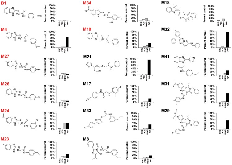
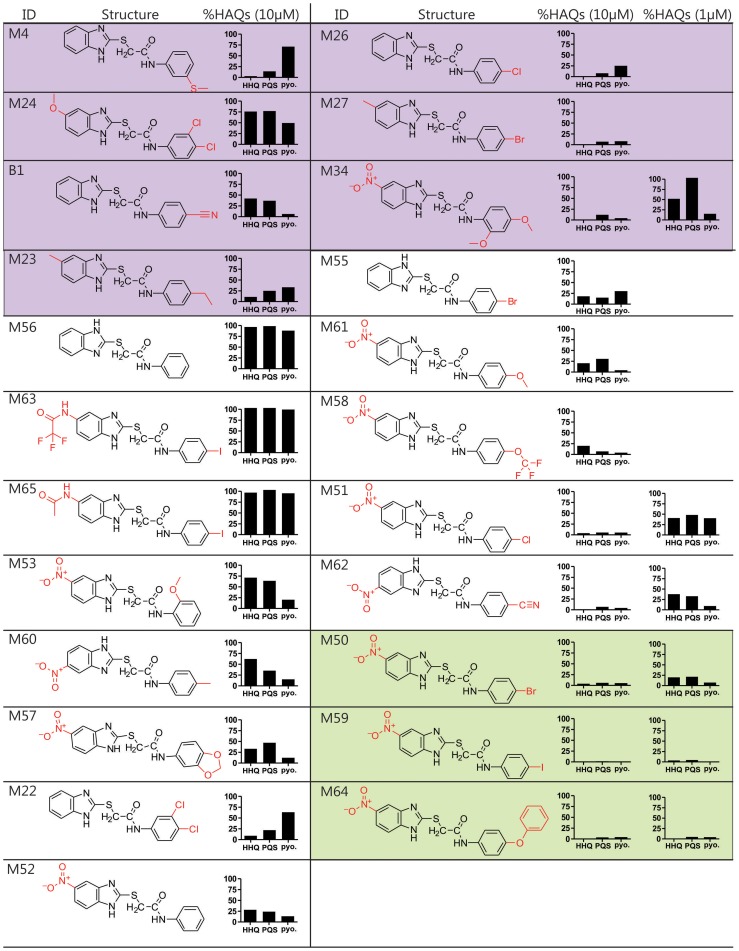
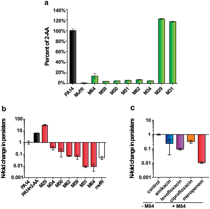
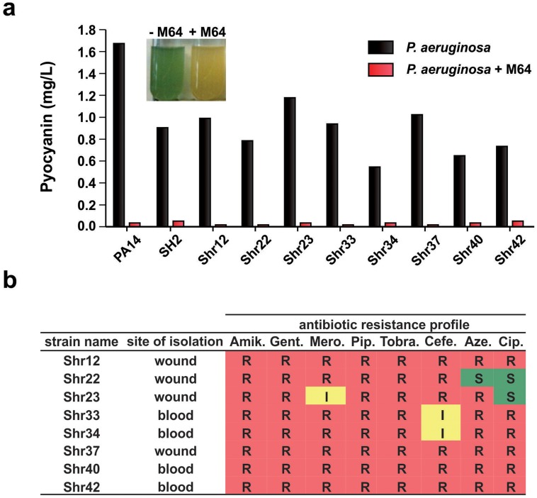
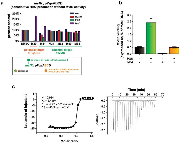
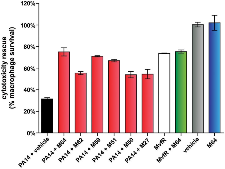
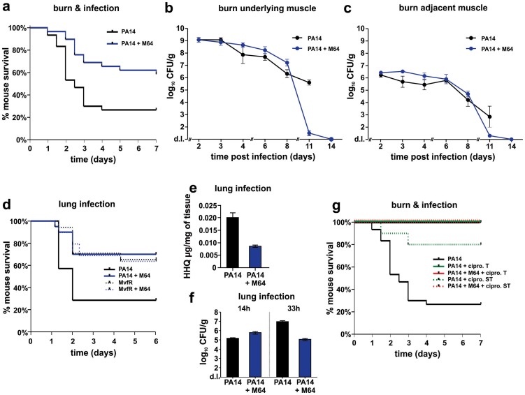
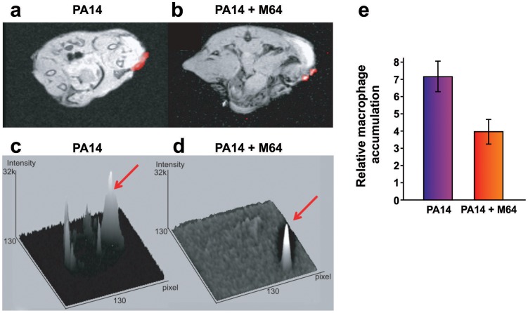
Etiological agents of acute, persistent, or relapsing clinical infections are often refractory to antibiotics due to multidrug resistance and/or antibiotic tolerance. Pseudomonas aeruginosa is an opportunistic Gram-negative bacterial pathogen that causes recalcitrant and severe acute chronic and persistent human infections. Here, we target the MvfR-regulated P. aeruginosa quorum sensing (QS) virulence pathway to isolate robust molecules that specifically inhibit infection without affecting bacterial growth or viability to mitigate selective resistance. Using a whole-cell high-throughput screen (HTS) and structure-activity relationship (SAR) analysis, we identify compounds that block the synthesis of both pro-persistence and pro-acute MvfR-dependent signaling molecules. These compounds, which share a benzamide-benzimidazole backbone and are unrelated to previous MvfR-regulon inhibitors, bind the global virulence QS transcriptional regulator, MvfR (PqsR); inhibit the MvfR regulon in multi-drug resistant isolates; are active against P. aeruginosa acute and persistent murine infections; and do not perturb bacterial growth. In addition, they are the first compounds identified to reduce the formation of antibiotic-tolerant persister cells. As such, these molecules provide for the development of next-generation clinical therapeutics to more effectively treat refractory and deleterious bacterial-human infections.


Conflict of interest statement

LR is the scientific founder, consultant and scientific advisory board member of Spero Therapeutics LLC. No funding from Spero Therapeutics was received. This does not alter our adherence to all PLOS policies on sharing data and materials.

Figures

Figure 1
Figure 1. Chemical structures of 17 MvfR-regulon inhibitors identified by whole cell HTS, and their corresponding inhibition of HAQ and pyocyanin production.
HHQ (dark grey bars), PQS (white bars), HQNO (light grey bars), and pyocyanin (black bars) levels were quantified plus or minus 50 µg/mL of each compound. Structures labelled in red share the common benzamide-benzimidazole core.
Figure 2
Figure 2. Structure and biological activity of benzamide-benzimidazole derivatives for inhibition of HAQ and pyocyanin production.
The HTS compounds are shaded purple, the 2nd generation commercially available derivatives are shaded white, and the 2nd generation synthetic derivatives are shaded green. Alterations to the M56 benzamide-benzamidazole core structure are marked in red. HHQ, PQS, and pyocyanin (pyo.) levels were quantified in response to 10 µM compounds, and 1 µM of the most potent compounds: M34, M51, M62, M50, M59, and M64.
Figure 3
Figure 3. The most potent inhibitors reduce 2-AA production and the formation of antibiotic tolerant persisters.
a. 2-AA levels in presence of 10 µM inhibitor. Error bars show mean +/− SD of at least 2 replicates. b. Observed fold change in persister cell concentrations of PA14 cultures with 10 µM inhibitor or with 0.75 mM 2-AA. Untreated PA14 cells and mvfR- cells were the positive and negative controls, respectively. Error bars show mean +/− SEM of at least 3 replicates. Differences between PA14 and the samples M34, M50, M62, M59, M51, M64 or mvfR- (p<0.01) as well as between PA14 and the samples PA14 + 2-AA or M29 (p<0.01) are statistically significant (one way ANOVA, Dunnett's test). c. Observed fold change in persister cell concentrations of PA14 plus 5 µM M64 in the presence of clinical antibiotics used to treat P. aeruginosa infections: amikacin (blue), levofloxacin (purple), ciprofloxacin (orange) and meropenem (red). All values were normalized to control cultures in 0.01% DMSO. Error bars show mean +/− SEM of at least 3 replicates. Differences between control and the samples amikacin, levofloxacin, ciprofloxacin or meropenem are statistically significant (p<0.01, one way ANOVA, Dunnett's test).
Figure 4
Figure 4. M64 reduces pyocyanin production in P. aeruginosa clinical multi-drug resistant strains.
a. Quantitative pyocyanin production in multi-drug resistant clinical P. aeruginosa isolates plus (red) and minus (black) 5 µM M64. A representative image of qualitative pyocyanin production, visible as green media, in PA14 culture +/− M64, is shown above the histogram. b. Antibiotic resistance profile of P. aeruginosa clinical strains and their respective isolation sites from infected patients. Amik. = amikacin, Gent. = gentamycin, Mero. = meropenem, Pip. = piperacin, Tobra. = tobramycin, Cefe. = cefepime, Aze. = azetromycin, Cip. = ciprofloxacin. R = resistant; I = intermediate; S = sensitive.
Figure 5
Figure 5. Identification of the molecular target and mode of action of the MvfR-regulon inhibitors.
a. The potential molecular target of an inhibitor is revealed by the pattern of HAQ and DHQ production in response to 100 µM of the inhibitor in mvfR mutant cells that constitutively express pqsABCD. Compounds that target MvfR should not impact HAQs production (e.g., M51, M34, M62, M50, M64); compounds that inhibit the PqsB or PqsC enzymes should result in increased DHQ, which is produced by PqsA/D, and reduced levels of HHQ, PQS, and HQNO, which require the PqsABCD enzymes (e.g., M29). b. 0.24 µM M64 blocks MvfR binding to the pqsA promoter of PA14, minus and plus 38 µM PQS. Binding was assessed by ChIP-qPCR and normalized to the rpoD promoter that lacks an MvfR binding site. MvfR bound DNA was expressed as the percent of total input DNA. Error bars represent mean +/− SEM of at least 3 replicates. c. Isothermal titration calorimetry analysis of the interaction between MvfRc91 (19 µM) and M64 (200 µM). Heat signals of the M64 titration into MvfRc91 are plotted against the molecular ratio between M64 and MvfRc91 (left panel), and against time (right panel). The best-fit curve corresponds to a single-site binding model. The stoichiometry of binding (N), association constant (KA), enthalpy (ΔH), and entropy of binding (ΔS) are presented.
Figure 6
Figure 6. MvfR-regulon inhibitors rescue PA14-macrophage cytotoxicity.
PA14-induced killing of Raw264.7 macrophage cells was determined minus and plus 100 µM inhibitor. Error bars represent mean +/− SEM of at least 3 replicates. Differences between PA14 + vehicle and the samples PA14 + M64, PA14 + M62, PA14 + M59, PA14 + M51, PA14 + M50, or PA14 + M27 are statistically significant (p<0.01, one way ANOVA, Dunnett's test). Differences between MvfR and MvfR + M64 (p>0.05) or vehicle and M64 (p>0.05) are not statistically significant (unpaired t test). Notably, M64 does not alter cytotoxicity of mvfR cells, and is itself non-cytotoxic.
Figure 7
Figure 7. M64 reduces PA14 virulence in mouse burn infection, and lung infection, models.
a. Survival curves of mice from the burn and infection model following PA14 infection, minus (black, n = 30), and plus (blue, n = 36), M64 (4 mg/kg). M64 was administered by intravenous injection 6 h post-burn and infection, and then twice a day for 6 days post-infection. Differences between PA14 and PA14 + M64 are statistically significant (p<0.001, Kaplan-Meier method). b., c. M64 does not significantly reduce PA14 bacterial load within the first 7 days post-infection, and alternatively promotes bacterial clearance over several days in the burn and infection model. PA14 CFU quantification in muscle underlying (b) or adjacent to (c) the abdominal infection site in mice infected with PA14, minus (black) and plus (blue) M64. Error bars show mean +/− SEM of at least 3 replicates. Animals that survived infection post-day 7 were used for CFU quantification at days 8 and 11. Differences between PA14 and PA14 + M64 are statistically significant at day 11 (p<0.001, unpaired t test) but not before day 11 (p>0.05, unpaired t test). d. Survival curves of mice from the lung infection model following PA14 infection, minus (solid black line, n≥10), and plus (solid blue line, n≥10) M64 (4 mg/kg); and mvfR infection, minus (interrupted black line, n≥10), and plus (interrupted blue line, n≥10) M64 (12 mg/kg). M64 was administered by intravenous injection at 2, 4, 8, and 12 h post infection, and then twice a day up to day 4. Differences between PA14 and PA14 + M64 (p<0.05) or between mvfR and mvfR + M64 (p<0.05) are statistically significant, while differences between mvfR and mvfR + M64 (p>0.05) or between PA14 + M64 and mvfR + M64 (p>0.05) are not statistically significant (Kaplan-Meier method). Animals were inoculated intranasally with 20 µL of 5×10⁶ PA14 cells and 20 µl of 8×10⁶ isogenic mvfR mutant cells, n≥10 mice. e. HHQ levels at 14 h post-infection from lung tissues in untreated mice, and from mice treated with M64. n = 7 for each experimental condition. Difference between PA14 and PA14 + M64 is statistically significant (p<0.001, unpaired t test). f. PA14 pulmonary bacterial load in mice infected with PA14 (black) and treated with M64 (blue) quantified at 14 or 33 h post infection. Error bars represent mean +/− SEM of at least 3 replicates. Difference between PA14 and PA14 + M64 at 33 h are statistically significant (p<0.05, unpaired t test) whereas difference at 14 h was not (p>0.05, unpaired t test). d.l., detection limit. NS, not significant. g. Survival curve of PA14-infected mice from the burn and infection models, untreated (black, n = 30), treated with ciprofloxacin (green, p<0.001), or treated with a combination of ciprofloxacin and M64 (red, p<0.001), using a 10 mg/kg therapeutic dose (T, straight line, n = 18–24, p<0.001) or a 4 mg/kg sub-therapeutic dose (ST, dashed line, n = 10, p<0.001) of ciprofloxacin. Ciprofloxacin was administered by intravenous (IV) injection twice a day for 4 days post-infection, and M64 was administered by IV injection 6 hours post-infection and then twice a day for 6 days post-infection. In all conditions the M64 dose was 4 mg/kg. Significance of survival rate differences compared to PA14 infected mice was determined using the Kaplan-Meier method.
Figure 8
Figure 8. Magnetic resonance imaging of M64 inhibition of macrophage recruitment at a burn and infection site.
a–e. In vivo positive contrast imaging of mice infected with PA14, plus and minus M64. The off resonance imaging transverse relaxation in the rotating frame (ORI-T2ρ) images were transformed to signal to noise ratio (SNR) images and thresholded in units of image standard deviation. a., b. The positive-contrast images are presented in pseudocolor, thresholded to signal greater than three in dimensionless SNR units, and superimposed on a FLASH image. For image processing, regions of interest (ROI) were drawn around the burn region and the total thresholded signal intensity was integrated within each ROI. Similar slices were chosen at the same anatomical location in all mice. c., d. 3-dimensional graphs of pixel intensities show an intense peak in the burn area for the PA14 control mouse, with this peak reduced by M64. e. Signal was measured in units of SNR, thresholded at three standard deviations, and measured within ROIs at the level of the burn and infection. The noise threshold was estimated by fitting the image background to a Rician distribution. Error bars depict standard error of the mean image intensity in the ROI. Error bars depict mean +/− SD of at least 3 replicates. Difference between PA14 and PA14 + M64 is statistically significant (p<0.05, unpaired t test).
Figure 9
Figure 9. M64 inhibits P. aeruginosa persistence in the mouse burn and infection models.
PA14 CFU quantification in muscle (a) underlying or (b) adjacent to the abdominal infection site in mice infected with PA14 and treated with ciprofloxacin (10 mg/kg), and minus (green) or plus (red) M64 (4 mg/kg). Ciprofloxacin and M64 were administered by intravenous injection 6 hours post-infection and then twice a day. Ciprofloxacin was administered for 4 days as described until no CFUs were detected in the muscle samples. Ciprofloxacin administration was stopped at day 4 to allow for the potential emergence and detection of antibiotic-tolerant cells. M64 was administered for 6 days, up until antibiotic-tolerant cells were detected in the PA14 + ciprofloxacin only group. Error bars represent mean +/− SEM of at least 3 replicates. d.l., detection limit.

References

    1. Boucher HW, Talbot GH, Bradley JS, Edwards JE, Gilbert D, et al. (2009) Bad bugs, no drugs: no ESKAPE! An update from the Infectious Diseases Society of America. Clin Infect Dis 48: 1–12. - PubMed
    1. Livermore DM (2012) Fourteen years in resistance. Int J Antimicrob Agents 39: 283–294. - PubMed
    1. Spellberg B, Guidos R, Gilbert D, Bradley J, Boucher HW, et al. (2008) The epidemic of antibiotic-resistant infections: a call to action for the medical community from the Infectious Diseases Society of America. Clin Infect Dis 46: 155–164. - PubMed
    1. Parker CT, Sperandio V (2009) Cell-to-cell signalling during pathogenesis. Cell Microbiol 11: 363–369. - PMC - PubMed
    1. Ng WL, Bassler BL (2009) Bacterial quorum-sensing network architectures. Annu Rev Genet 43: 197–222. - PMC - PubMed

Publication types

MeSH terms

Substances