. 2014 Aug 21;5(8):e1386.
doi: 10.1038/cddis.2014.359.

Effects of acute versus post-acute systemic delivery of neural progenitor cells on neurological recovery and brain remodeling after focal cerebral ischemia in mice

Affiliations

Effects of acute versus post-acute systemic delivery of neural progenitor cells on neurological recovery and brain remodeling after focal cerebral ischemia in mice

T R Doeppner et al. Cell Death Dis. .

Abstract

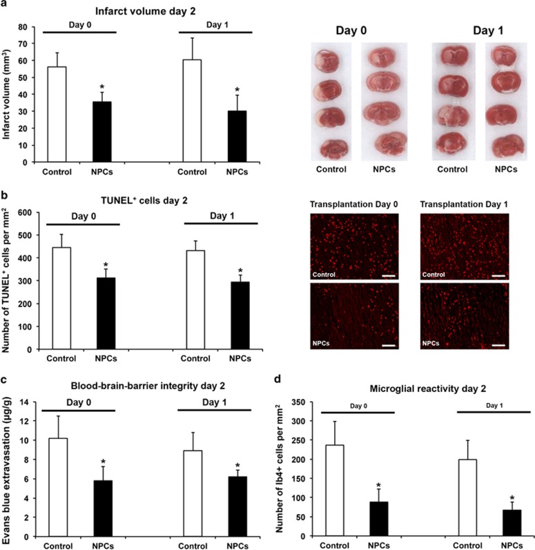
Intravenous transplantation of neural progenitor cells (NPCs) induces functional recovery after stroke, albeit grafted cells are not integrated into residing neural networks. However, a systematic analysis of intravenous NPC delivery at acute and post-acute time points and their long-term consequences does not exist. Male C57BL6 mice were exposed to cerebral ischemia, and NPCs were intravenously grafted on day 0, on day 1 or on day 28. Animals were allowed to survive for up to 84 days. Mice and tissues were used for immunohistochemical analysis, flow cytometry, ELISA and behavioral tests. Density of grafted NPCs within the ischemic hemisphere was increased when cells were transplanted on day 28 as compared with transplantation on days 0 or 1. Likewise, transplantation on day 28 yielded enhanced neuronal differentiation rates of grafted cells. Post-ischemic brain injury, however, was only reduced when NPCs were grafted at acute time points. On the contrary, reduced post-ischemic functional deficits due to NPC delivery were independent of transplantation paradigms. NPC-induced neuroprotection after acute cell delivery was due to stabilization of the blood-brain barrier (BBB), reduction in microglial activation and modulation of both peripheral and central immune responses. On the other hand, post-acute NPC transplantation stimulated post-ischemic regeneration via enhanced angioneurogenesis and increased axonal plasticity. Acute NPC delivery yields long-term neuroprotection via enhanced BBB integrity and modulation of post-ischemic immune responses, whereas post-acute NPC delivery increases post-ischemic angioneurogenesis and axonal plasticity. Post-ischemic functional recovery, however, is independent of NPC delivery timing, which offers a broad therapeutic time window for stroke treatment.


Figures

Figure 1
Figure 1
Acute transplantation of neural progenitor cells (NPCs) is neuroprotective. NPCs or PBS (control) was intravenously administered during reperfusion after induction of stroke. (a) Different numbers of NPCs were transplanted intravenously (solved in 200 μl of PBS) and infarct volumes were assessed on day 2 post stroke using cresyl violet staining. (b) Representative photographs for each condition shown in (a). (c) Analysis of grafted GFP+ NPCs within the ischemic hemisphere 2 days after stroke. (d) Representative photographs of GFP+ NPCs within the ischemic lesion site as exemplarily taken from mice that received 107 NPCs. All data are given as mean±S.D. *Significantly different from the corresponding control, P<0.05. Scale bars: 50 μm
Figure 2
Figure 2
Post-acute transplantation of neural progenitor cells (NPCs) yields high neuronal differentiation rates. NPCs were intravenously transplanted at the time points given (i.e., day 0, day 1, and day 28 post stroke) and animals were killed on day 56 or on day 84 followed by immunohistochemical analyses. (a) Assessment of GFP+ NPCs within the ischemic hemisphere depending on cell delivery time points. (b) Differentiation analysis of GFP+ NPCs from (a) regarding co-expression of Nestin, GFAP, CNPase, Dcx and NeuN. All data are given as mean±S.D. *Significantly different from controls, P<0.05
Figure 3
Figure 3
Sustained reduction of post-ischemic brain injury requires acute cell transplantation. Neural progenitor cells (NPCs) were grafted at the time points given, whereas control animals received PBS only. Post-stroke brain injury was assessed via analysis of neuronal density both on day 56 (a) and on day 84 (b). All data are given as mean±S.D. *Significantly different from controls, P<0.05
Figure 4
Figure 4
Improved post-ischemic functional recovery is independent of cell delivery timing. Assessment of post-stroke functional recovery was analyzed on days 35, 42, 56 and 84 using the rota rod (a), the tight rope (b), the corner turn (c) and the water maze test (d). Neural progenitor cells (NPCs) were intravenously transplanted at the time points given, whereas controls received PBS only. Maximal testing time was 300 s for the rota rod test (a). The tight rope test (b) was analyzed using a validated score from 0 (min) to 20 (max). For the corner turn test (c), the laterality index was calculated using the following formula: (number of left turns−number of right turns)/10. Water maze test performance (d) was analyzed at the time points given. Mice were tested for maximal 90 s per run. Data are given as means out of eight runs. If animals did not reach the platform, then they were scored 90 s. Behavioral data from all tests are given as mean±S.D. *Significantly different from controls, P<0.05
Figure 5
Figure 5
Acute transplantation of neural progenitor cells (NPCs) induces neuroprotection via stabilization of blood–brain barrier (BBB) and reduction in post-ischemic inflammation. Mice received intravenous injections of either PBS (control) or NPCs on day 0 or on day 1 post stroke. Brain injury was assessed using TTC staining (a) and TUNEL staining (b) on day 2 after induction of stroke. Stability of the BBB (c) was analyzed measuring extravasation of Evans blue photometrically, which was injected 2 hours before killing of animals. (d) Microglial activation on day 2 post stroke was immunohistochemically analyzed within defined regions of interest as described in the Materials and Methods. All data are given as mean±S.D. *Significantly different from controls, P<0.05. Scale bars: 50 μm
Figure 6
Figure 6
Acute delivery of neural progenitor cells (NPCs) modulates peripheral and central immune response. Mice were treated with PBS (control) or NPCs on day 0 or on day 1, respectively. Absolute amounts of CD45+ leukocytes were measured within the blood (a) and the ischemic hemisphere of the brain (b) on day 2 after induction of stroke. All data are given as mean±S.D. *Significantly different from controls, P<0.05
Figure 7
Figure 7
Post-acute transplantation of neural progenitor cells (NPCs) induces increased post-ischemic angioneurogenesis and stimulation of neuroplasticity. All mice received intravenous transplantation of NPCs at the time points given followed by killing of animals on day 84 post stroke. Control animals received PBS only. (a) Analysis of endogenous new-born GFP cells was performed within the ischemic hemisphere using bromodeoxyuridine (BrdU) labeling. Photographs depicted show representative arrangement of BrdU+ cells within the lesion site taken from mice that were treated with PBS or NPCs on day 28 post stroke. (b) Neuronal differentiation analysis of endogenous GFP BrdU+ cells regarding co-expression of doublecortin (blue columns) and NeuN (red columns) within ischemic hemispheres. Representative photographs of co-localization between BrdU and Dcx/NeuN were taken from mice treated with NPCs on day 28. (c) Assessment of vascular density was performed within ischemic hemispheres after treatment of mice as described above using CD31 staining. Photographs depicted show representative stainings of mice that were treated with either PBS or NPCs on day 28 post stroke. (d) Axonal density as one means indicating post-ischemic neuroplasticity was measured via stereotactic injection of the anterograde tract tracer biotinylated dextran amine (BDA) into the contralateral cortex 70 days after stroke. Axon labeling was performed within the ipsilateral hemisphere using 3,3'-diaminobenzidine (DAB) staining on day 84 post stroke. Axonal densities within the cortex of the ischemic hemisphere were measured from six fields per section out of eight sections of each mouse divided by total mean densities of all mice. Results are given as percentage of proportional areas. Representative photographs were taken from mice that were treated with either PBS or NPCs. All data are given as mean±S.D. *Significantly different from controls, P<0.05. Scale bars: 50 μm (a, c and d) and 20 μM (b)

References

    1. Leong WK, Henshall TL, Arthur A, Kremer KL, Lewis MD, Helps SC, et al. Human adult dental pulp stem cells enhance poststroke functional recovery through non-neural replacement mechanisms. Stem Cells Transl Med. 2012;1:177–187. - PMC - PubMed
    1. Bacigaluppi M, Pluchino S, Martino G, Kilic E, Hermann DM. Neural stem/precursor cells for the treatment of ischemic stroke. J Neurol Sci. 2008;265:73–77. - PubMed
    1. Bliss TM, Andres RH, Steinberg GK. Optimizing the success of cell transplantation therapy for stroke. Neurobiol Dis. 2010;37:275–283. - PMC - PubMed
    1. Doeppner TR, Ewert TA, Tonges L, Herz J, Zechariah A, Elali A, et al. Transduction of neural precursor cells with TAT-heat shock protein 70 chaperone: therapeutic potential against ischemic stroke after intrastriatal and systemic transplantation. Stem cells. 2012;30:1297–1310. - PubMed
    1. Zheng W, Honmou O, Miyata K, Harada K, Suzuki J, Liu H, et al. Therapeutic benefits of human mesenchymal stem cells derived from bone marrow after global cerebral ischemia. Brain Res. 2010;1310:8–16. - PubMed