Review
. 2014 Sep 21;59(18):R233-302.
doi: 10.1088/0031-9155/59/18/R233. Epub 2014 Aug 21.

An exponential growth of computational phantom research in radiation protection, imaging, and radiotherapy: a review of the fifty-year history

Affiliations
Review

An exponential growth of computational phantom research in radiation protection, imaging, and radiotherapy: a review of the fifty-year history

X George Xu. Phys Med Biol. .

Abstract

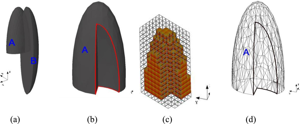
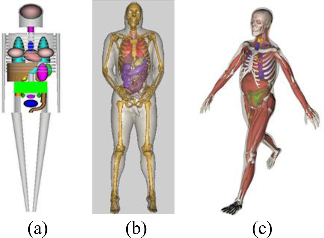
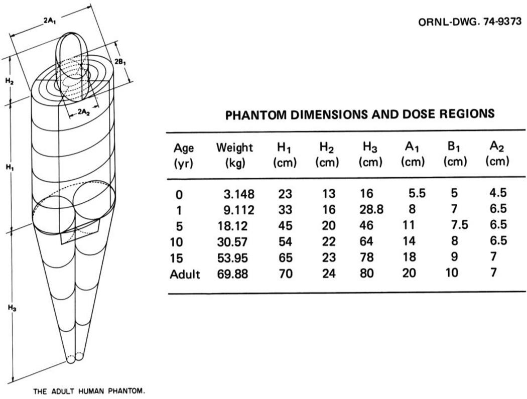
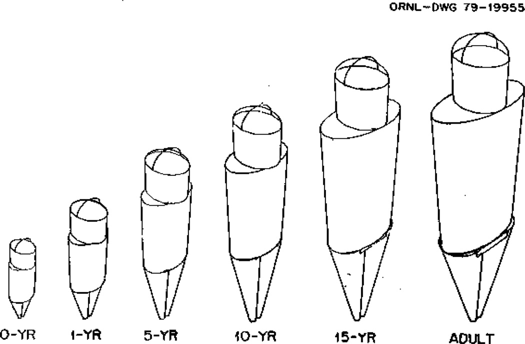
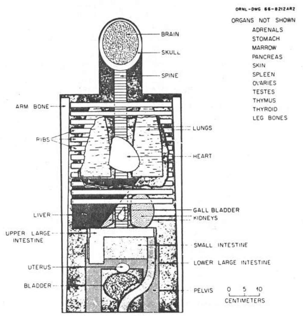
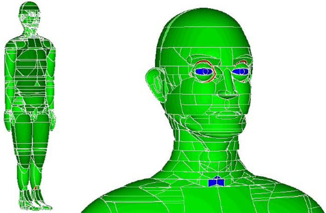
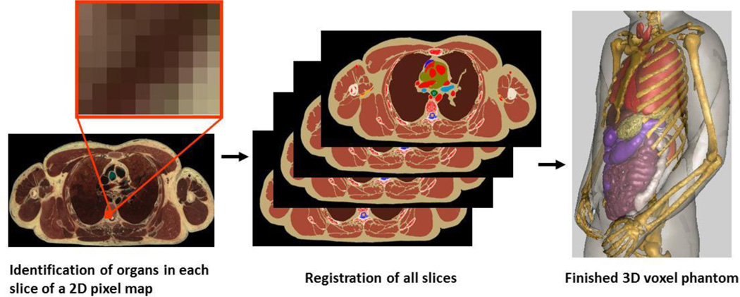
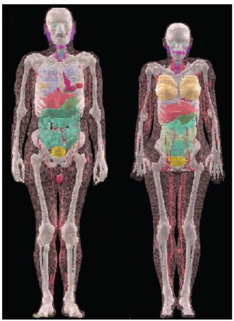
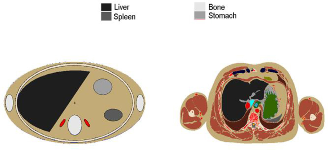
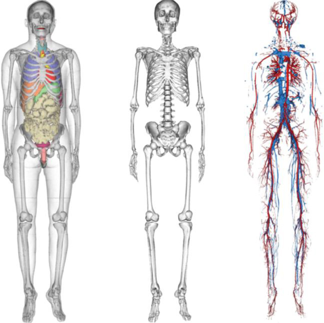
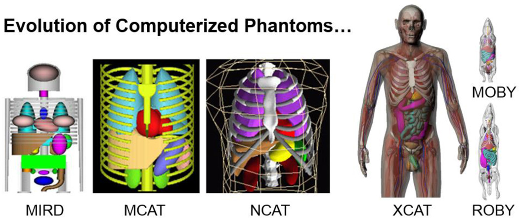
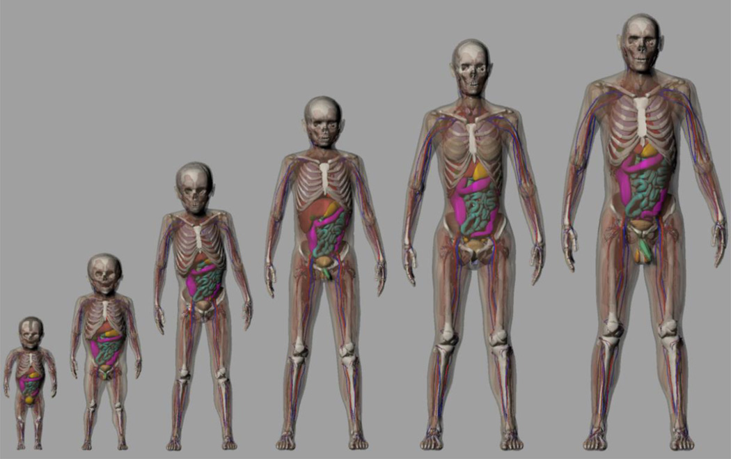
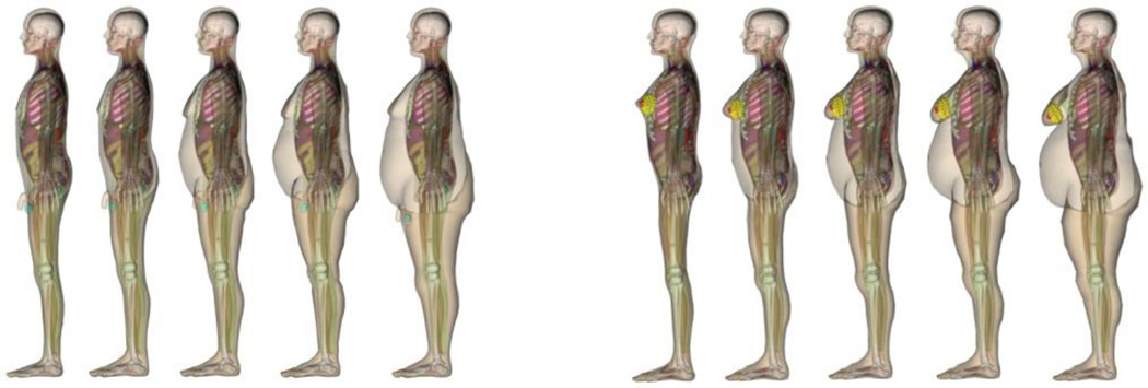
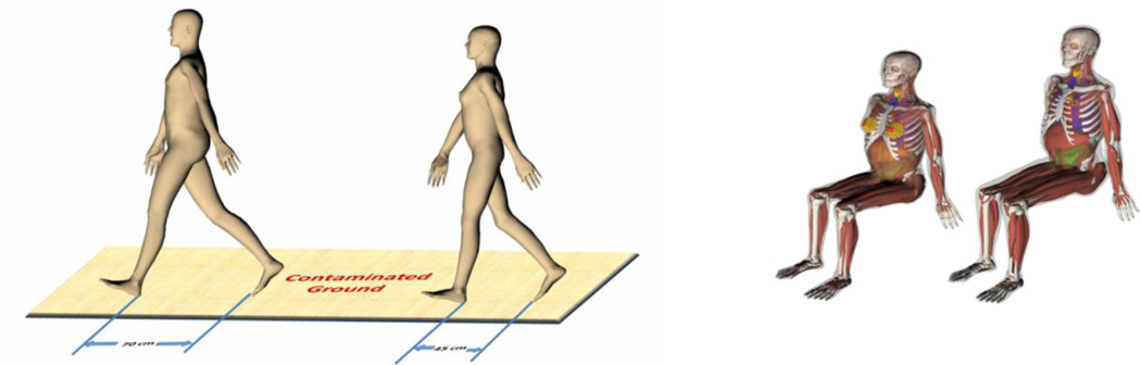
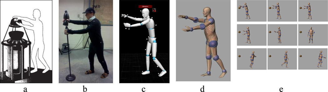
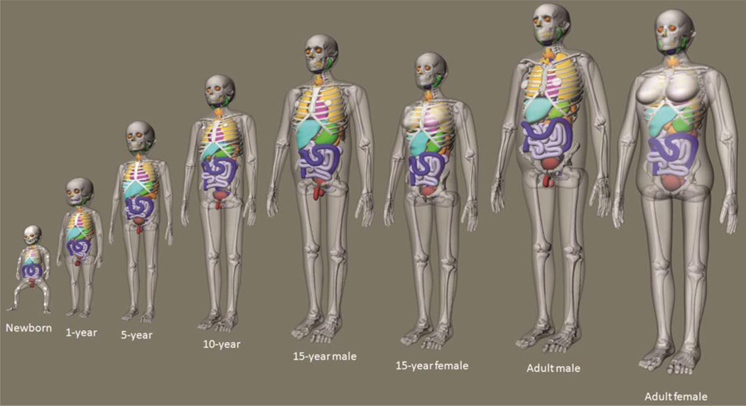
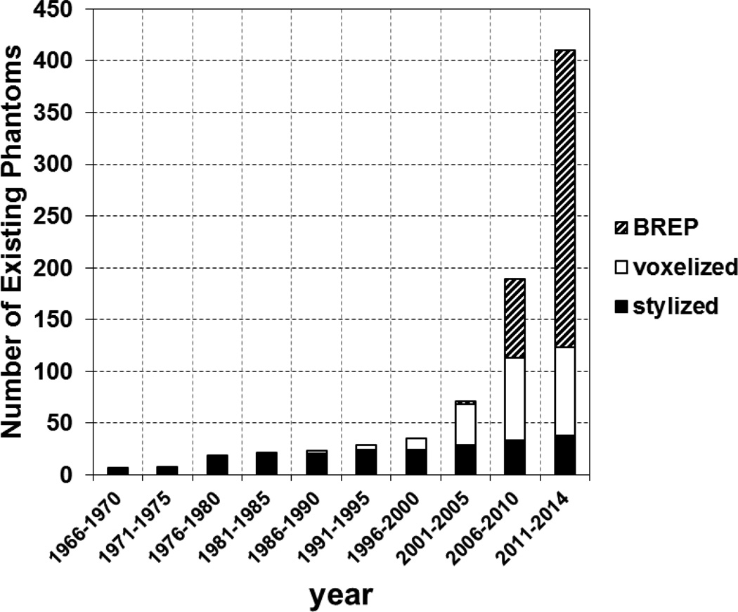
Radiation dose calculation using models of the human anatomy has been a subject of great interest to radiation protection, medical imaging, and radiotherapy. However, early pioneers of this field did not foresee the exponential growth of research activity as observed today. This review article walks the reader through the history of the research and development in this field of study which started some 50 years ago. This review identifies a clear progression of computational phantom complexity which can be denoted by three distinct generations. The first generation of stylized phantoms, representing a grouping of less than dozen models, was initially developed in the 1960s at Oak Ridge National Laboratory to calculate internal doses from nuclear medicine procedures. Despite their anatomical simplicity, these computational phantoms were the best tools available at the time for internal/external dosimetry, image evaluation, and treatment dose evaluations. A second generation of a large number of voxelized phantoms arose rapidly in the late 1980s as a result of the increased availability of tomographic medical imaging and computers. Surprisingly, the last decade saw the emergence of the third generation of phantoms which are based on advanced geometries called boundary representation (BREP) in the form of Non-Uniform Rational B-Splines (NURBS) or polygonal meshes. This new class of phantoms now consists of over 287 models including those used for non-ionizing radiation applications. This review article aims to provide the reader with a general understanding of how the field of computational phantoms came about and the technical challenges it faced at different times. This goal is achieved by defining basic geometry modeling techniques and by analyzing selected phantoms in terms of geometrical features and dosimetric problems to be solved. The rich historical information is summarized in four tables that are aided by highlights in the text on how some of the most well-known phantoms were developed and used in practice. Some of the information covered in this review has not been previously reported, for example, the CAM and CAF phantoms developed in 1970s for space radiation applications. The author also clarifies confusion about 'population-average' prospective dosimetry needed for radiological protection under the current ICRP radiation protection system and 'individualized' retrospective dosimetry often performed for medical physics studies. To illustrate the impact of computational phantoms, a section of this article is devoted to examples from the author's own research group. Finally the author explains an unexpected finding during the course of preparing for this article that the phantoms from the past 50 years followed a pattern of exponential growth. The review ends on a brief discussion of future research needs (a supplementary file '3DPhantoms.pdf' to figure 15 is available for download that will allow a reader to interactively visualize the phantoms in 3D).


Figures

Figure 1
Figure 1
A model of the left lung defined by different modeling methods. (a) The CSG-type modeling before the Boolean operation (subtraction) is performed involving two ellipsoids A and B. (b) After the subtraction of B from A. (c) A voxel representation of the lung. (d) A BREP-type of modeling of the same lung using polygon mesh.
Figure 2
Figure 2
Three phantom generations: (1) Stylized phantom; (2) Voxel phantom (but displayed in smooth surfaces); (3) BREP phantom.
Figure 3
Figure 3
The adult male phantom and its dimensions. Similar descriptions and diagrams were purposely followed in a series of ORNL technical reports by Snyder et al (1978), Cristy (1980), and Cristy and Eckerman (1987).
Figure 4
Figure 4
External views of the age-specific phantom phantoms representing an adult male and children at 15-year old (adult female), 10-year old, 5-year old, 1-year old, and 0-year old (newborn) (From Cristy and Eckerman 1987). When used for an adult female, the 15-year old phantom has breasts appropriate for a reference adult female, which are not shown.
Figure 5
Figure 5
Anterior view of the principal organs in the head and trunk of the adult phantom developed by Snyder et al (1978). Although the heart and head have been modified, this schematic illustrates the crude nature of the geometric modeling by today’s standards. At the time, however, this was important work represented the state of the science.
Figure 6
Figure 6
Diagram of the uterus of the 9-month gestation model in the Stabin et al (1995) pregnant female phantom series.
Figure 7
Figure 7
The CAM phantom. (Left) The whole body view showing arms separated from the trunk. (Right)The close-up view of the facial details (http://cmpwg.ans.org).
Figure 8
Figure 8
Steps to create a voxel phantom using the Visible Human cadaver image dataset as an example.
Figure 9
Figure 9
ICRP adult Reference Male and Female that are based on earlier work at the GSF (ICRP 2009).
Figure 10
Figure 10
Comparison of stylized adult phantom (left) and VIP-Man phantom (Xu et al 2000) (right) showing profound differecnes in anatomical detail. Such anatomical differences were believed effect the accuracy of radiation dose estimates.
Figure 11
Figure 11
One of the Chinese phantoms—VCH phantom showing (left) internal organs, (middle) whole-body skeleton, and (right) vascular system (Zhang et al 2008b).
Figure 12
Figure 12
Original MIRD phantom is shown with MCAT, NCAT, XCAT, MOBY and ROBY phantoms (Courtesy of Paul Segars).
Figure 13
Figure 13
A portion of the XCAT Phantom Family representing ages between newborn and 12 years-old. The phantoms can be adjusted to patient-specific information (Courtesy of Paul Segars).
Figure 14
Figure 14
RPI-P phantoms for pregnant women. (a) BREP-type geometry of a 9-month old fetus in mesh format. (b) The mother and fetus after assembly showing the 3-, 6- and 9-month gestational periods (from left to right).
Figure 15
Figure 15
The triangle mesh based RPI Adult Male and Adult Female phantoms (A supplementary file “3DPhantoms.pdf” to this figure is available for download that will allow a reader to interactively visualize the phantoms in 3D).
Figure 16
Figure 16
The RPI Adult Male (top) and Adult Female (bottom) phantoms representing the 5th, 25th, 50th, 75th, and 95th weight percentiles (from left to right) (Na et al 2010).
Figure 17
Figure 17
Illustration of the method to develop overweight and obese individuals by adding adipose tissues: (a) abdominal organs (surface rendering mode) and VAT (wireframe rendering mode) which surrounds the abdominal organs, (b) SAT layer beneath the skin, defined as the region between the body surface and internal body cavity.
Figure 18
Figure 18
Phantoms for overweight and obese individuals. (Left) males, and (right) females. The phantoms have the same height (1.76 m for the male and 1.63 m for the female) but differ in weight. From left to right, the weight classifications are, normal-weight, overweight, obese level-I, obese level-II and morbidly obese (Ding et al 2012).
Figure 19
Figure 19
(Left) Walking adult male and adult female phantoms on contaminated ground with a step size of 70 cm and 45 cm, respectively. (Right) Sitting phantoms on a floor above nuclear medicine clinic.
Figure 20
Figure 20
Motion capture technology was used to develop realistic posture sequence for a criticality accident. (a) A worker was exposed to criticality excursion and died 66 hours later. (b) An actor reconstructs the postures using motion capture. (c) The postures are recorded sequentially. (d) The CHAD phantom recreates the same sequential postures. (e) A total of 9 postures used for Monte Carlo dose calculations.
Figure 21
Figure 21
UF family phantoms developed from the BREP methods (Bolch et al 2010).
Figure 22
Figure 22
The anthropomorphic MASH phantom organized by weight and height percentiles (Cassola et al 2011)
Figure 23
Figure 23
Male phantoms of different body types based on the CAESAR database (Broggio et al 2011).
Figure 24
Figure 24
The Virtual Family: Duke, Ella, Billie, Thelonious (from left to right) by IT’IS (Christ et al 2010).
Figure 25
Figure 25
The number of phantoms in existence since 1966, showing a somewhat surprising exponential growth due to the rapid increase in voxel and BREP phantoms in recent decades (Note: once a phantom is reported in the literature, it is counted in subsequent years when plotting this figure).

References

    1. Akkurt H, Bekar KB, Eckerman KF. VOXMAT: Phantom model with combination of voxel and mathematical geometry. Health Phys. 2008;95(1):S100.
    1. Alderson SW, Lanzl LH, Rollins M, Spira J. An instrumented phantom system for analog computation of treatment plans. Am. J. Roentgenol. Radium. Ther. Nucl. Med. 1962;87:185–195. - PubMed
    1. Allison J, et al. Geant4 developments and applications. IEEE Trans. Nucl. Sci. 2006;53:270–278.
    1. Alziar I, et al. Individual radiation therapy patient whole-body phantoms for peripheral dose evaluations: method and specific software. Phys. Med. Biol. 2009;54:N375–N383. - PubMed
    1. Andreo P. Monte Carlo techniques in medical radiation physics. Phys. Med. Biol. 1991;36:861–920. - PubMed

Publication types