. 2014 Sep;24(9):1121-36.
doi: 10.1038/cr.2014.112. Epub 2014 Aug 22.

Blocking autocrine VEGF signaling by sunitinib, an anti-cancer drug, promotes embryonic stem cell self-renewal and somatic cell reprogramming

Affiliations

Blocking autocrine VEGF signaling by sunitinib, an anti-cancer drug, promotes embryonic stem cell self-renewal and somatic cell reprogramming

Guofang Chen et al. Cell Res. 2014 Sep.

Abstract

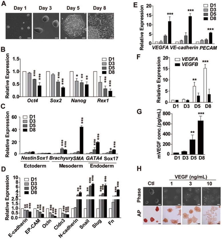
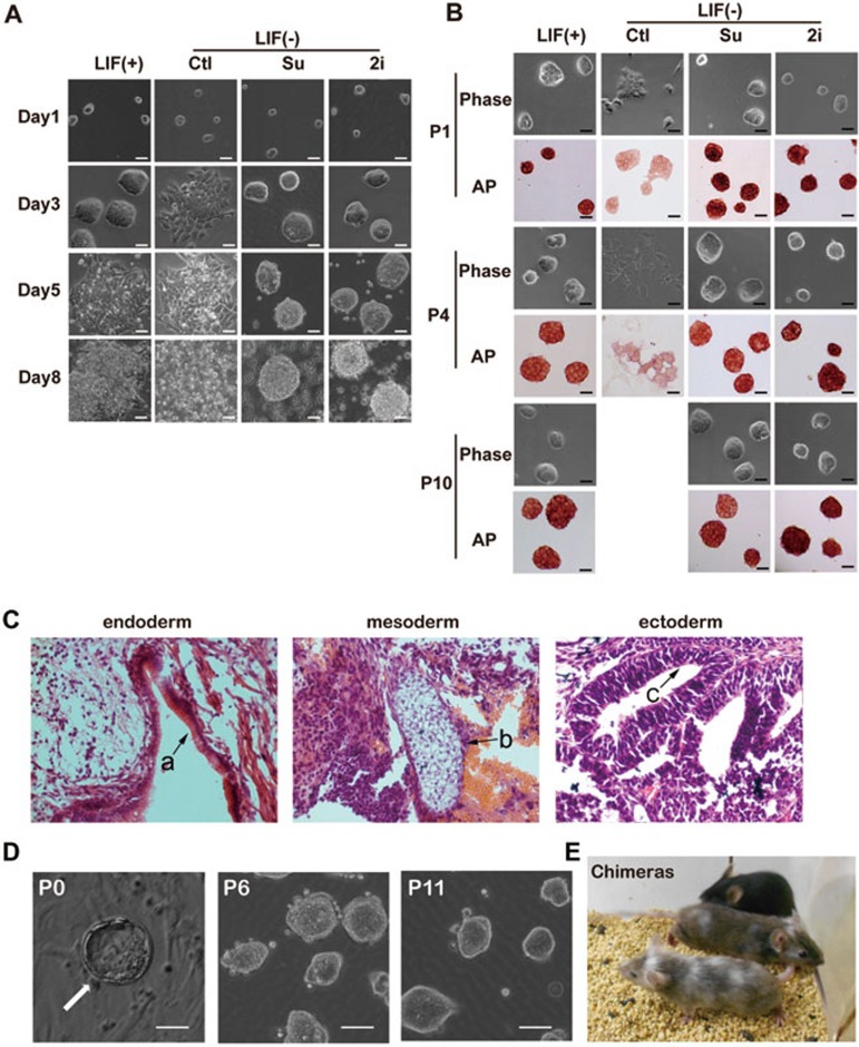
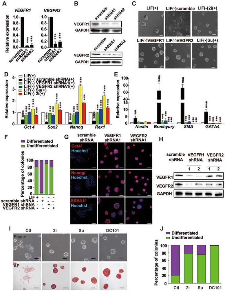
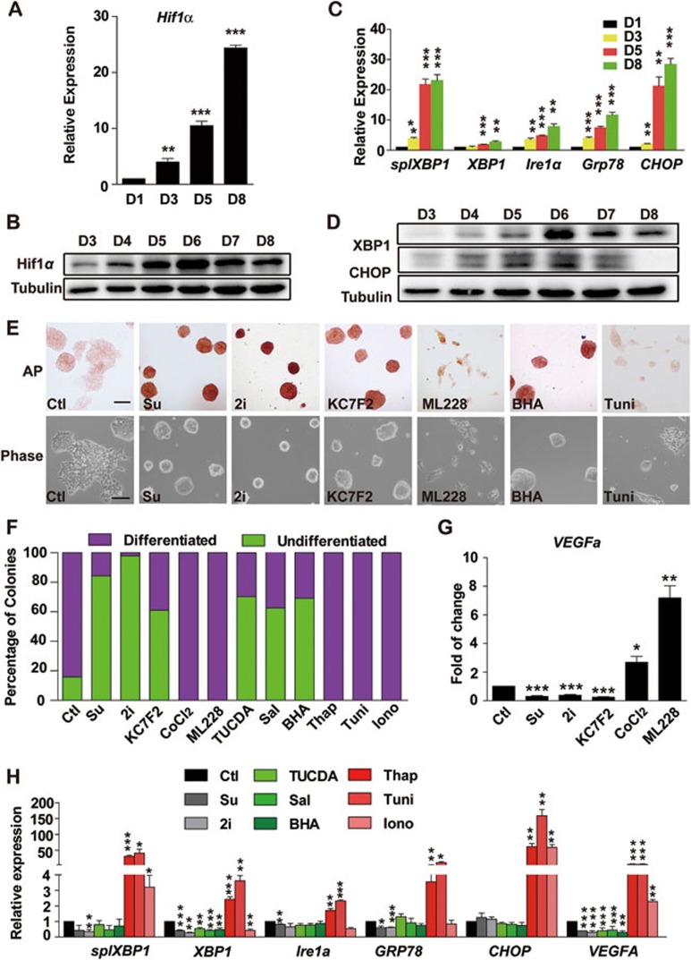
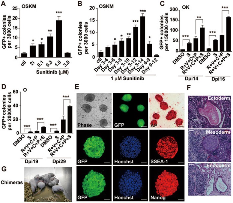
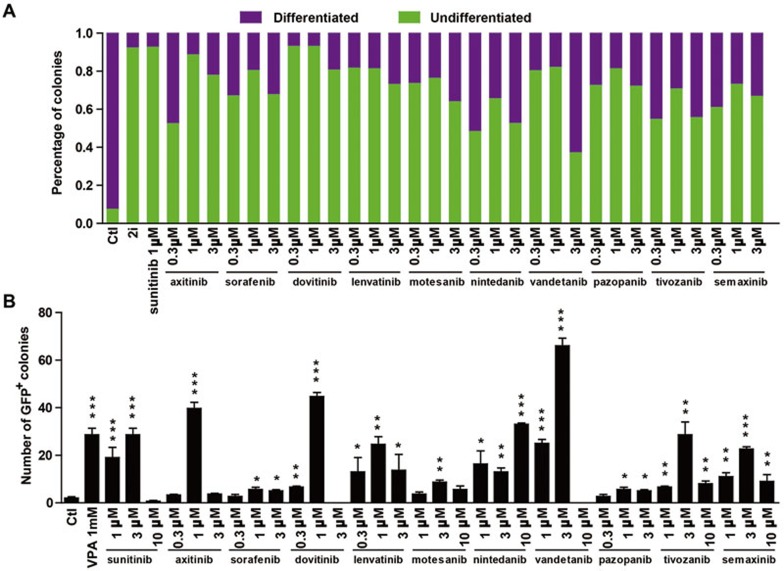
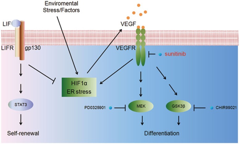
Maintaining the self-renewal of embryonic stem cells (ESCs) could be achieved by activating the extrinsic signaling, i.e., the use of leukemia inhibitory factor (LIF), or blocking the intrinsic differentiation pathways, i.e., the use of GSK3 and MEK inhibitors (2i). Here we found that even in medium supplemented with LIF, mESCs still tend to differentiate toward meso-endoderm lineages after long-term culture and the culture spontaneously secretes vascular endothelial growth factors (VEGFs). Blocking VEGF signaling with sunitinib, an anti-cancer drug and a receptor tyrosine kinase (RTK) inhibitor mainly targeting VEGF receptors (VEGFRs), is capable of maintaining the mESCs in the undifferentiated state without the need for feeder cells or LIF. Sunitinib facilitates the derivation of mESCs from blastocysts, and the mESCs maintained in sunitinib-containing medium remain pluripotent and are able to contribute to chimeric mice. Sunitinib also promotes iPSC generation from MEFs with only Oct4. Knocking down VEGFR2 or blocking it with neutralizing antibody mimicks the effect of sunitinib, indicating that blocking VEGF/VEGFR signaling is indeed beneficial to the self-renewal of mESCs. We also found that hypoxia-inducible factor alpha (HIF1α) and endoplasmic reticulum (ER) stress are involved in the production of VEGF in mESCs. Blocking both pathways inhibits the expression of VEGF and prevents spontaneous differentiation of mESCs. Interestingly, LIF may also exert its effect by downregulating HIF1α and ER stress pathways and subsequent VEGF expression. These results indicate the existence of an intrinsic differentiation pathway in mESCs by activating the autocrine VEGF signaling. Blocking VEGF signaling with sunitinib or other small molecules help to maintain the mESCs in the ground state of pluripotency.


Figures

Figure 1
Figure 1
mESCs differentiate toward meso-endoderm lineage spontaneously during long-term culture. (A) Morphology of E14 cells cultured in mES medium containing 1 000 U/ml LIF without passage for 1-8 days. Medium was changed every day. Scale bar, 50 μm. (B-F) qRT-PCR analysis of pluripotency genes (B), lineage-specific genes (mesoderm: Brachyury and SMA, endoderm: GATA4, Sox17, ectoderm: Nestin and Sox1) (C), genes involved in EMT (D), vascular marker genes (E) and VEGF genes (F) in mESCs described in A on days 1, 3, 5 and 8. (G) ELISA analysis of VEGF secretion from mESCs described in A. (H) VEGF-induced morphology and alkaline phosphatase (AP) change in E14 cells. Scale bar, 50 μm. E14 cells were cultured in mES medium containing 1 000 U/ml LIF for 48 h, then vehicle or 1, 3, 10 ng/ml mVEGF164 was added into the medium for another 48 h prior to analysis. Data are mean ± SEM (n = 3). *P < 0.05, **P < 0.01, ***P < 0.001 vs day 1. Independent experiments were repeated at least three times.
Figure 2
Figure 2
Sunitinib maintains the self-renewal and pluripotency of mESCs. (A) Morphology of E14 cells cultured in mES media containing 1 000 U/ml LIF, or in mES media without LIF but containing 2i (3 μM PD0325901 and 3 μM CHIR99021) or sunitinib (Su, 1 μM), for 1-8 days. Media were changed every day. Scale bar, 50 μm. (B) Morphology and alkaline phosphatase (AP) staining of E14 cells at passages 1, 4 and 10 maintained in mES media containing 1 000 U/ml LIF, or in mES media without LIF but containing 2i (3 μM PD0325901 and 3 μM CHIR99021) or sunitinib (1 μM). Scale bar, 50 μm. (C) H&E staining of teratomas generated with E14 cells maintained with sunitinib (passage 11). Typical structures of the three embryonic germ layers are shown as follows: a, gastrointestinal-like epithelium (endoderm); b, cartilage-like structure (mesoderm); c, neural tube-like structure (ectoderm). Scale bars, 20 μm. (D) Blastocyst outgrowth (P0, the arrow) on feeder cells and mES medium containing sunitinib and 2 000 U/ml LIF. mESCs were subsequently maintained in mES media containing sunitinib for multiple passages. Scale bar, 20 μm. (E) Chimeric mice generated with mESCs derived and maintained with sunitinib (passage 6).
Figure 3
Figure 3
VEGFR knockdown recapitulates the effect of sunitinib. (A, B) Validation of the shRNAs targeting VEGFRs by qRT-PCR and western blot. (C) Morphology of E14 cells expressing the indicated shRNA cultured in mES media without LIF. Cells treated with sunitinib or 2i were used as positive control. Scale bar, 50 μm. (D, E) qRT-PCR analysis of pluripotency genes (Oct4, Sox2, Nanog, Rex1) (D) and lineage-specific genes (mesoderm: Brachyury and SMA, endoderm: GATA4, ectoderm: Nestin) (E) in cells expressing the indicated shRNA cultured in mES media without LIF (passage 4). Cells treated with sunitinib or LIF were used as positive control. Data are mean SEM (n = 3). ##P < 0.01, ###P < 0.001 vs LIF (+) condition; *P < 0.05, **P < 0.01, ***P < 0.001 vs scramble shRNA. (F) E14 cells expressing the indicated shRNA were cultured in LIF-free mES media for 3 days at an initial density of 10 000 cells/well in 24-well plate. The percent of undifferentiated and differentiated colonies were calculated according to AP staining and morphology. Independent experiments were repeated at least three times. (G) Immunofluorescence staining of pluripotency markers (Oct4, Nanog, SSEA1) in cells described in E (passage 4). Cell nuclei were stained with Hoechst. Scale bar, 80 μm. (H) Western blot analysis of VEGFR1 and 2 in cells transfected with scramble shRNA, or shRNAs targeting VEGFR1 or 2 for 48 h. (I) Morphology and AP staining of E14 cells cultured in mES medium without LIF but containing 2i (3 μM PD0325901 and 3 μM CHIR99021), sunitinib (1 μM) or DC101 (20 μg/ml) for 3 days. (J) The percent of undifferentiated and differentiated colonies corresponding to I.
Figure 4
Figure 4
Role of HIF1α and ER stress in VEGF expression and pluripotency of mESCs. (A, B) qRT-PCR (A) and western blot analysis (B) of HIF1α in E14 cells cultured in mES media containing 1 000 U/ml LIF for 1-8 days. Tubulin served as a loading control for western blot. (C, D) qRT-PCR (C) and western blot analysis (D) of genes involved in ER stress in E14 cells cultured in mES medium containing 1 000 U/ml LIF for 1-8 days. (E) Morphology (bottom) and AP staining (top) of E14 cells cultured in mES medium without LIF but containing sunitinib (1 μM), 2i, ML228 (1 μM), KC7F2 (1 μM), BHA (10 μM) or tunicamycin (Tuni, 2.5 μg/ml) for 48 h. Scale bar, 50 μm. (F) Statistical data of the percent of undifferentiated and differentiated colonies in E14 cells cultured in LIF-free mES medium containing the indicated compounds for 3 days, including sunitinib, 2i, KC7F2 (1 μM), CoCl2 (30 μM), ML228 (1 μM), TUCDA (0.1 mM), salubrinal (Sal, 10 μM), BHA (10 μM), thapsigargin (Thap, 0.4 μM), tunicamycin (Tuni, 2.5 μg/ml) and ionomycin (Iono, 3.6 μM). (G) qRT-PCR analysis of VEGFA in E14 cells cultured in LIF-free mES medium containing sunitinib, 2i or the indicated HIF1α regulators (same concentrations as in F) for 3 days. Data are mean ± SEM (n = 3). *P < 0.05, **P < 0.01, ***P < 0.001 vs control. (H) qRT-PCR analysis of the expression of ER stress-related genes and VEGFA in E14 cells cultured in LIF-free mES medium containing sunitinib, 2i or the indicated ER stress regulators (same concentrations as in F) for 3 days. Data are mean ± SEM (n = 3), *P < 0.05, **P < 0.01, ***P < 0.001 vs control. Independent experiments were repeated at least three times.
Figure 5
Figure 5
Sunitinib promotes reprogramming of somatic cells. (A) Dose response of sunitinib in 4F-mediated iPSC induction. Sunitinib at the indicated concentration was added into 3 000 4F-transduced MEFs from day 3 to day 14. (B) Time response of sunitinib in iPSC induction. Sunitinib (1 μM) was added at the indicated time periods, and GFP+ colonies were counted on day 14. Data are mean SEM (n = 3). *P < 0.05, **P < 0.01, ***P < 0.001 vs control. Independent experiments were repeated at least three times. (C) Sunitinib facilitated iPSC generation from 2F (OK)-transduced MEFs alone or in combination with several known chemicals. For OK-mediated reprogramming, 150 000 OK-infected cells in 6-well plates were treated with 1 μM sunitinib (S) alone or various combinations of compounds (including 1 μM R, RepSox; 0.5 mM V, VPA; 3 μM C, CHIR and 2 μM P, Parnate) from day 3 to day 14. (D) Sunitinib facilitated iPSC generation from Oct4-transduced MEFs alone or in combination with several known chemicals. For Oct4-mediated reprogramming, 200 000 Oct4-infected cells in 6-well plates were treated with 1 μM sunitinib (S) alone or the indicated combinations of compounds from day 3 to day 14. GFP+ colonies were counted on day 19 and day 29. (E) Top: morphology, GFP expression and AP staining in iPSC clones induced with 1F (Oct4) plus sunitinib (1 μM). Scale bar, 50 μm. Middle and bottom: immunofluorescent staining of SSEA-1 and Nanog in the same clone. Scale bar, 10 μm. (F) HE-stained sections of teratomas formed with iPSC clone described in E. Typical structures of the three embryonic germ layers are shown: epidermis (ectoderm), muscle and cartilage (mesoderm) and epithelium (endoderm). Scale bar, 20 μm. (G) chimeric mice produced with iPSC clone described in E.
Figure 6
Figure 6
Other RTK inhibitors targeting VEGFRs facilitate the self-renewal of mESCs and the generation of iPSCs. (A) E14 cells were cultured in LIF-free mES media containing sunitinib or other inhibitors at the indicated concentrations for 3 days. The initial cell density was 1 000 cells/well in 96-well plate. The percent of AP+ (undifferentiated) and AP (differentiated) colonies were calculated. 2i culture was used as a positive control. (B) MEFs transduced with Yamanaka 4 factors were treated with sunitinib or the indicated inhibitors (added from day 3 to day 14) and GFP+ colonies were counted on day 14. VPA (1 mM, added from day 3 to day 8) treatment was used as a positive control. Data are mean ± SEM (n = 3). *P < 0.05, **P < 0.01, ***P < 0.001 vs vehicle control.
Figure 7
Figure 7
Schematic representation of the autocrine VEGF signaling in mESCs. Production of VEGF is activated via HIF1α and/or ER stress, presumably due to environmental stress in the in vitro culture. VEGF activates VEGFRs expressed on the surface of the mESCs, inducing the activation of MEK, GSK3β and possibly other pathways that promote differentiation and lineage commitment of mESCs. Blocking VEGFRs with sunitinib inhibits both the MEK and GSK3 pathways and the feed-forward loop between HIF1α/ER stress and VEGF/VEGFR, and thus prevents the spontaneous differentiation of mESCs.

Similar articles

Cited by

References

    1. Ulloa-Montoya F, Verfaillie CM, Hu WS. Culture systems for pluripotent stem cells. J Biosci Bioeng. 2005;100:12–27. - PubMed
    1. Williams RL, Hilton DJ, Pease S, et al. Myeloid leukaemia inhibitory factor maintains the developmental potential of embryonic stem cells. Nature. 1988;336:684–687. - PubMed
    1. Ying QL, Nichols J, Chambers I, Smith A. BMP induction of Id proteins suppresses differentiation and sustains embryonic stem cell self-renewal in collaboration with STAT3. Cell. 2003;115:281–292. - PubMed
    1. Kunath T, Saba-El-Leil MK, Almousailleakh M, Wray J, Meloche S, Smith A. FGF stimulation of the Erk1/2 signalling cascade triggers transition of pluripotent embryonic stem cells from self-renewal to lineage commitment. Development. 2007;134:2895–2902. - PubMed
    1. Ying QL, Wray J, Nichols J, et al. The ground state of embryonic stem cell self-renewal. Nature. 2008;453:519–523. - PMC - PubMed

Publication types

MeSH terms