. 2014 Aug 22;46(8):e110.
doi: 10.1038/emm.2014.49.

Therapeutic effect of hepatocyte growth factor-secreting mesenchymal stem cells in a rat model of liver fibrosis

Affiliations

Therapeutic effect of hepatocyte growth factor-secreting mesenchymal stem cells in a rat model of liver fibrosis

Myung-Deok Kim et al. Exp Mol Med. .

Abstract

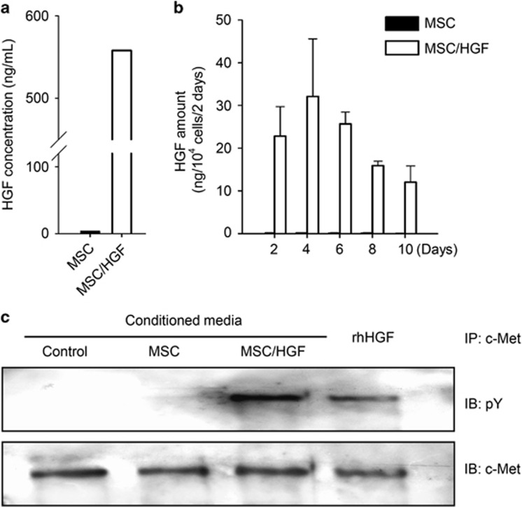
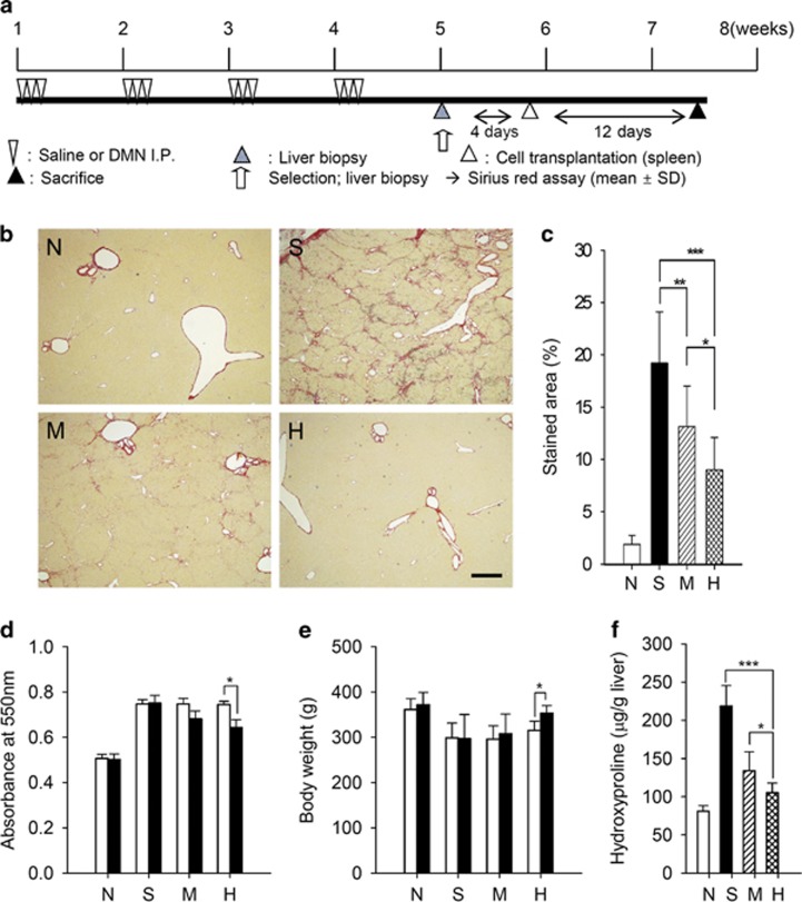
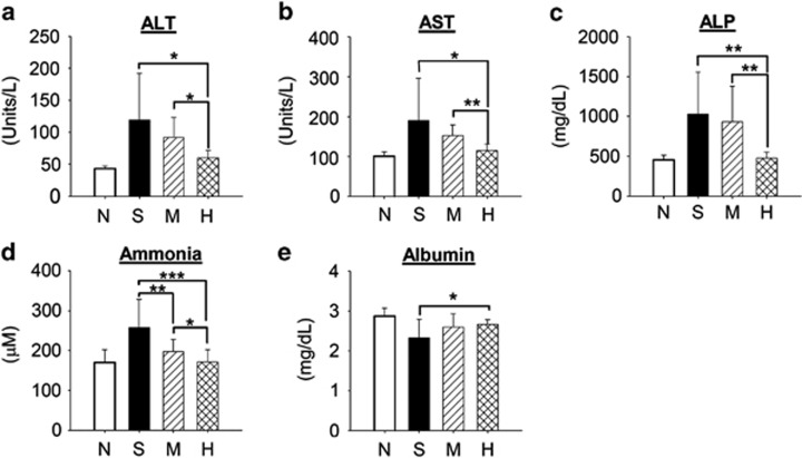
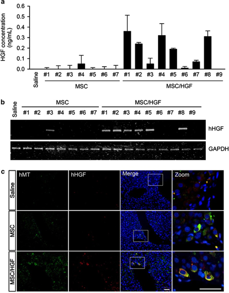
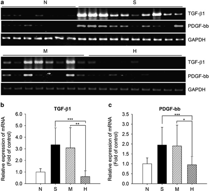
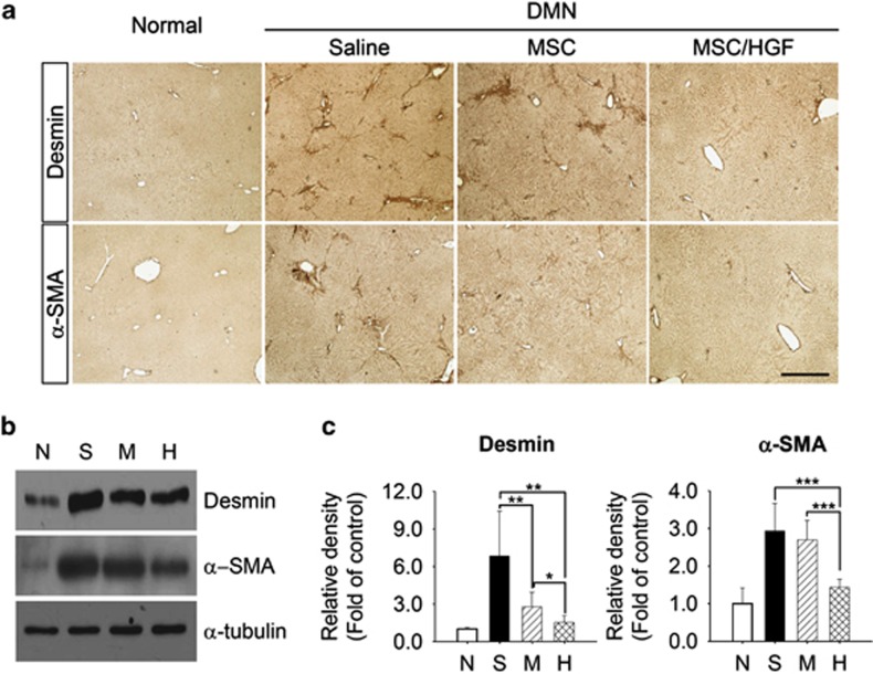
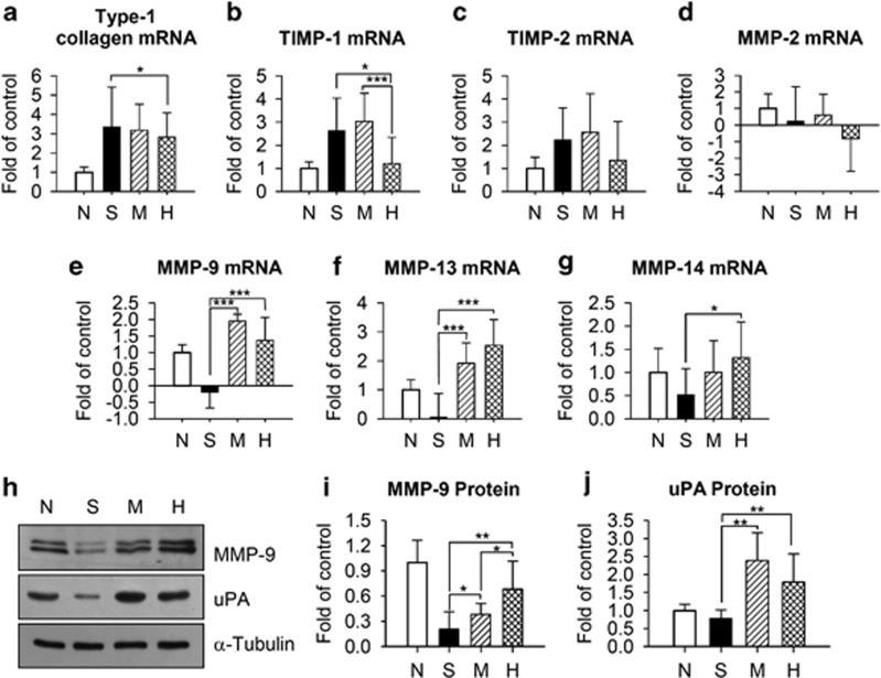
Bone marrow-derived mesenchymal stromal cells (MSCs) have been reported to be beneficial for the treatment of liver fibrosis. Here, we investigated the use of genetically engineered MSCs that overexpress hepatocyte growth factor (HGF) as a means to improve their therapeutic effect in liver fibrosis. Liver fibrosis was induced by intraperitoneal injection of dimethylnitrosamine. HGF-secreting MSCs (MSCs/HGF) were prepared by transducing MSCs with an adenovirus carrying HGF-encoding cDNA. MSCs or MSCs/HGF were injected directly into the spleen of fibrotic rats. Tissue fibrosis was assessed by histological analysis 12 days after stem cell injection. Although treatment with MSCs reduced fibrosis, treatment with MSCs/HGF produced a more significant reduction and was associated with elevated HGF levels in the portal vein. Collagen levels in the liver extract were decreased after MSC/HGF therapy, suggesting recovery from fibrosis. Furthermore, liver function was improved in animals receiving MSCs/HGF, indicating that MSC/HGF therapy resulted not only in reduction of liver fibrosis but also in improvement of hepatocyte function. Assessment of cell and biochemical parameters revealed that mRNA levels of the fibrogenic cytokines PDGF-bb and TGF-β1 were significantly decreased after MSC/HGF therapy. Subsequent to the decrease in collagen, expression of matrix metalloprotease-9 (MMP-9), MMP-13, MMP-14 and urokinase-type plasminogen activator was augmented following MSC/HGF, whereas tissue inhibitor of metalloprotease-1 (TIMP-1) expression was reduced. In conclusion, therapy with MSCs/HGF resulted in an improved therapeutic effect compared with MSCs alone, probably because of the anti-fibrotic activity of HGF. Thus, MSC/HGF represents a promising approach toward a cell therapy for liver fibrosis.


Figures

Figure 1
Figure 1
Analysis of HGF secreted by MSCs/HGF. (a) The amount of human HGF after 4 days of culture. (b) Secretion of HGF by MSCs or MSCs/HGF at 2, 4, 6, 8 and 10 days after Ad-HGF transduction. (c) Immunoblot showing c-Met phosphorylation in MDCK2 cells treated with conditioned media obtained from indicated cells. Human recombinant HGF protein (rhHGF, 100 unit ml−1) was used as a positive control. IB, immunoblotting; IP, immunoprecipitation; pY, phospho-tyrosine.
Figure 2
Figure 2
Assessment of liver fibrosis in MSC/HGF-transplanted rats and controls. (a) Diagram of the treatment protocol. (b) Extracellular deposition of collagen fibers stained with Sirius Red. Scale bar=0.5 mm. (c) Quantification of collagen by image analysis. (d, e) Collagen content and body weight quantified before (white bar) and 12 days after transplantation (black bar). (f) Hydroxyproline content. Normal rats (N), control fibrotic animals (S), fibrotic animals transplanted with MSCs (M) or MSCs/HGF (H). Data represent mean±s.d. for each group (*P<0.05, **P<0.01, ***P<0.005).
Figure 3
Figure 3
Examination of serum parameters. (a, b) Transaminases (ALT and AST), (c) alkaline phosphatase (ALP), (d) ammonia and (e) albumin. Normal rats, N; fibrotic control animals, S; fibrotic animals transplanted with MSCs, M, or MSCs/HGF, H. Data represent the mean±s.d. for each group (*P<0.05, **P<0.01, ***P<0.005).
Figure 4
Figure 4
Analysis of human HGF expression in rats transplanted with MSCs/HGF. (a) Human HGF proteins. Values represent the mean±s.d. of triplicate assays of individual rats. (b) Human HGF mRNA. (c) Immunostaining for human mitochondria-specific antigen (hMT) and human HGF (hHGF). Blue: bis-benzamide, green: hMT, red: hHGF, Scale bar, 50 μm. Samples are fibrotic livers from control animals or from animals transplanted with MSCs or MSCs/HGF except in (a), where analysis was performed on serum collected from the portal vein 12 days after transplantation.
Figure 5
Figure 5
Analysis of profibrogenic cytokine expression. (a) Detection of TGF-β1 and PDGF-bb mRNA in individual rats by RT–PCR. (b, c) Mean relative expression levels of TGF-β1 and PDGF-bb in liver tissues from normal rats, N; control fibrotic animals, S; or fibrotic animals transplanted with MSCs, M, or MSC/HGF, H. Data are normalized to GAPDH expression levels and represent the mean±s.d. for each group (*P<0.05, **P<0.01, ***P<0.005).
Figure 6
Figure 6
Effect of MSC/HGF transplantation on Ito cell activation. (a) α-SMA and desmin were detected by immunohistochemistry in liver sections. Scale bar, 500 μm. (b) Liver extracts from each group were subjected to western blot analysis using antibodies against α-SMA and desmin. (c) The relative amount of α-SMA and desmin proteins was calculated by densitometry. Normal rats, N; control fibrotic animals, S; and fibrotic animals transplanted with MSCs, M, or MSCs/HGF, H. Data represent the mean±s.d. for each group. α-SMA; α-smooth muscle actin (*P<0.05, **P<0.01, ***P<0.005).
Figure 7
Figure 7
Analysis of the expression of ECM remodeling factors. (a) Type 1 collagen; (b, c) TIMP-1 and -2; and (dg) MMP-2, -9, -13 and -14 mRNAs were quantified by semiquantitative RT-PCR. (h) MMP-9 and uPA protein levels were evaluated by western blotting and (i, j) quantified by densitometric analysis. Samples were obtained from normal rats, N; control fibrotic animals, S; fibrotic animals transplanted with MSCs, M, or MSCs/HGF, H. Data represent the mean±s.d. for each group. MMP, matrix metalloproteinase; TIMP, tissue inhibitor of matrix metalloproteinase, uPA, urokinase type plasminogen activator (*P<0.05, **P<0.01, ***P<0.005).

References

    1. Bataller R, Brenner DA. Liver fibrosis. J Clin Invest. 2005;115:209–218. - PMC - PubMed
    1. Sakaida I, Terai S, Yamamoto N, Aoyama K, Ishikawa T, Nishina H, et al. Transplantation of bone marrow cells reduces CCl4-induced liver fibrosis in mice. Hepatology. 2004;40:1304–1311. - PubMed
    1. Fang B, Shi M, Liao L, Yang S, Liu Y, Zhao RC. Systemic infusion of FLK1(+) mesenchymal stem cells ameliorate carbon tetrachloride-induced liver fibrosis in mice. Transplantation. 2004;78:83–88. - PubMed
    1. Oyagi S, Hirose M, Kojima M, Okuyama M, Kawase M, Nakamura T, et al. Therapeutic effect of transplanting HGF-treated bone marrow mesenchymal cells into CCl4-injured rats. J Hepatol. 2006;44:742–748. - PubMed
    1. Ong SY, Dai H, Leong KW. Hepatic differentiation potential of commercially available human mesenchymal stem cells. Tissue Eng. 2006;12:3477–3485. - PubMed

Publication types

Substances