Review
. 2014 Sep 15;462(3):397-413.
doi: 10.1042/BJ20140400.

Controlling gene networks and cell fate with precision-targeted DNA-binding proteins and small-molecule-based genome readers

Affiliations
Review

Controlling gene networks and cell fate with precision-targeted DNA-binding proteins and small-molecule-based genome readers

Asuka Eguchi et al. Biochem J. .

Abstract

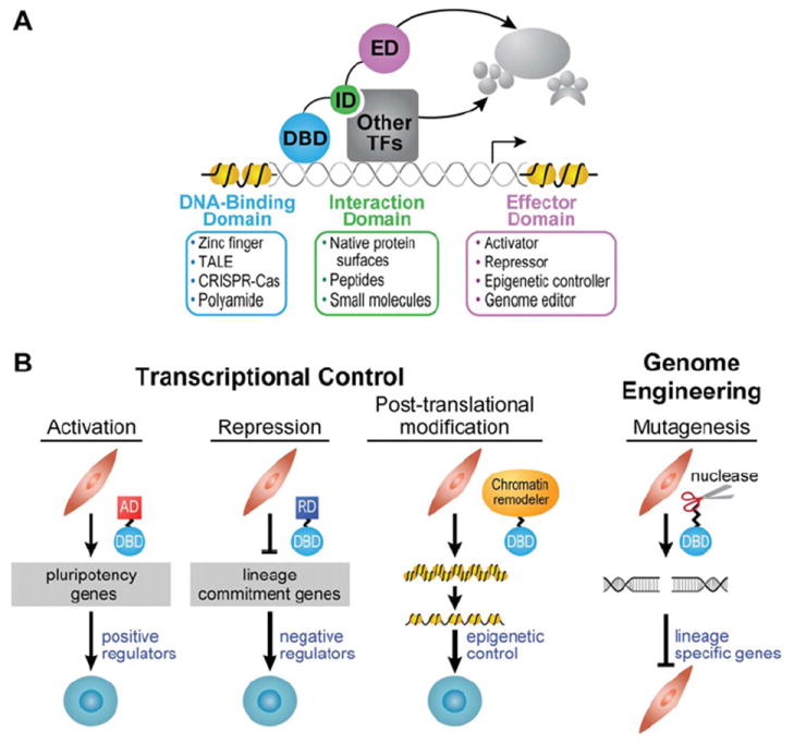
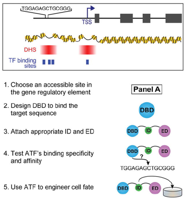
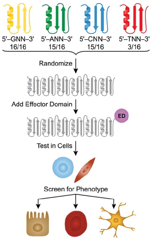
Transcription factors control the fate of a cell by regulating the expression of genes and regulatory networks. Recent successes in inducing pluripotency in terminally differentiated cells as well as directing differentiation with natural transcription factors has lent credence to the efforts that aim to direct cell fate with rationally designed transcription factors. Because DNA-binding factors are modular in design, they can be engineered to target specific genomic sequences and perform pre-programmed regulatory functions upon binding. Such precision-tailored factors can serve as molecular tools to reprogramme or differentiate cells in a targeted manner. Using different types of engineered DNA binders, both regulatory transcriptional controls of gene networks, as well as permanent alteration of genomic content, can be implemented to study cell fate decisions. In the present review, we describe the current state of the art in artificial transcription factor design and the exciting prospect of employing artificial DNA-binding factors to manipulate the transcriptional networks as well as epigenetic landscapes that govern cell fate.


Figures

Figure 1
Figure 1. Toolbox and modular design
(A) Modularity of DNA-binding factors and small molecules. The DBD makes specific contacts with DNA and identifies preferred sites within the genome. An ID can be added to allow the molecule to partner with other proteins of interest. The ED provides the molecule with function. (B) Applications toward controlling gene networks. Regulatory control: a gene-activating ATF can positively regulate gene expression. Conversely, a gene-repressing ATF could negatively regulate its targets. A chromatin-remodelling enzyme fused to a DBD could reorganize the chromatin structure at specific loci where epigenetic control would have important effects in triggering the transcriptional circuitry for a particular phenotype. Genome engineering: attaching a nuclease domain to a DBD would enable genome editing for the purposes of altering transcriptional regulatory elements. In the Figure, reprogramming of fibroblasts to iPS cells is depicted; however, these versatile tools can be applied to a variety of biological contexts. AD, activation domain; RD, repression domain.
Figure 2
Figure 2. Co-operative assembly of TFs
(A) Eukaryotic TFs bind genomic DNA by co-operative assembly. The factors have a different dissociation constant (KD) depending on whether they bind as monomers or as a larger complex. As a complex, affinity to DNA increases, and binding towards its target site is highly specific. Pol II, RNA polymerase II; TBP, TATA-box-binding protein. (B) A polyamide attached to the YPWM tetrapeptide allows the small molecule DBD to interact with Exd. (C) This interaction with Exd increases affinity for the cognate site three orders of magnitude [99].
Figure 3
Figure 3. Activation domains
To activate genes, a helical peptide with acidic and hydrophobic residues or a small-molecule mimic bearing amphipathic surfaces can be effective in recruiting general transcriptional factors [27]. The transactivator of the herpes simplex virus, VP16, is one such acidic protein that has been sown to activate transcription across eukaryotes [144]. VP64, a tetrameric repeat of the 11-amino-acid core of the activation region from VP16 (DALDDFDLDML) is a widely used activation domain (AD) in mammalian systems [142]. In contexts where VP64 has not been effective, RelA (p65) has been used to recruit the general transcriptional machinery [145]. RelA, a subunit of the NF-κB (nuclear factor κB) complex, also contains acidic residues interspersed with hydrophobic residues [146]. Pollock et al. [147] created a chimaera of RelA and human HSF1, called S3H, which was discovered to be a stronger activator than RelA by itself. Similarly, eleven repeats of the 5-amino-acid minimal activation region of β-catenin (FDTDL) was shown to be a functional activation domain [143]. The amphipathic nature of activation domains led to the design of an AH (amphipathic helix) that displays a negatively charged face and a hydrophobic face [148]. These same principles were applied to the creation of an RNA-based activation domain as well as the isoxazoladine small molecule activation domains [149,150]. Other synthetic activation domains include the small molecule ligand of Sur-2, a subunit of the mammalian Mediator complex (Med), which led to the use of wrenchnolol as an activation module [151]. Kodadek and colleagues also identified multiple peptoids that bind to CREB (cAMP-response-element-binding protein)-binding protein and function as activation domains in human cells [152]. Rather than develop molecules that attempt to block co-operative assembly, one can tether small molecules to a DBD in order to harness co-operative assembly to associate natural TFs at specific sites across the genome. An activation domain could be developed from small molecules that recruit the transcriptional machinery, TFs, chromatin-modifying enzymes as well as chromatin readers. For example, this strategy could be used to repurpose ligands that bind nuclear hormone receptors such as 17-β-oestradiol and testosterone, or ligands that target the super-elongation complex such as I-BET151 and JQ1 [153,154]. By fusing a small molecule to a DBD, the small molecule can recruit its protein target to activate a gene of interest. Moreover, tethering these molecules to specific loci across the genome via an engineered DBD would also reduce the off-target actions of ligands.
Figure 4
Figure 4. Repressor domains
If the DBD has a high affinity for its binding site, it can function as a competitive inhibitor by occupying potential genomic binding sites of transcriptional activators or interacting with activation domains and preventing them from recruiting the transcriptional machinery. The more effective approach that does not rely on precise placement of competitors at target sites relies on the use of repression modules that recruit repressor complexes or machinery that place transcriptional silencing marks on histone tails. The most widely used repression domain (RD) is the KRAB domain, the 75-amino-acid region at the N-terminus of KOX1 [155]. Extensive investigations of a leukaemia-inducing avian virus led to the finding of v-ErbA as a constitutive repressor [156]. v-ErbA is a variant of the chicken thyroid hormone receptor α, and its mutations render it incapable of activating transcription [157]. Instead, the mutated v-ErbA represses transcription by recruiting HDAC3 and N-CoR-SMRT in human cells [158]. Furthermore, studies of the Mad family of repressor complexes in yeast led to the discovery of the ID of Mad1 (residues 1–36) which functions as a potent repression domain by recruiting co-repressor Sin3A/B [159]. In a screen for factors that bind the Ets2 promoter, Sgouras et al. [160] screened for a novel ETS protein, which came from a family of transcriptional activators. To their surprise, they stumbled upon a transcriptional repressor, ERF (Ets2 repressor factor), which consists of a repressor domain capable of conferring its repressive function to heterologous DBDs [160]. Even plant repression domains such as the one from the Arabidopsis thaliana protein SUPERMAN [161] can function as a repressor in mammalian cells, but, interestingly, they only do so under hypoxic conditions [143]. (KRAB, v-ErbA and MAD1 under PDB codes 1V65, 3N00 and 1S5Q respectively).
Figure 5
Figure 5. Designing an ATF for a targeted approach
(1)Choose an accessible site in the gene regulatory element as shown in panel A. The location of the TSS (transcription start site) should be taken into consideration. Within the promoter and enhancer elements, DHSs (DNase I hypersensitive sites) shown in red identify regions of accessible chromatin. The most proximal DHS downstream of the TSS (not shown in this example) can also be used for gene regulation [145]. The binding sites for endogenous TFs, depicted as blue boxes, can be considered for competitive binding effects. DHS and TF binding site information is available for some cell types on ENCODE or modENCODE. Some DHSs are better for targeting than others, but selection has to be evaluated experimentally. (2) Design a DBD to bind target sequence. The length of the sequence as well as the nucleotide content should be considered. The rules for DBD design are unique for each class. (3) Attach appropriate ID and ED. The ID is an optional component for co-operative assembly with other TFs. The ED is important for providing the ATF with function (activation or repression). An NLS should also be included to ensure uptake into the nucleus. (4) Test ATF’s binding specificity and affinity. Specificity and affinity can be tested in vitro by EMSA (electrophoretic mobility shift assay) or more comprehensively by CSI (cognate site identifier) methods and can be inferred in cells by luciferase assay with the target site cloned upstream of luciferase [162]. (5) Use ATF in cells for experiments. The ATF can be expressed in cells by a delivery method of choice to induce changes in cell fate.
Figure 6
Figure 6. ZF library design
ZF modules designed to target GNN, ANN, CNN and TNN sequences are randomized to create polydactyl ZF proteins (depicted is a six-ZF library). Each grey motif represents a module that can target any given triplet. More modules exist for targeting triplets beginning with purines than those beginning with pyrimidines. An ED, such as an AD (activation domain), is used to regulate genes. The library is tested in cells (depicted as fibroblasts or pluripotent cells) by transduction, and hits are selected by screening for a desired phenotype or activation of a lineage-specific gene. ATFs are recovered by sequencing integrated transgenes. Positive ATFs can then be validated for their cell fate-defining effects.

Similar articles

Cited by

References

    1. Takahashi K, Yamanaka S. Induction of pluripotent stem cells from mouse embryonic and adult fibroblast cultures by defined factors. Cell. 2006;126:663–676. - PubMed
    1. Davis RL, Weintraub H, Lassar AB. Expression of a single transfected cDNA converts fibroblasts to myoblasts. Cell. 1987;51:987–1000. - PubMed
    1. Cohen DE, Melton D. Turning straw into gold: directing cell fate for regenerative medicine. Nat Rev Genet. 2011;12:243–252. - PubMed
    1. Graf T, Enver T. Forcing cells to change lineages. Nature. 2009;462:587–594. - PubMed
    1. Yu J, Vodyanik MA, Smuga-Otto K, Antosiewicz-Bourget J, Frane JL, Tian S, Nie J, Jonsdottir GA, Ruotti V, Stewart R, et al. Induced pluripotent stem cell lines derived from human somatic cells. Science. 2007;318:1917–1920. - PubMed

Publication types

MeSH terms