. 2014 Aug 22;18(4):484.
doi: 10.1186/s13054-014-0484-2.

Early changes of muscle membrane properties in porcine faecal peritonitis

Early changes of muscle membrane properties in porcine faecal peritonitis

Karin A Ackermann et al. Crit Care. .

Abstract

Introduction: Sepsis-induced myopathy and critical illness myopathy (CIM) are possible causes of muscle weakness in intensive care patients. They have been attributed to muscle membrane dysfunction. The aim of this study was to investigate membrane properties in the early stage of experimental sepsis by evaluating muscle excitability.

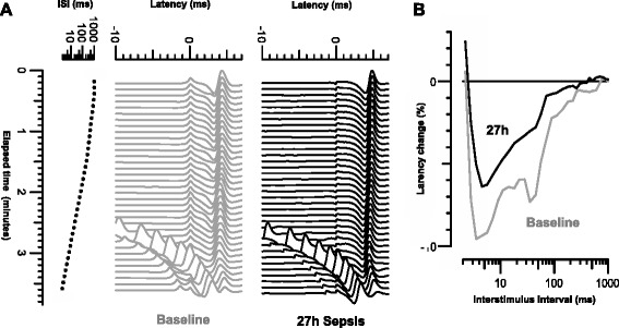
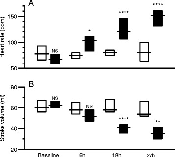
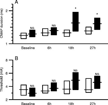
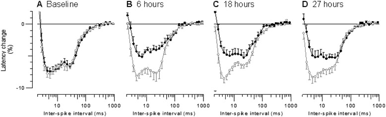
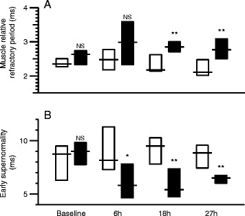
Methods: In total, 20 anesthetized and mechanically ventilated pigs were randomized to either faecal peritonitis (n = 10) or to non-septic controls (n = 10). Resuscitation with fluids and vasoactive drugs was started 3 hours after peritonitis induction. Muscle membrane properties were investigated by measuring muscle velocity recovery cycles before induction of peritonitis as well as 6, 18 and 27 hours thereafter. Muscle relative refractory period (MRRP) and early supernormality (ESN) were assessed.

Results: Peritonitis lasting 27 hours was associated with an increase of MRRP by 28% from 2.38 ± 0.18 ms (mean ± SD) to 3.47 ± 1.79 ms (P <0.01) and a decrease of ESN by 31% from 9.64 ± 2.82% to 6.50 ± 2.64% (P <0.01). ESN reduction was already apparent 6 hours after induction of peritonitis. Values in controls did not show any significant alterations.

Conclusions: Muscle membrane abnormalities consistent with membrane depolarization and/or sodium channel inactivation occurred within 6 hours of peritonitis induction. This indicates that changes that have been described in established sepsis-induced myopathy and/or CIM start early in the course of sepsis. Muscle excitability testing facilitates evaluation of the time course of these changes.


Figures

Figure 1
Figure 1
Recordings of multi-fibre action potentials in pig extensor digitorum at baseline (grey) and 27 hours after induction of sepsis (black). (A) Recordings of multi-fibre action potentials in pig extensor digitorum at baseline (grey) and 27 hours after induction of sepsis (black). Test stimulus is at latency 0 ms, and conditioning stimuli applied at intervals varied from 1,000 to 1.4 ms. Response latencies were measured to peak after subtraction of response to conditioning stimulus alone. (B) Velocity recovery cycles at the two times plotted as percentage changes in latency against the inter-stimulus interval (logarithmic scale).
Figure 2
Figure 2
Differences between control animals (white) and animals with peritonitis (black) at baseline, 6 hours, 18 hours and 27 hours after induction of sepsis. (A) Heart rate, (B) stroke volume. Boxes indicate interquartile ranges and lines indicate median values. Comparisons between control and peritonitis animals were made with Welch rank test (NS = P >0.05, * = P <0.05, ** = P <0.01, **** = P <0.0001).
Figure 3
Figure 3
Differences between control animals (white) and animals with peritonitis (black) at baseline, 6 hours, 18 hours and 27 hours after induction of sepsis. (A) CMAP duration, (B) stimulus intensity to evoke an amplitude of 50% maximal CMAP. Boxes indicate interquartile ranges and lines indicate median values. Comparisons between control and peritonitis animals were made with Welch rank test (NS = P >0.05, * = P <0.05). CMAP, compound muscle action potential.
Figure 4
Figure 4
Mean muscle velocity recovery cycles for the 10 control animals (open grey circles) compared with those for the 10 animals with peritonitis (filled black circles) at (A) baseline, (B) 6 hours, (C) 18 hours and (D) 27 hours after peritonitis induction. Error bars indicate SEM.
Figure 5
Figure 5
Differences between control animals (white) and animals with peritonitis (black) at baseline, 6 hours, 18 hours and 27 hours after induction of sepsis. (A) Muscle relative refractory period, (B) early supernormality. Boxes indicate interquartile ranges and lines indicate median values. Comparisons between control and peritonitis animals were made with Welch rank test (NS = P >0.05, * = P <0.05, ** = P <0.01).

References

    1. Bolton CF. Neuromuscular manifestations of critical illness. Muscle Nerve. 2005;32:140–163. doi: 10.1002/mus.20304. - DOI - PubMed
    1. Howard RS, Tan SV, Z’Graggen WJ. Weakness on the intensive care unit. Pract Neurol. 2008;5:280–295. doi: 10.1136/jnnp.2008.157263. - DOI - PubMed
    1. Latroncio N, Bolton CF. Critical illness polyneuropathy and myopathy: a major cause of muscle weakness and paralysis. Lancet Neurol. 2011;10:931–941. doi: 10.1016/S1474-4422(11)70178-8. - DOI - PubMed
    1. Koch S, Spuler S, Deja M, Bierbrauer J, Dimroth A, Behse F, Spies CD, Wernecke KD, Weber-Carstens S. Critical illness myopathy is frequent: accompanying neuropathy protracts ICU discharge. J Neurol Neurosurg Psychiatry. 2011;82:287–293. doi: 10.1136/jnnp.2009.192997. - DOI - PubMed
    1. Latronico N, Fenzi F, Recupero D, Guarneri B, Tomelleri G, Tonin P. Critical illness myopathy and neuropathy. Lancet. 1996;347:1579–1582. doi: 10.1016/S0140-6736(96)91074-0. - DOI - PubMed

Publication types

MeSH terms