. 2015 Apr;26(4):797-804.
doi: 10.1681/ASN.2013090961. Epub 2014 Aug 21.

Urinary tract effects of HPSE2 mutations

Collaborators, Affiliations

Urinary tract effects of HPSE2 mutations

Helen M Stuart et al. J Am Soc Nephrol. 2015 Apr.

Abstract

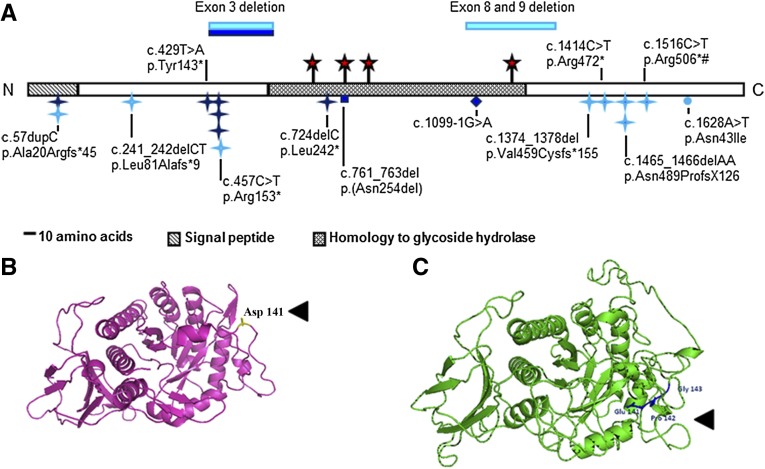
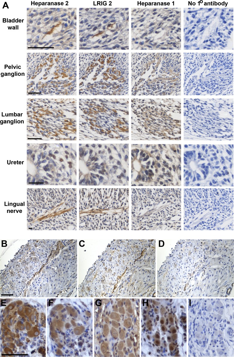
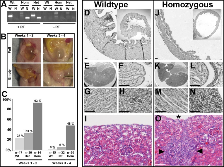
Urofacial syndrome (UFS) is an autosomal recessive congenital disease featuring grimacing and incomplete bladder emptying. Mutations of HPSE2, encoding heparanase 2, a heparanase 1 inhibitor, occur in UFS, but knowledge about the HPSE2 mutation spectrum is limited. Here, seven UFS kindreds with HPSE2 mutations are presented, including one with deleted asparagine 254, suggesting a role for this amino acid, which is conserved in vertebrate orthologs. HPSE2 mutations were absent in 23 non-neurogenic neurogenic bladder probands and, of 439 families with nonsyndromic vesicoureteric reflux, only one carried a putative pathogenic HPSE2 variant. Homozygous Hpse2 mutant mouse bladders contained urine more often than did wild-type organs, phenocopying human UFS. Pelvic ganglia neural cell bodies contained heparanase 1, heparanase 2, and leucine-rich repeats and immunoglobulin-like domains-2 (LRIG2), which is mutated in certain UFS families. In conclusion, heparanase 2 is an autonomic neural protein implicated in bladder emptying, but HPSE2 variants are uncommon in urinary diseases resembling UFS.

Keywords: genetics and development; human genetics; molecular genetics; pediatric nephrology.


Figures

Figure 1.
Figure 1.
HPSE2 mutations in UFS and the variant in primary VUR. (A) Schematic of heparanase 2 showing location of mutations. Dark blue indicates mutations described in this report. Light blue indicates mutations from previous reports. Blue stars, nonsense or frameshift mutations; circle, missense mutation; diamond, splice-site mutation; red stars, predicted N-glycosylation sites; #, founder mutation in Ochoa’s Columbian cohort. Domains were predicted by Pfam and SignalP. N and C, the proteins amino and carboxy terminals, respectively. (B and C) Wild-type full length wild-type heparanase 2 protein (B) and the c.422_423insGCCCGG-p.Asp141delinsGluProGly variant (C). The heparanase 2 sequence was aligned to 51 α-L-arabinofuranosidase with ClustalW, and the structural model was generated using PHYRE2 (http://www.sbg.bio.ic.ac.uk/phyre2/html/page.cgi?id=index).
Figure 2.
Figure 2.
Immunohistochemistry in wild-type mice detects UFS proteins. Sections were counterstained (blue) with hematoxylin. Positive immunodetection signals are brown. (A) Embryonic day 14. Note the similar patterns for heparanase 2 and LRIG2 in a bladder wall nerve, a nascent pelvic ganglion, a nascent lumber ganglion, mesenchymal-like cells in the wall of the proximal (i.e., top of the) ureter, and the lingual nerve. Heparanase 1 was also detected in the ganglia cell bodies but not in the nerve trunks themselves. (B–D) Serial sections of 1-week postnatal bladder immunostained for heparanase 2 (B) or LRIG2 (C). (D) The primary antibody was omitted. Note signals for both proteins in the nerve (running from top right to bottom left). (E–I) Immunohistochemistry of pelvic ganglia flanking the bladder outflow tract 2 weeks after birth: β3-tubulin, a neuronal marker (E); heparanase 2 (F); heparanase 1 (G); LRIG2 (H); negative control with no primary antibody (I). Scale bars are 50 μm.
Figure 3.
Figure 3.
Hpse2 mutant mouse bladder and kidney phenotypes. (A) RT-PCR of RNA from postnatal bladders. Note the abnormal Hpse2 transcript (N, 158 base pairs) in gene trap homozygous (Hom) and heterozygous (Het) tissues. The wild-type Hpse2 transcript (W, 364 base pairs) was the only one present in wild-type (Wt) mice but was not detected in homozygous (Hom) tissue. The right side of the gel shows the experiment when RT was not used. (B) Examples of “full” and “empty” bladder phenotypes in autopsies of mice in the first and second postnatal fortnights. (C) Frequencies of “full” bladder phenotypes in the three genotypes, in the first and second postnatal fortnights (total numbers analyzed are also shown). (D–O) Histology of mutant mouse bladders and kidneys; D–I are wild-type organs and J–O are homozygous mutant organs. All sections were stained with hematoxylin; I and O were also stained with eosin. Wild-type (D) and homozygous Hpse2 mutant (J) bladders at 2 weeks. Note muscle in walls of both organs. When bladders are harvested, urine can escape and organs tend to deflate. Nevertheless, an impression of the difference between homozygous and wild-type bladders is shown in the low power insets (upper right of each frame). Wild-type kidneys at 2 weeks showing sagittal section overview (E) and higher powers of papilla (F), medullary tubules (G), and glomeruli (H). Counterpart zones in littermate homozygous kidney (K–N) are grossly similar to those in wild-type organ. An area of glomerular crowding in a mutant kidney is shown in N (from area marked by asterisk in K). High-power views of outer cortex in 3-week-old kidneys from wild-type (I) and mutant (O) littermate mice. Note the small area (demarcated by arrowheads in O) of glomerular crowding and tubulo-interstitial changes next to a concavity (asterisk) in the organ’s surface. Scale bars are 50 µm.

References

    1. Online Mendelian Inheritance in Man. Available at: http://www.ncbi.nlm.nih.gov/omim. Accessed April 25, 2014
    1. Ochoa B: Can a congenital dysfunctional bladder be diagnosed from a smile? The Ochoa syndrome updated. Pediatr Nephrol 19: 6–12, 2004 - PubMed
    1. Newman WG, Woolf AS, Stuart HM: Urofacial syndrome. GeneReviews 1993–2013, 2013. Available at: http://www.ncbi.nlm.nih.gov/books/NBK154138/. Accessed April 24, 2014
    1. Daly SB, Urquhart JE, Hilton E, McKenzie EA, Kammerer RA, Lewis M, Kerr B, Stuart H, Donnai D, Long DA, Burgu B, Aydogdu O, Derbent M, Garcia-Minaur S, Reardon W, Gener B, Shalev S, Smith R, Woolf AS, Black GC, Newman WG: Mutations in HPSE2 cause urofacial syndrome. Am J Hum Genet 86: 963–969, 2010 - PMC - PubMed
    1. Pang J, Zhang S, Yang P, Hawkins-Lee B, Zhong J, Zhang Y, Ochoa B, Agundez JAG, Voelckel MA, Fisher RB, Gu W, Xiong WC, Mei L, She JX, Wang CY: Loss-of-function mutations in HPSE2 cause the autosomal recessive urofacial syndrome. Am J Hum Genet 86: 957–962, 2010 - PMC - PubMed

Publication types

Supplementary concepts

LinkOut - more resources