

Visualizing life with ambient mass spectrometry
- PMID: 25146170
- PMCID: PMC4297699
- DOI: 10.1016/j.copbio.2014.07.005
Visualizing life with ambient mass spectrometry
Abstract
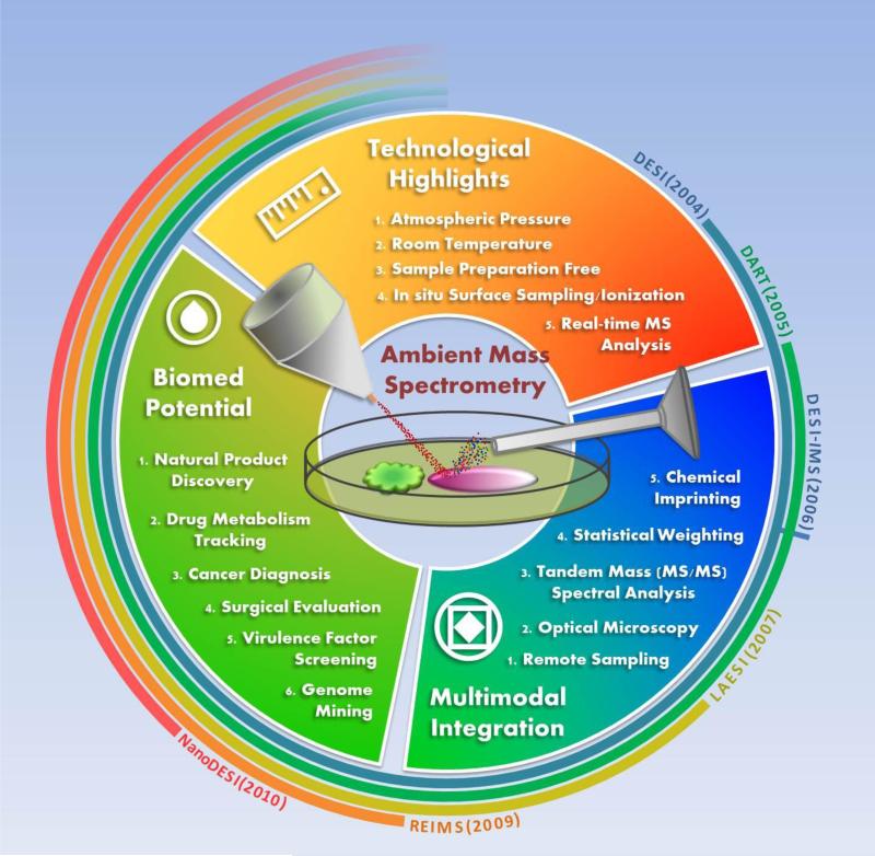
Since the development of desorption electrospray ionization (DESI), many other ionization methods for ambient and atmospheric pressure mass spectrometry have been developed. Ambient ionization mass spectrometry has now been used for a wide variety of biological applications, including plant science, microbiology, neuroscience, and cancer pathology. Multimodal integration of atmospheric ionization sources with the other biotechnologies, as well as high performance computational methods for mass spectrometry data processing is one of the major emerging area's for ambient mass spectrometry. In this opinion article, we will highlight some of the most influential technological advances of ambient mass spectrometry in recent years and their applications to the life sciences.
Copyright © 2014 Elsevier Ltd. All rights reserved.
Figures
Similar articles
-
Mass spectrometry imaging under ambient conditions.Mass Spectrom Rev. 2013 May-Jun;32(3):218-43. doi: 10.1002/mas.21360. Epub 2012 Sep 20. Mass Spectrom Rev. 2013. PMID: 22996621 Free PMC article. Review.
-
Analysis of lipids with desorption atmospheric pressure photoionization-mass spectrometry (DAPPI-MS) and desorption electrospray ionization-mass spectrometry (DESI-MS).J Mass Spectrom. 2012 May;47(5):611-9. doi: 10.1002/jms.2992. J Mass Spectrom. 2012. PMID: 22576874
-
Surface analysis of lipids by mass spectrometry: more than just imaging.Prog Lipid Res. 2013 Oct;52(4):329-53. doi: 10.1016/j.plipres.2013.04.005. Epub 2013 Apr 24. Prog Lipid Res. 2013. PMID: 23623802 Review.
-
Automated ambient desorption-ionization platform for surface imaging integrated with a commercial Fourier transform ion cyclotron resonance mass spectrometer.Anal Chem. 2009 Oct 15;81(20):8479-87. doi: 10.1021/ac901368q. Anal Chem. 2009. PMID: 19761221
-
Clinical Application of Ambient Ionization Mass Spectrometry.Mass Spectrom (Tokyo). 2017;6(Spec Iss):S0060. doi: 10.5702/massspectrometry.S0060. Epub 2017 Feb 24. Mass Spectrom (Tokyo). 2017. PMID: 28337399 Free PMC article.
Cited by
-
Toward a new focus in antibiotic and drug discovery from the Streptomyces arsenal.Front Microbiol. 2015 May 13;6:461. doi: 10.3389/fmicb.2015.00461. eCollection 2015. Front Microbiol. 2015. PMID: 26029195 Free PMC article. Review.
-
Cardiolipins Are Biomarkers of Mitochondria-Rich Thyroid Oncocytic Tumors.Cancer Res. 2016 Nov 15;76(22):6588-6597. doi: 10.1158/0008-5472.CAN-16-1545. Epub 2016 Sep 22. Cancer Res. 2016. PMID: 27659048 Free PMC article.
-
Mass spectrometry tools and workflows for revealing microbial chemistry.Analyst. 2015 Aug 7;140(15):4949-66. doi: 10.1039/c5an00171d. Analyst. 2015. PMID: 25996313 Free PMC article. Review.
-
Deep-ultraviolet laser ablation electrospray ionization mass spectrometry.J Mass Spectrom. 2019 Mar;54(3):281-287. doi: 10.1002/jms.4338. J Mass Spectrom. 2019. PMID: 30675964 Free PMC article.
-
High resolution laser mass spectrometry bioimaging.Methods. 2016 Jul 15;104:118-26. doi: 10.1016/j.ymeth.2016.03.002. Epub 2016 Mar 10. Methods. 2016. PMID: 26972785 Free PMC article. Review.
References
-
- Fenn JB, Mann M, Meng CK, Wong SF, Whitehouse CM. Electrospray ionization for mass spectrometry of large biomolecules. Science. 1989;246:64–71. - PubMed
-
- Hillenkamp F, Karas M, Beavis RC, Chait BT. Matrix-assisted laser desorption/ionization mass spectrometry of biopolymers. Anal. Chem. 1991;63:A1193–A1202. - PubMed
-
- Cooks RG, Ouyang Z, Takáts Z, Wiseman JM. Ambient mass spectrometry. Science. 2006;311:1566–1570. - PubMed
-
- Takáts Z, Wiseman JM, Gologan B, Cooks RG. Mass spectrometry sampling under ambient conditions with desorption electrospray ionization. Science. 2004;306:471–473. - PubMed
Publication types
MeSH terms
Grants and funding
LinkOut - more resources
Full Text Sources
Other Literature Sources
