

Heterogeneous expression pattern of interleukin 17A (IL-17A), IL-17F and their receptors in synovium of rheumatoid arthritis, psoriatic arthritis and osteoarthritis: possible explanation for nonresponse to anti-IL-17 therapy?
- PMID: 25146432
- PMCID: PMC4292832
- DOI: 10.1186/s13075-014-0426-z
Heterogeneous expression pattern of interleukin 17A (IL-17A), IL-17F and their receptors in synovium of rheumatoid arthritis, psoriatic arthritis and osteoarthritis: possible explanation for nonresponse to anti-IL-17 therapy?
Abstract
Introduction: Accumulating evidence suggests an important role for interleukin 17 (IL-17) in the pathogenesis of several inflammatory diseases, including rheumatoid arthritis (RA) and psoriatic arthritis (PsA). Accordingly, clinical trials aimed at blocking IL-17 have been initiated, but clinical results between patients and across different diseases have been highly variable. The objective was to determine the variability in expression of IL-17A, IL-17F and their receptors IL-17RA and IL-17RC in the synovia of patients with arthritis.
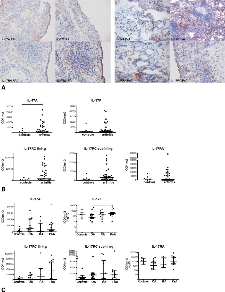
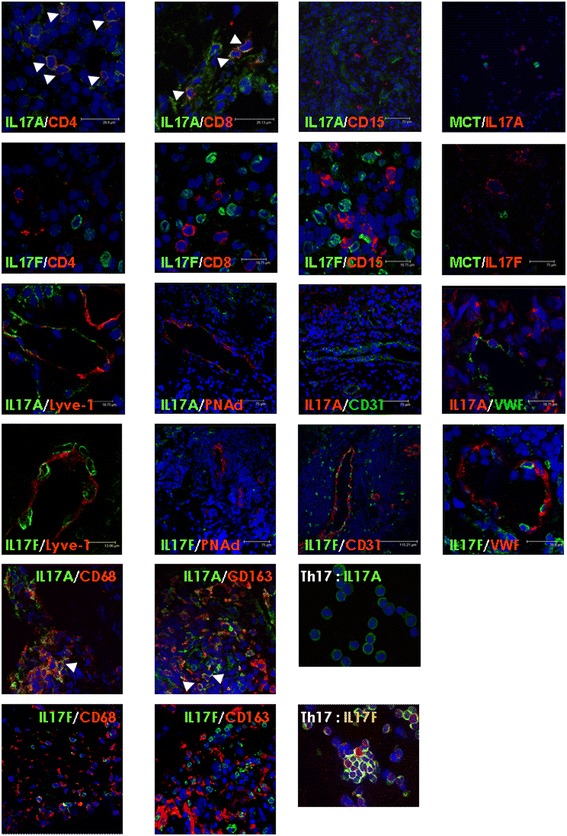
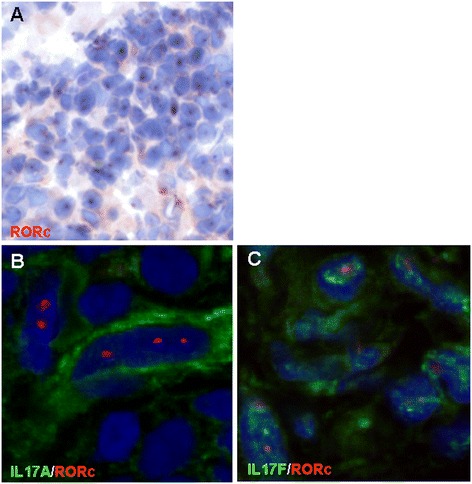
Methods: Synovial biopsies were obtained from patients with RA (n = 11), PsA (n = 15) and inflammatory osteoarthritis (OA, n = 14). For comparison, synovia from noninflamed knee joints (n = 7) obtained from controls were included. Frozen sections were stained for IL-17A, IL-17F, IL-17RA and IL-17RC and evaluated by digital image analysis. We used confocal microscopy to determine which cells in the synovium express IL-17A and IL-17F, double-staining with CD4, CD8, CD15, CD68, CD163, CD31, von Willebrand factor, peripheral lymph node address in, lymphatic vessel endothelial hyaluronan receptor 1, mast cell tryptase and retinoic acid receptor-related orphan receptor γt (RORγt).
Results: IL-17A, IL-17F, IL-17RA and IL-17RC were abundantly expressed in synovial tissues of all patient groups. Whereas IL-17RA was present mostly in the synovial sublining, IL-17RC was abundantly expressed in the intimal lining layer. Digital image analysis showed a significant (P < 0.05) increase of only IL-17A in arthritis patients compared to noninflamed control tissues. The expression of IL-17A, IL-17F and their receptors was similar in the different patient groups, but highly variable between individual patients. CD4+ and CD8+ cells coexpressed IL-17A, and few cells coexpressed IL-17F. IL-17A and IL-17F were not expressed by CD15+ neutrophils. Mast cells were only occasionally positive for IL-17A or IL-17F. Interestingly, IL-17A and IL-17F staining was also observed in macrophages, as well as in blood vessels and lymphatics. This staining probably reflects receptor-bound cytokine staining. Many infiltrated cells were positive for the transcription factor RORγt. Colocalisation between RORγt and IL-17A and IL-17F indicates local IL-17 production.
Conclusions: Increased expression of IL-17A is not restricted to synovial tissues of RA and PsA patients; it is also observed in inflammatory OA. The heterogeneous expression levels may explain nonresponse to anti-IL-17 therapy in subsets of patients.
Figures
References
-
- Nistala K, Adams S, Cambrook H, Ursu S, Olivito B, de Jager W, Evans JG, Cimaz R, Bajaj-Elliott M, Wedderburn LR. Th17 plasticity in human autoimmune arthritis is driven by the inflammatory environment. Proc Natl Acad Sci U S A. 2010;107:14751–14756. doi: 10.1073/pnas.1003852107. - DOI - PMC - PubMed
Publication types
MeSH terms
Substances
LinkOut - more resources
Full Text Sources
Other Literature Sources
Medical
Research Materials
Miscellaneous
