Comment
. 2015 Jan;138(Pt 1):e321.
doi: 10.1093/brain/awu234. Epub 2014 Aug 21.

'Behr syndrome' with OPA1 compound heterozygote mutations

Affiliations
Comment

'Behr syndrome' with OPA1 compound heterozygote mutations

Valerio Carelli et al. Brain. 2015 Jan.
No abstract available


Figures

Figure 1
Figure 1
(A) Reconstruction of the paternal and maternal genealogies of the proband with Behr syndrome (IV-4). The maternal side is remarkable for segregating isolated optic neuropathy in a dominant fashion associated with the heterozygous c.1705+1G>T splicing mutation. (B–D) Ophthalmological investigations. The fundus picture of the proband (IV-4) reveals severe optic atrophy with bilateral pale optic disc (B). The fundus picture of the mother (III-4) shows only a mild optic atrophy with temporal pallor of the optic discs bilaterally (C). Optical coherence tomography examination of retinal nerve fibre layer (RNFL) thickness in the proband shows a diffuse reduction of thickness compatible with the severe optic atrophy (D). (E–G) Semithin cross-section of the sural nerve biopsy. A marked depletion of large myelinated axons affects the sural nerve (E and F). At higher magnification some axons appear to have a thin myelin sheath related to the axonal diameter (G). (H–L) Brain MRI and 1HMR spectroscopy studies. On the left (H) the sagittal FSPGR (Fast SPoiled GRadient echo) T1 shows a mild cerebellar atrophy in the proband (IV-4), and on the right (I) a mild pathological accumulation of lactate (rectangle) was detected in the CSF of lateral ventriculi. On the axial FLAIR T2 sequence no signal changes were detected in the proband (IV-4) (J), whereas few focal hyperintensities, related to not specific areas of gliosis, in the cerebral white matter of the proband’s father (III-3) (K) and mother (III-4) (L) were present. TEMP = temporal; SUP = superior; NAS = nasal; INF = inferior; MRI = magnetic resonance imaging; HMR = proton magnetic resonance; CSF = cerebrospinal fluid; FLAIR = Fluid attenuated inversion recovery.
Figure 2
Figure 2
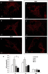
(A–F) Mitochondrial network morphology. Control (A) and patient (B–F) fibroblasts were grown in Dulbecco’s modified Eagle’s medium-glucose and loaded with Mitotracker Red as described in Zanna et al. (2008). Representative out of eight similar images are shown for each individual. (G) ATP synthesis. Fibroblasts were treated with 50 µg/ml digitonin, the rate of ATP synthesis was measured as described in Zanna et al. (2008) and was normalized for citrate synthase (CS) activity. Data (mean ± SEM) were obtained from five controls and the three OPA1 mutated fibroblasts. The experiment was performed at least in triplicate. Asterisk denotes values significantly different (P < 0.05) by Kruskal-Wallis one-way ANOVA on Ranks. GPD = Glyceraldehyde 3-phosphate dehydrogenase.

Comment in

Comment on

  • Multi-system neurological disease is common in patients with OPA1 mutations.
    Yu-Wai-Man P, Griffiths PG, Gorman GS, Lourenco CM, Wright AF, Auer-Grumbach M, Toscano A, Musumeci O, Valentino ML, Caporali L, Lamperti C, Tallaksen CM, Duffey P, Miller J, Whittaker RG, Baker MR, Jackson MJ, Clarke MP, Dhillon B, Czermin B, Stewart JD, Hudson G, Reynier P, Bonneau D, Marques W Jr, Lenaers G, McFarland R, Taylor RW, Turnbull DM, Votruba M, Zeviani M, Carelli V, Bindoff LA, Horvath R, Amati-Bonneau P, Chinnery PF. Yu-Wai-Man P, et al. Brain. 2010 Mar;133(Pt 3):771-86. doi: 10.1093/brain/awq007. Epub 2010 Feb 15. Brain. 2010. PMID: 20157015 Free PMC article.

References

    1. Amati-Bonneau P, Valentino ML, Reynier P, Gallardo ME, Bornstein B, Boissière A, et al. OPA1 mutations induce mitochondrial DNA instability and optic atrophy ‘plus' phenotypes. Brain. 2008;131:338–51. - PubMed
    1. Anikster Y, Kleta R, Shaag A, Gahl WA, Elpeleg O. Type III 3-methylglutaconic aciduria (optic atrophy plus syndrome, or Costeff optic atrophy syndrome): identification of the OPA3 gene and its founder mutation in Iraqi Jews. Am J Hum Genet. 2001;69:1218–24. - PMC - PubMed
    1. Behr C. Die komplizierte, hereditar-familiare Optikusatrophie des Kindesalters – ein bisher nicht beschriebener Symptomkomplex. Klin Mbl Augenheilkd. 1909;47:138–60.
    1. Bonifert T, Karle KN, Tonagel F, Batra M, Wilhelm C, Theurer Y, et al. Pure and syndromic optic atrophy explained by deep intronic OPA1 mutations and an intralocus modifier. Brain. 2014;197:2164–77. - PMC - PubMed
    1. Costeff H, Gadoth N, Apter N, Prialnic M, Savir H. A familial syndrome of infantile optic atrophy, movement disorder, and spastic paraplegia. Neurology. 1989;39:595–7. - PubMed

Substances